. 2001 Oct;75(19):9357-66.
doi: 10.1128/JVI.75.19.9357-9366.2001.

Human immunodeficiency virus type 1 N-terminal capsid mutants that exhibit aberrant core morphology and are blocked in initiation of reverse transcription in infected cells

Affiliations

Human immunodeficiency virus type 1 N-terminal capsid mutants that exhibit aberrant core morphology and are blocked in initiation of reverse transcription in infected cells

S Tang et al. J Virol. 2001 Oct.

Abstract

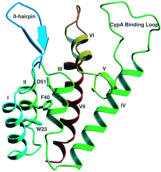
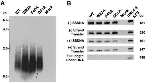
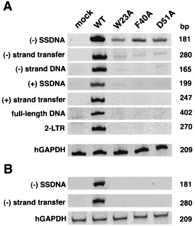
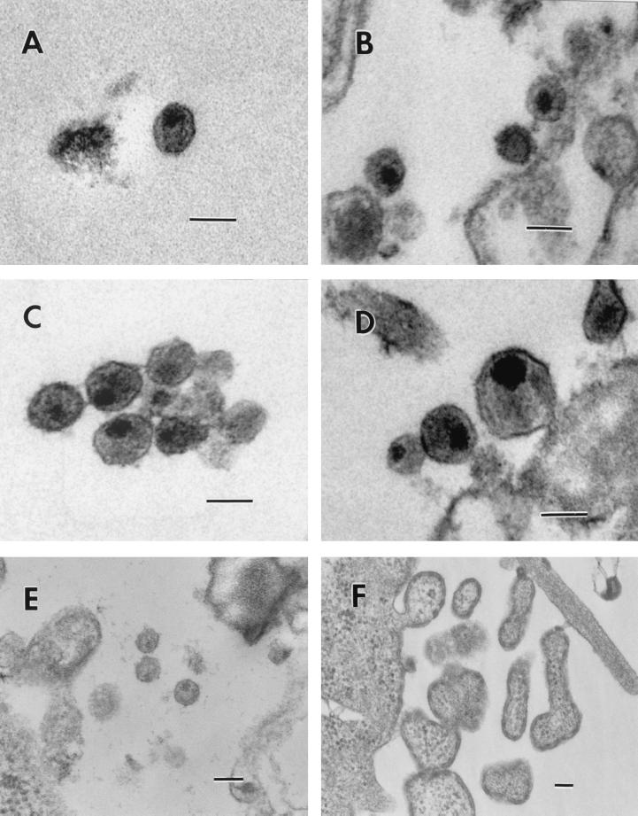
A group of conserved hydrophobic residues faces the interior of the coiled-coil-like structure within the N-terminal domain of the human immunodeficiency virus type 1 (HIV-1) capsid protein (CA). It has been suggested that these residues are important for maintaining stable structure and functional activity. To investigate this possibility, we constructed two HIV-1 clones, in which Trp23 or Phe40 was changed to Ala. We also constructed a third mutant, D51A, which has a mutation that destroys a salt bridge between Pro1 and Asp51. All three mutants are replication defective but produce virus particles. Mutant virions contain all of the viral proteins, although the amount and stability of CA are decreased and levels of virion-associated integrase are reduced. The mutations do not affect endogenous reverse transcriptase activity; however, the mutants are blocked in their ability to initiate reverse transcription in infected cells and no minus-strand strong-stop DNA is detected. The defect in reverse transcription is associated with striking defects in the morphology of mutant virus cores, as determined by transmission electron microscopy. Our data indicate that the mutations made in this study disrupt CA structure and prevent proper maturation of virus cores. We propose that this results in a defect in core stability or in an early postentry event preceding reverse transcription.


Figures

FIG. 1
FIG. 1
Ribbon representation showing a front view of the N-terminal core domain of HIV-1 CA. The β-hairpin structure, CypA binding loop, and seven α-helices are labeled (20, 38). Note that the helices are numbered as described by Gitti et al. (20). The three residues mutated in this study, W23, F40, and D51, are located in helices I, II, and III, respectively. The diagram was generated with Molscript (32).
FIG. 2
FIG. 2
Analysis of mutant and wild-type (WT) cell- and virion-associated proteins. HeLa cells were transfected with the indicated pNL4-3 molecular clones (2). (A and B, top) Detection of [35S]Cys-labeled cell- and virion-associated proteins, respectively. Lysates were immunoprecipitated with AIDS patient serum and separated by SDS-PAGE in 10% gels; radioactive bands were visualized by fluorography. The positions of the precursor and mature viral proteins are indicated. (B, bottom) Detection of unlabeled HIV-1 CA bands by Western blot analysis. Unlabeled viral lysates were subjected to SDS-PAGE in a 10% minigel and were then analyzed by Western blotting (see Materials and Methods). Blots were sequentially probed with primary rabbit antibodies against HIV-1 CA and with secondary anti-rabbit IgG antibodies; the protein bands were visualized by chemiluminescence. Note that, since a minigel was used for the Western blot analysis, the CA and CA-related bands were not separated as well as in the gel of the virion-associated immunoprecipitated proteins (B, top).
FIG. 3
FIG. 3
Endogenous RT activities of pNL4-3KFS wild-type (WT) and mutant virions. (A) 32P-labeled viral DNA products made by wild-type and mutant virus particles. Incubation conditions and methods for the analysis of the DNA products by electrophoresis in a denaturing agarose gel are given in Materials and Methods. 32P-labeled λ HindIII size markers are shown on the left. (B) PCR analysis of unlabeled DNA products made by mutant and wild-type virus particles. The DNA products were amplified using appropriate primer sets, as described in Materials and Methods. The negative control (Mock) is a reaction mixture containing the reaction buffer but not virus. One nanogram of pNL4-3KFS DNA (10, 13) was used as a positive control. The size of each PCR product is shown on the right; the specific product amplified is indicated on the left.
FIG. 4
FIG. 4
PCR analysis of viral DNA in infected cells. Virus stocks obtained by cotransfection of 293T cells with pNL4-3KFS (10, 13) molecular clones and pSV-A-MLVenv (33) were normalized for RT activity and used to infect H9 cells (A) and CD4-positive HeLa indicator cells (29) (B). Isolation of viral DNA from each cell line including trypsin treatment to remove bound virions from the HeLa cells, PCR analysis, and electrophoretic separation of the products are described in Materials and Methods. To control for the amount of total DNA in each sample, hGAPDH sequences (46) were amplified from the same samples used to analyze viral DNA products. The size of each PCR product is shown on the right; the specific product amplified is indicated on the left. Mock, DNA from uninfected cells; WT, wild type.
FIG. 5
FIG. 5
Electron micrographs of wild-type and CA mutant viral particles. (A and B) Wild type; (C and D) D51A; (E) F40A; (F) W23A. Scale bars, 100 nm (all panels).

References

    1. Accola M A, Öhagen Å, Göttlinger H G. Isolation of human immunodeficiency virus type 1 cores: retention of Vpr in the absence of p6gag. J Virol. 2000;74:6198–6202. - PMC - PubMed
    1. Adachi A, Gendelman H E, Koenig S, Folks T, Willey R, Rabson A, Martin M A. Production of acquired immunodeficiency syndrome-associated retrovirus in human and nonhuman cells transfected with an infectious molecular clone. J Virol. 1986;59:284–291. - PMC - PubMed
    1. Alin K, Goff S P. Amino acid substitutions in the CA protein of Moloney murine leukemia virus that block early events in infection. Virology. 1996;222:339–351. - PubMed
    1. Bowzard J B, Wills J W, Craven R C. Second-site suppressors of Rous sarcoma virus CA mutations: evidence for interdomain interactions. J Virol. 2001;75:6850–6856. - PMC - PubMed
    1. Braaten D, Franke E K, Luban J. Cyclophilin A is required for an early step in the life cycle of human immunodeficiency virus type 1 before the initiation of reverse transcription. J Virol. 1996;70:3551–3560. - PMC - PubMed

Publication types

Substances