. 2001 Oct;75(19):9407-14.
doi: 10.1128/JVI.75.19.9407-9414.2001.

Identification of a central DNA flap in feline immunodeficiency virus

Affiliations

Identification of a central DNA flap in feline immunodeficiency virus

T Whitwam et al. J Virol. 2001 Oct.

Abstract

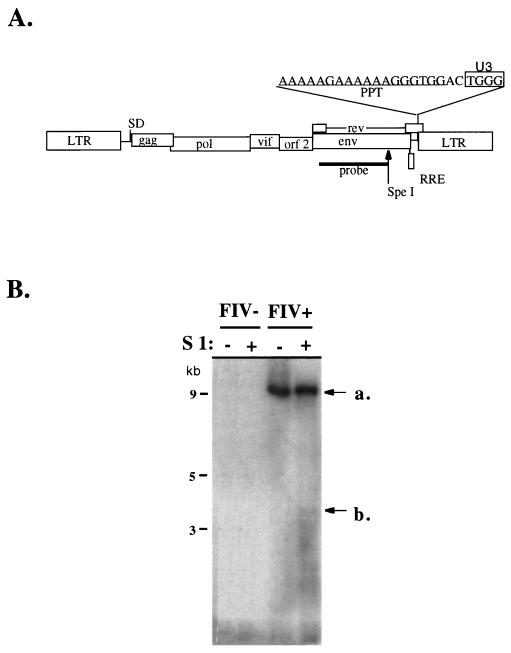
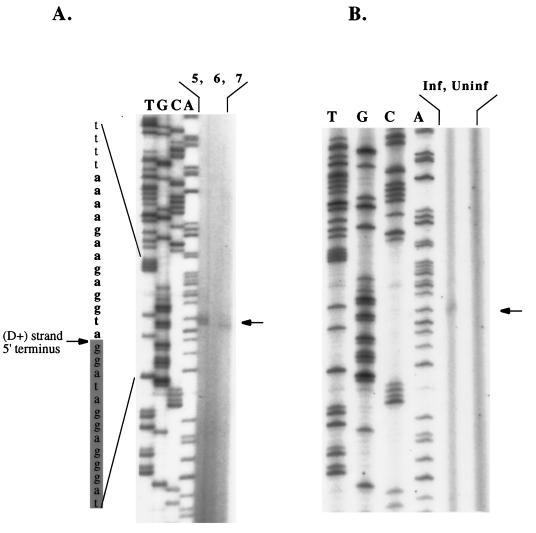
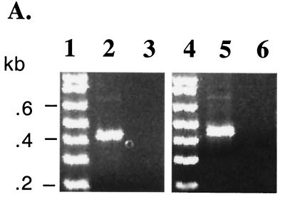
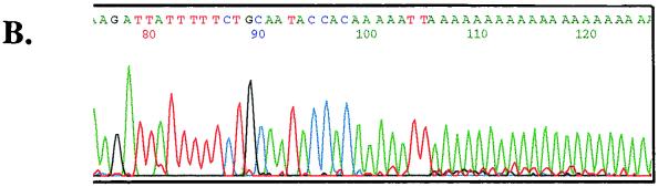
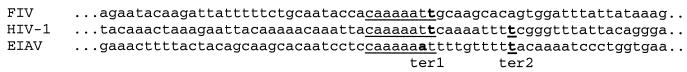
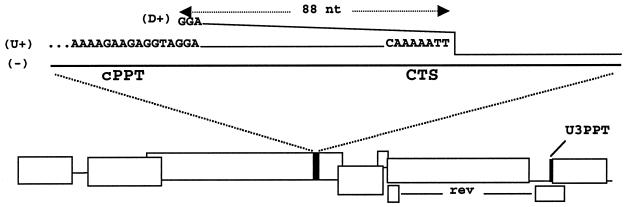
A duplication of the polypurine tract (PPT) at the center of the human immunodeficiency virus type 1 (HIV-1) genome (the cPPT) has been shown to prime a separate plus-strand initiation and to result in a plus-strand displacement (DNA flap) that plays a role in nuclear import of the viral preintegration complex. Feline immunodeficiency virus (FIV) is a lentivirus that infects nondividing cells, causes progressive CD4(+) T-cell depletion, and has been used as a substrate for lentiviral vectors. However, the PPT sequence is not duplicated elsewhere in the FIV genome and a central plus-strand initiation or strand displacement has not been identified. Using Southern blotting of S1 nuclease-digested FIV preintegration complexes isolated from infected cells, we detected a single-strand discontinuity at the approximate center of the reverse-transcribed genome. Primer extension analyses assigned the gap to the plus strand, and mapped the 5' terminus of the downstream (D+) segment to a guanine residue in a purine-rich tract in pol (AAAAGAAGAGGTAGGA). RACE experiments then mapped the 3' terminus of the upstream plus (U+)-strand segment to a T nucleotide located 88 nucleotides downstream of the D+ strand 5' terminus, thereby identifying the extent of D+ strand displacement and the central termination sequence of this virus. Unlike HIV, the FIV cPPT is significantly divergent in sequence from its 3' counterpart (AAAAAAGAAAAAAGGGTGG) and contains one and in some cases two pyrimidines. An invariant thymidine located -2 to the D+ strand origin is neither required nor optimal for codon usage at this position. Although the mapped cPPTs of FIV and HIV-1 act in cis, they encode homologous amino acids in integrase.


Figures

FIG. 1
FIG. 1
S1 nuclease analysis of LMW DNA from FIV-infected and uninfected cells. (A) Structure of provirus showing the sequence of the U3PPT (underlined) and the nearby sequence. U3 element nucleotides present in integrated proviruses are boxed. The Southern blot probe used in the S1 nuclease analysis (Fig. 1B) is illustrated. The SpeI site is unique. (B) Southern blot of S1 nuclease-digested and undigested unintegrated DNA from infected and uninfected cells. A band is detectable at ca. 3.3 kb in S1 nuclease-digested Hirt DNA from infected cells. The band is not present in the absence of S1 nuclease treatment in Hirt DNAs from infected cells or in either uninfected cell Hirt DNA.
FIG. 2
FIG. 2
Primer extension analysis identifies the 5′ terminus of the D+ strand. The deduced D+ strand is shaded gray at left. The stop was detected in each of three separate primer extension reactions, which are shown in lanes 5 and 7 in panel A and in the second lane from right in panel B. See the text for discussion of the doublet in panel A, lane 5. A termination band was not seen in control, uninfected cell LMW DNAs analyzed in parallel (A, lane 6, and B, right lane).
FIG. 3
FIG. 3
Identification of the CTS and alignment with other lentiviruses. (A) Heminested PCRs. Hirt DNA from infected cells (lanes 2 and 5) and uninfected cells (lanes 3 and 6) was homopolymerically tailed with dATP and terminal deoxynucleotide transferase prior to the PCR. PCR was performed with a 90-s extension time using an antisense oligo(dT)-containing primer and the following 5′ nesting primers upstream of the identified cPPT sequence (nt 4959 to 4974): an outer sense primer beginning at nt 4576 in the first round and an inner primer at either nt 4702 (left panel) or nt 4674 (right panel) in the second round. The size of the single bands produced places the site of joining of the oligo(dA) tail ca. 80 to 120 nt downstream of the cPPT depending on the exact site of oligo(dT) annealing to the tail. A very faint larger band of between 600 and 700 bp, possibly representing a minor U+ strand termination, can be seen in both panels but was not detected in any sequenced clones. (B) The site of attachment of the poly(dA) tail by terminal deoxynucleotide transferase unambiguously identifies the 3′ terminus of the U+ strand. An electropherogram from 1 of the 24 clones sequenced is shown. The CTS in the pol gene is CA5T2. No other terminations were present in 24 clones (12 were sequenced from each of the two separate amplifications shown in left and right panels of Fig. 3A).
FIG. 3
FIG. 3
Identification of the CTS and alignment with other lentiviruses. (A) Heminested PCRs. Hirt DNA from infected cells (lanes 2 and 5) and uninfected cells (lanes 3 and 6) was homopolymerically tailed with dATP and terminal deoxynucleotide transferase prior to the PCR. PCR was performed with a 90-s extension time using an antisense oligo(dT)-containing primer and the following 5′ nesting primers upstream of the identified cPPT sequence (nt 4959 to 4974): an outer sense primer beginning at nt 4576 in the first round and an inner primer at either nt 4702 (left panel) or nt 4674 (right panel) in the second round. The size of the single bands produced places the site of joining of the oligo(dA) tail ca. 80 to 120 nt downstream of the cPPT depending on the exact site of oligo(dT) annealing to the tail. A very faint larger band of between 600 and 700 bp, possibly representing a minor U+ strand termination, can be seen in both panels but was not detected in any sequenced clones. (B) The site of attachment of the poly(dA) tail by terminal deoxynucleotide transferase unambiguously identifies the 3′ terminus of the U+ strand. An electropherogram from 1 of the 24 clones sequenced is shown. The CTS in the pol gene is CA5T2. No other terminations were present in 24 clones (12 were sequenced from each of the two separate amplifications shown in left and right panels of Fig. 3A).
FIG. 4
FIG. 4
The identified FIV CTS. The CTS is aligned with those of the two other viruses for which a CTS has been established, i.e., HIV-1 (9) and EIAV (32). Termination nucleotides are in boldface. There is no significant nucleotide sequence homology downstream of the FIV cPPT except at the CTS (CA5T2, underlined), which corresponds to ter1. FIV lacks a sequence equivalent to the ter2 sites reported for HIV-1 and EIAV, a finding which is consistent with the single U+ strand 3′ terminus established in the present study.
FIG. 5
FIG. 5
Structure of the central DNA flap in FIV. The minus, U+, and D+ strands are labeled, and relevant nucleotides are shown. The region of overlap is 88 nt, extending from the 5′ end of the D+ strand to the 3′ terminus of the U+ strand.

Similar articles

Cited by

References

    1. Barr M C, Zou L, Holzschu D L, Phillips L, Scott F W, Casey J W, Avery R J. Isolation of a highly cytopathic lentivirus from a nondomestic cat. J Virol. 1995;69:7371–7374. - PMC - PubMed
    1. Blum H E, Harris J D, Ventura P, Walker D, Staskus K, Retzel E, Haase A T. Synthesis in cell culture of the gapped linear duplex DNA of the slow virus visna. Virology. 1985;142:270–277. - PubMed
    1. Bouyac-Bertoia M, Dvorin J, Fouchier R, Jenkins Y, Meyer B, Wu L, Emerman M, Malim M H. HIV-1 infection requires a functional integrase NLS. Mol Cell. 2001;7:1025–1035. - PubMed
    1. Bukrinsky M I, Sharova N, Dempsey M P, Stanwick T L, Bukrinskaya A G, Haggerty S, Stevenson M. Active nuclear import of human immunodeficiency virus type 1 preintegration complexes. Proc Natl Acad Sci USA. 1992;89:6580–6584. - PMC - PubMed
    1. Burns J C, Friedmann T, Driever W, Burrascano M, Yee J K. Vesicular stomatitis virus G glycoprotein pseudotyped retroviral vectors: concentration to very high titer and efficient gene transfer into mammalian and nonmammalian cells. Proc Natl Acad Sci USA. 1993;90:8033–8037. - PMC - PubMed

Publication types