. 2001 Oct;75(19):9415-26.
doi: 10.1128/JVI.75.19.9415-9426.2001.

RING finger Z protein of lymphocytic choriomeningitis virus (LCMV) inhibits transcription and RNA replication of an LCMV S-segment minigenome

Affiliations

RING finger Z protein of lymphocytic choriomeningitis virus (LCMV) inhibits transcription and RNA replication of an LCMV S-segment minigenome

T I Cornu et al. J Virol. 2001 Oct.

Abstract

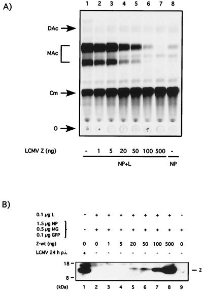
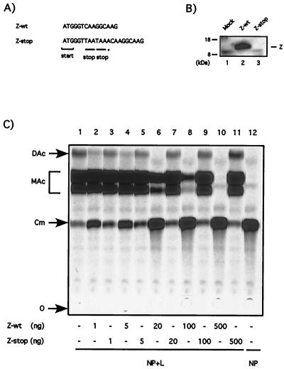
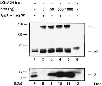
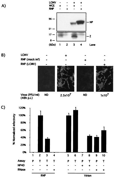
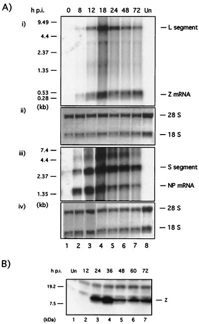
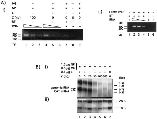
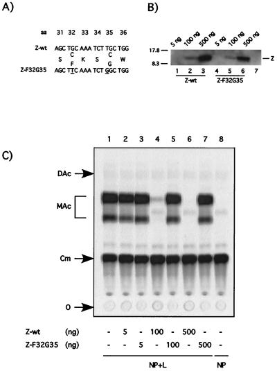
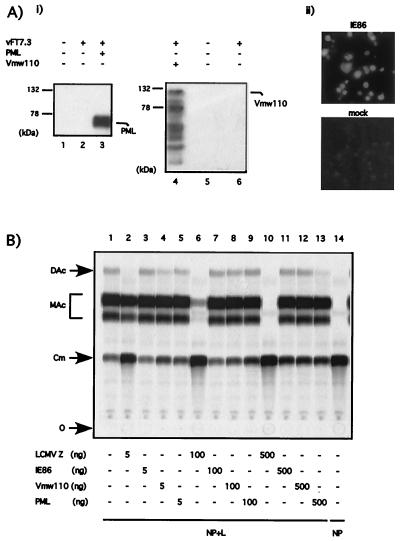
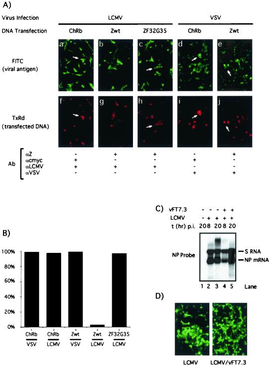
Arenaviruses have a bisegmented negative-strand RNA genome whose proteomic capability is limited to only four polypeptides, namely, nucleoprotein (NP), surface glycoprotein (GP) that is proteolytically processed into GP1+GP2, polymerase (L), and a small (11-kDa) RING finger protein (Z). The role of Z during the Lymphocytic choriomeningitis virus (LCMV) life cycle is poorly understood. We investigated the function of Z in virus transcription and replication by using a reverse genetic system for the prototypic arenavirus LCMV. This system involves an LCMV minigenome and the minimal viral trans-acting factors (NP and L), expressed from separated cotransfected plasmids. Cotransfection of the Z cDNA strongly inhibited LCMV minigenome expression. The effect required synthesis of Z protein; its magnitude was dose dependent and occurred with levels of Z protein substantially lower than those observed in LCMV-infected cells. Coexpression of Z did not prevent the encapsidation of plasmid supplied minigenome, but it affected both transcription and RNA replication similarly. Mutations in Z that unfolded its RING finger domain eliminated its inhibitory activity, but RING proteins not related to Z did not affect LCMV minigenome expression. Consistent with the minigenome results, cells transiently expressing Z exhibited decreased susceptibility to infection with LCMV.


Figures

FIG. 1
FIG. 1
LCMV Z inhibits CAT expression by the LCMVSCAT2 minigenome in a dose-dependent manner. BHK-21 cells were infected with vTF7.3 (MOI = 3) and subsequently cotransfected with 0.5 μg of pLCMVSCAT2, 1.5 μg of pCITE-NP, 0.1 μg of pGEM-L, 0.1 μg of pTMI-GFP, and increasing amounts of pUCIRES-Z as indicated. Plasmid pGEM-L was not added in lane 8. In all of the samples the total amount of DNA was kept constant (2.7 μg) by adding the appropriate amount of plasmid pTMI. The use of pTMI-GFP allowed us to determine the efficiency of transfection, based on number of GFP-positive cells, prior harvesting of the cells for CAT assays, and protein expression analysis. (A) LCMVSCAT2 minigenome expression. At 24 h after transfection, cell lysates were prepared for measuring CAT activity as described in Material and Methods. (B) Z protein expression. Cell lysates were subjected to SDS-PAGE, and the levels of Z protein expression were determined by Western blot using a rabbit antibody to Z. Lysate applied in lane 1 was prepared from cells infected with LCMV (MOI = 3) at 24 h p.i. The position of the Z protein is indicated on the right, and the molecular size markers are indicated on the left. O, origin; Cm, chloramphenicol; MAc, monoacetylated chloramphenicol; DAc, diacetylated chloramphenicol.
FIG. 2
FIG. 2
Inhibitory effect of the Z plasmid requires synthesis of the Z protein. (A) Nucleotide sequence differences between Z-wt and the Z-stop mutant. The Z-stop mutant contains two stop codons (underlined) inserted downstream of the ATG start codon. The insertion of an additional A (✽) into the Z-stop construct created a frameshift of the downstream reading frame. (B) The Z-stop mutant is unable to produce the Z protein. BHK-21 cells were infected with vTF7.3 (MOI = 3) and transfected with 0.5 μg of pUCIRES-Z or 0.5 μg of pUCIRESZ-stop. Lysates were prepared 24 h after transfection and subjected to SDS-PAGE. Z protein expression was detected by Western blotting with a rabbit antiserum to Z. The position of the Z protein is indicated on the right, and the molecular size markers are indicated on the left. (C) The Z-stop mutant does not inhibit CAT activity by the LCMVSCAT2 minigenome. BHK-21 cells were infected with vTF7.3 (MOI = 3) and cotransfected with 0.5 μg of pLCMVSCAT2, 1.5 μg of pCITE-NP, 0.1 μg of pGEM-L, 0.1 μg of pTMI-GFP, and increasing amount of pUCIRES-Z or pUCIRESZ-stop as indicated. Plasmid pGEM-L was not added in lane 12. In all of the samples the amount of DNA was kept constant by adding plasmid pTMI to 2.7 μg. At 24 h after transfection, the samples were assayed for CAT activity as described in Materials and Methods. O, origin; Cm, chloramphenicol; MAc, monoacetylated chloramphenicol; DAc, diacetylated chloramphenicol.
FIG. 3
FIG. 3
Expression of Z protein does not affect plasmid-derived expression of NP or L. Equal amounts of cell lysates were separated either on an SDS–10% PAGE gel for the detection of NP and L protein expression or on an SDS–16% PAGE gel to detect Z protein expression. Lysates in lanes 2 to 5 and lanes 8 to 11 were prepared from cells infected with vTF7.3 (MOI = 3) and transfected with 1 μg of pCITE-NP and 1 μg of pGEM-L. The amount of cotransfected pUCIRES-Z is indicated at the top. Lysate prepared from mock-infected cells was loaded in lanes 6 and 12, whereas lysate from LCMV-infected cells was loaded in lanes 1 and 7. The positions of the L, NP, and Z protein are indicated on the right, and the molecular size markers are indicated on the left. Consistent with previous findings, L protein was not detected by Western blotting in the cell lysates from LCMV-infected cells (33).
FIG. 4
FIG. 4
Biochemical and biological characterization of LCMV RNP. (A) Levels of Z associated with LCMV RNP. Whole-cell extracts (lanes 1 and 3) and RNP (lanes 2 and 4) were prepared from mock (lanes 1 and 2) and LCMV-infected (lanes 3 and 4) cells and analyzed by Western blotting using a guinea pig antiserum to LCMV and a rabbit antiserum to Z. The amount of RNP loaded in lane 4 corresponded to a sixfold-higher amount (cell equivalent) of that loaded in lane 3. (B) Infectivity of LCMV RNP. Cells were transfected with RNP prepared from mock- and LCMV-infected cells. As controls, cells were also either LCMV or mock infected. At 48 h p.i. or posttransfection, cells were fixed and analyzed by immunofluorescence by using a guinea pig polyclonal antiserum to LCMV. Levels of infectious LCMV in the corresponding supernatants (48 h p.i.) were determined by plaque assay (18). (C) Characterization of infectivity associated with LCMV RNP. LCMV RNP (lanes 1 to 4) or virions (lanes 5 to 10) were treated with NP-40 (0.05% for 15 min on ice) or pancreatic RNase (25 μg for 30 min at 20°C) or were left untreated. Infectivity associated with treated and untreated virions and RNP was assayed by direct plaque assay (P) or by transfection (T). RNP-associated infectivity was normalized to that obtained by transfection with untreated RNP. Virion-associated infectivity was normalized to that obtained by direct plaque assay with untreated virions. Three independent assays were done. Average values and standard deviations are shown.
FIG. 5
FIG. 5
Kinetic of Z expression during the natural course of LCMV infection. (A) Kinetic of Z mRNA synthesis. BHK-21 cells were infected with LCMV (MOI = 3), and total cell RNA isolated at the indicated time points was analyzed by Northern blot hybridization. For each time point, equal amounts of RNA were loaded in two different gels. Lane 7, in both gels, corresponds to RNA from mock-infected cells. Both gels were identically processed for Northern blot analysis. One membrane was used to detect Z mRNA and L segment by using a [32P]dCTP labeled Z-DNA probe (panel i). The other membrane was used to detect NP mRNA and the S segment using a [32P]dCTP labeled NP DNA probe (panel iii). Molecular markers are indicated on the right. Prior to hybridization, each membrane was stained with methylene blue to assess the total amount of RNA in each sample based on levels of 28S and 18S RNA (panels ii and iv). (B) Kinetics of Z protein expression. BHK-21 cells were infected with LCMV (MOI = 3) and harvested at the indicated time points. Cell lysates were subjected to SDS-PAGE and analyzed for the expression of Z protein by Western blotting with a rabbit antibody to Z. Lane 1 corresponds to mock-infected cells.
FIG. 6
FIG. 6
Effect of Z on encapsidation and RNA synthesis of LCMV S-segment minigenome. (A) Z does not inhibit encapsidation of plasmid supplied LCMVSCAT2 minigenome. (i) BHK-21 cells (106 cells/well, six-well plate) were infected with vTF7.3 (MOI = 3) and cotransfected with 0.5 μg of pLCMVSCAT2, 1.5 μg of pCITE-NP, 0.1 μg of pGEM-L, and 0.1 μg of pTMI-GFP. Plasmids expressing L and NP were omitted in lanes 7 and 8, respectively. Samples from lanes 1 to 3 were also transfected with 100 ng of pUCIRES-Z. In all of the samples the amount of DNA was kept constant (2.7 μg) by adding plasmid pTMI. At 24 h after transfection, cells (three wells/sample) were harvested and RNP was obtained as described in Materials and Methods. Encapsidated RNA was isolated and subjected to semiquantitative RT-PCR by using decreasing amounts of input RNA: 1/4, 1/8, and 1/16 of the total RNA were used in lanes 1 to 3 and lanes 4 to 6. In lanes 7 and 8, 1/8 of the total RNA was amplified. In lane 9, 1/4 of total RNA was analyzed, omitting the RT step as a control for plasmid DNA contamination in the RNP preparation. Molecular size markers are indicated on the left. MG refers to minigenome LCMVSCAT2. (ii) Bona fide LCMV RNPs were prepared from LCMV-infected BHK-21 cells (3 × 10⁶ cells) at 48 h p.i. (MOI = 3). Encapsidated RNA was isolated and analyzed by semiquantitative RT-PCR as described in Materials and Methods. In this case, primers used for PCR amplified a segment (ca. 350 bp) of the S segment corresponding to NP sequences. (B) Effect of Z protein on RNA synthesis mediated by the LCMV polymerase. BHK-21 cells were infected with vTF7.3 (MOI = 3) and cotransfected with 0.5 μg of pLCMVSCAT2, 1.5 μg of pCITE-NP, 0.1 μg of pGEM-L, 0.1 μg of pTMI-GFP, and increasing amounts of pUCIRES-Z as indicated. Plasmid pGEM-L was not added in lane 7. Total cellular RNA was isolated, and equal amounts (5 μg) were analyzed by Northern blot hybridization. (i) Subgenomic CAT mRNA and full-length antigenomic RNA species were detected with a [32P]UTP AS CAT riboprobe. Molecular size markers are indicated on the left. (ii) Methylene blue staining of the membrane was done to determine the positions and levels of the 28S and 18S RNA.
FIG. 7
FIG. 7
Disruption of the RING finger motif annuls the inhibitory effect of the Z protein. (A) Amino acid changes introduced in the RING domain of mutant Z-F32G35. The RING finger mutant F32G35 contains two nucleotide changes at codon positions 32 and 35 that disrupt the RING finger motif by changing amino acid residues from C to F and from C to G, respectively. (B) The RING finger mutant Z F32G35 is stably expressed. BHK-21 cells were infected with vTF7.3 (MOI = 3) and transfected with the indicated amounts of pUCIRES-Z or pUCIRESZ-F32G35 plasmid DNA. Lane 7 shows results for mock-transfected cells. Lysates were subjected to SDS-PAGE, and immunodetection of Z proteins was performed with a rabbit anti-Z antibody. The position of Z is indicated on the right, and the molecular size markers are indicated on the left. (C) Mutant Z-F32G35 does not inhibit CAT activity mediated by the LCMVSCAT2 minigenome. Monolayers of BHK-21 cells were infected with vTF7.3 (MOI = 3) and cotransfected with 0.5 μg of pLCMVSCAT2, 1.5 μg of pCITE-NP, 0.1 μg of pGEM-L, 0.1 μg of pTMI-GFP, and increasing amounts of pUCIRES-Z and pUCIRESZ-F32G35, as indicated at the bottom. Plasmid pGEM-L was not added in lane 8. In all of the samples, the amount of DNA was kept constant by adding plasmid pTMI to 2.7 μg. Samples were assayed for CAT activity as described in Materials and Methods. O, origin; Cm, chloramphenicol; MAc, monoacetylated chloramphenicol; DAc, diacetylated chloramphenicol.
FIG. 8
FIG. 8
LCMVSCAT2 minigenome expression is not inhibited by RING finger proteins unrelated to Z. (A) Expression of viral (Vmw110 and IE86) and cellular (PML) RING finger proteins in BHK cells. BHK-21 cells were infected with vTF7.3 (MOI = 3) and transfected with 0.5 μg of RMX-PML (panel i, lane 3), 0.5 μg of pT7-110 (panel ii, lane 4), or 0.5 μg of pGEM-IE86 (panel ii). Protein expression was assayed by Western blotting with either a rabbit anti-PML antibody (panel i, lanes 1 to 3) or a mouse anti-Vmw110 antibody (panel i, lanes 4 to 6) or by immunofluorescence with a mouse anti-IE86 antibody (panel ii). The positions of PML and Vmw110 are indicated on the right, and the molecular size markers are indicated on the left. (B) Inhibition of CAT expression by the LCMVSCAT2 minigenome is not observed with RING finger proteins unrelated to Z. BHK-21 cells were cotransfected with 0.5 μg of pLCMVSCAT2, 1.5 μg of pCITE-NP, 0.1 μg of pGEM-L, 0.1 μg of pTMI-GFP, and increasing amount of pUCIRES-Z, pGEM-IE86, pT7-110, or RMX-PML as indicated at the bottom of the figure. Plasmid pGEM-L was omitted in lane 14. In all of the samples the amount of DNA was kept constant by adding vector pTMI to 2.7 μg. Samples were assayed for CAT activity as described in Materials and Methods. O, origin; Cm, chloramphenicol; MAc, monoacetylated chloramphenicol; DAc, diacetylated chloramphenicol.
FIG. 9
FIG. 9
Effect of vTF7.3-mediated expression of Z on LCMV infection. (A) Effect of vTF7.3-mediated Z and ChRb expression on LCMV and VSV infections. BHK-21 cells were infected with vTF7.3 (MOI = 3) and subsequently transfected with ChRb (a, f, d, and i), Zwt (b, g, e, and j), or ZF32G35 (c and h) and infected with either LCMV (MOI = 3) (a to c and f to h) or VSV (MOI = 3) (d, e, i, and j). Cells were fixed at 12 and 24 h p.i. with VSV and LCMV, respectively, and analyzed by immunofluorescence by using the indicated primary antibodies, together with the appropriate secondary antibodies coupled to either FITC or TXR. (B) Quantitation of immunofluorescence results shown in panel A. In each case a minimum of 100 positive transfected cells were counted, and the percentage of cells also expressing the corresponding viral antigen was determined. (C) Effect of vTF7.3 infection on LCMV RNA synthesis. Cells were infected with LCMV alone (lanes 2 and 3) or with both LCMV and vTF7.3 (lanes 4 and 5). At the indicated times p.i., RNA was prepared and analyzed by Northern blotting using an LCMV NP probe. (D) Effect of vTF7.3 infection on LCMV antigen expression. Cells were infected with LCMV or coinfected with LCMV plus vTF7.3. After 24 h the cells were fixed and examined by immunofluorescence using a guinea pig polyclonal antiserum to LCMV.

References

    1. Atreya P L, Peeples M E, Collins P L. The NS1 protein of human respiratory syncytial virus is a potent inhibitor of minigenome transcription and RNA replication. J Virol. 1998;72:1452–1461. - PMC - PubMed
    1. Borden K L, Boddy M N, Lally J, O'Reilly N J, Martin S, Howe K, Solomon E, Freemont P S. The solution structure of the RING finger domain from the acute promyelocytic leukaemia proto-oncoprotein PML. EMBO J. 1995;14:1532–1541. - PMC - PubMed
    1. Borden K L, Campbell-Dwyer E J, Salvato M S. An arenavirus RING (zinc-binding) protein binds the oncoprotein promyelocyte leukemia protein (PML) and relocates PML nuclear bodies to the cytoplasm. J Virol. 1998;72:758–766. - PMC - PubMed
    1. Borden K L, Campbell-Dwyer E J, Carlile G W, Djavani M, Salvato M S. Two RING finger proteins, the oncoprotein PML and the arenavirus Z protein, colocalize with the nuclear fraction of the ribosomal P proteins. J Virol. 1998;72:3819–3826. - PMC - PubMed
    1. Borden K L, Campbell-Dwyer E J, Salvato M S. The promyelocytic leukemia protein PML has a pro-apoptotic activity mediated through its RING domain. FEBS Lett. 1997;418:30–34. - PMC - PubMed

Publication types

Substances

LinkOut - more resources