. 2001 Oct;75(19):9458-69.
doi: 10.1128/JVI.75.19.9458-9469.2001.

Molecular basis for the relative substrate specificity of human immunodeficiency virus type 1 and feline immunodeficiency virus proteases

Affiliations

Molecular basis for the relative substrate specificity of human immunodeficiency virus type 1 and feline immunodeficiency virus proteases

Z Q Beck et al. J Virol. 2001 Oct.

Abstract

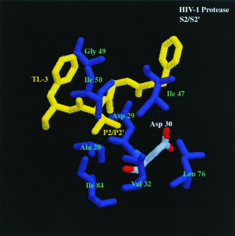
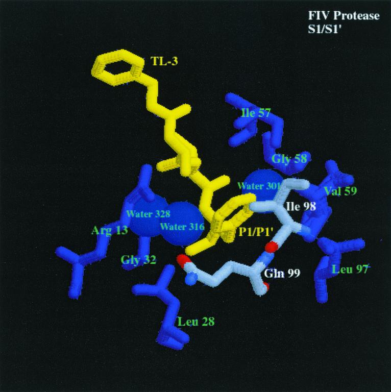
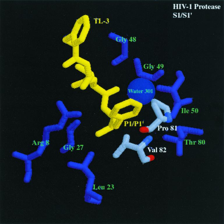
We have used a random hexamer phage library to delineate similarities and differences between the substrate specificities of human immunodeficiency virus type 1 (HIV-1) and feline immunodeficiency virus (FIV) proteases (PRs). Peptide sequences were identified that were specifically cleaved by each protease, as well as sequences cleaved equally well by both enzymes. Based on amino acid distinctions within the P3-P3' region of substrates that appeared to correlate with these cleavage specificities, we prepared a series of synthetic peptides within the framework of a peptide sequence cleaved with essentially the same efficiency by both HIV-1 and FIV PRs, Ac-KSGVF/VVNGLVK-NH(2) (arrow denotes cleavage site). We used the resultant peptide set to assess the influence of specific amino acid substitutions on the cleavage characteristics of the two proteases. The findings show that when Asn is substituted for Val at the P2 position, HIV-1 PR cleaves the substrate at a much greater rate than does FIV PR. Likewise, Glu or Gln substituted for Val at the P2' position also yields peptides specifically susceptible to HIV-1 PR. In contrast, when Ser is substituted for Val at P1', FIV PR cleaves the substrate at a much higher rate than does HIV-1 PR. In addition, Asn or Gln at the P1 position, in combination with an appropriate P3 amino acid, Arg, also strongly favors cleavage by FIV PR over HIV PR. Structural analysis identified several protease residues likely to dictate the observed specificity differences. Interestingly, HIV PR Asp30 (Ile-35 in FIV PR), which influences specificity at the S2 and S2' subsites, and HIV-1 PR Pro-81 and Val-82 (Ile-98 and Gln-99 in FIV PR), which influence specificity at the S1 and S1' subsites, are residues which are often involved in development of drug resistance in HIV-1 protease. The peptide substrate KSGVF/VVNGK, cleaved by both PRs, was used as a template for the design of a reduced amide inhibitor, Ac-GSGVF Psi(CH(2)NH)VVNGL-NH(2.) This compound inhibited both FIV and HIV-1 PRs with approximately equal efficiency. These findings establish a molecular basis for distinctions in substrate specificity between human and feline lentivirus PRs and offer a framework for development of efficient broad-based inhibitors.


Figures

FIG. 1
FIG. 1
(A) Phage selected with wt HIV-1 PR, as described by Beck et al. (2) and cleaved with (from left to right) 160, 40, and 0 nM HIV-1 PR and 156, 78, and 0 nM FIV PR for 1 h at pH 6.7 in 0.2 M NaCl–0.1 M MES buffer. Cleavage of the phage at the random hexamer region results in loss of antibody epitope and loss of chemiluminescent signal on blots. (B) Phage selected with FIV PR and wt HIV-1 PR (see Materials and Methods). Phage were incubated according to the conditions described in the text.
FIG. 2
FIG. 2
JEB inhibitor core structure.
FIG. 3
FIG. 3
Comparison of the S2/S2′ and S1/S1′ subsites of HIV-1 PR and FIV PR bound to the dihydroxyl-containing inhibitor TL-3 (yellow) (pdb file TL3H for HIV-1 PR and pdb file 1b11 for FIV PR). The first panel shows S2/S2′ subsites of FIV PR bound to TL-3. Residues composing the S2/S2′ pocket are labeled in green and white, and the corresponding residues are enlarged. The differences highlighted between FIV PR and HIV-1 PR (shown in the second panel), bound to TL-3 with structurally homologous amino acids highlighted similar to FIV PR, are residues Ile-35 (FIV PR)/Asp-30 (HIV-1 PR). The differences in Ile-35 (FIV PR) and structurally homologous Asp-30 (HIV-1 PR) may create the ability for HIV-1 PR to cleave substrates with Asn at the P2 position and with Glu or Gln at the P2′ position at a higher rate than FIV PR cleaves the same substrates. In the third panel, S1/S1′ subsite of FIV PR bound to TL-3. Residues composing the S1/S1′ pocket are labeled in green and white, and the corresponding residues are enlarged. The highlighted amino acids in the binding pockets of FIV PR and HIV-1 PR (shown in the fourth panel), bound to TL-3 with the structurally homologous amino acids to FIV PR highlighted in the same fashion as FIV PR, are residues Ile-98 (FIV PR)/Pro-81 (HIV-1 PR) and Gln-99 (FIV PR)/Val-82 (HIV-1 PR) (oxygen, red; nitrogen, blue; carbon, gray). The difference in the charge and structure of these residues is proposed to create the ability of FIV PR to cleave substrates with Asn and Gln in the S1 pocket and Ser in the S1′ pocket better than HIV-1 PR.
FIG. 3
FIG. 3
Comparison of the S2/S2′ and S1/S1′ subsites of HIV-1 PR and FIV PR bound to the dihydroxyl-containing inhibitor TL-3 (yellow) (pdb file TL3H for HIV-1 PR and pdb file 1b11 for FIV PR). The first panel shows S2/S2′ subsites of FIV PR bound to TL-3. Residues composing the S2/S2′ pocket are labeled in green and white, and the corresponding residues are enlarged. The differences highlighted between FIV PR and HIV-1 PR (shown in the second panel), bound to TL-3 with structurally homologous amino acids highlighted similar to FIV PR, are residues Ile-35 (FIV PR)/Asp-30 (HIV-1 PR). The differences in Ile-35 (FIV PR) and structurally homologous Asp-30 (HIV-1 PR) may create the ability for HIV-1 PR to cleave substrates with Asn at the P2 position and with Glu or Gln at the P2′ position at a higher rate than FIV PR cleaves the same substrates. In the third panel, S1/S1′ subsite of FIV PR bound to TL-3. Residues composing the S1/S1′ pocket are labeled in green and white, and the corresponding residues are enlarged. The highlighted amino acids in the binding pockets of FIV PR and HIV-1 PR (shown in the fourth panel), bound to TL-3 with the structurally homologous amino acids to FIV PR highlighted in the same fashion as FIV PR, are residues Ile-98 (FIV PR)/Pro-81 (HIV-1 PR) and Gln-99 (FIV PR)/Val-82 (HIV-1 PR) (oxygen, red; nitrogen, blue; carbon, gray). The difference in the charge and structure of these residues is proposed to create the ability of FIV PR to cleave substrates with Asn and Gln in the S1 pocket and Ser in the S1′ pocket better than HIV-1 PR.

References

    1. Alewood P, Alewood D, Miranda L, Love S, Meutermans W, Wilson D. Rapid in situ neutralization protocols for Boc and Fmoc solid-phase chemistries. Methods Enzymol. 1997;289:14–29. - PubMed
    1. Beck Z Q, Hervio L, Dawson P E, Elder J H, Madison E L. Identification of efficiently cleaved substrates for HIV-1 protease using a phage display library and use in inhibitor development. Virology. 2000;274:391–401. - PubMed
    1. Billich A, Winkler G. Analysis of subsite preferences of HIV-1 proteinase using MA/CA junction peptides substituted at the P3–P1′ positions. Arch Biochem Biophys. 1991;290:186–190. - PubMed
    1. Doyon L, Croteau G, Thibeault D, Poulin F, Pilote L, Lamarre D. Second locus involved in human immunodeficiency virus type 1 resistance to protease inhibitors. J Virol. 1996;70:3763–3769. - PMC - PubMed
    1. Doyon L, Payant C, Brakier-Gingras L, Lamarre D. Novel Gag-Pol frameshift site in human immunodeficiency virus type 1 variants resistant to protease inhibitors. J Virol. 1998;72:6146–6150. - PMC - PubMed

Publication types