. 2001 Oct;75(19):9470-82.
doi: 10.1128/JVI.75.19.9470-9482.2001.

A cellular J-domain protein modulates polyprotein processing and cytopathogenicity of a pestivirus

Affiliations

A cellular J-domain protein modulates polyprotein processing and cytopathogenicity of a pestivirus

G Rinck et al. J Virol. 2001 Oct.

Abstract

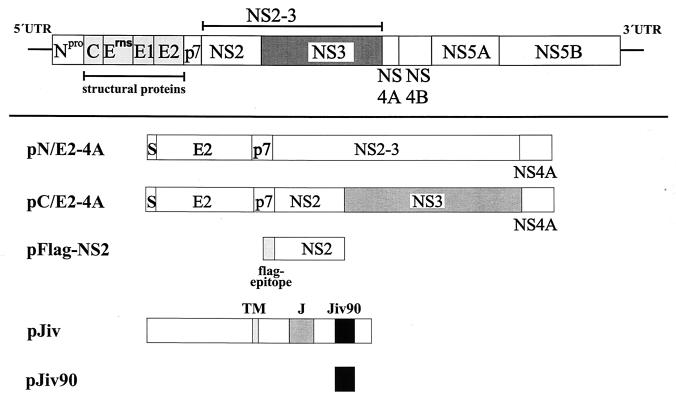
Pestiviruses are positive-strand RNA viruses closely related to human hepatitis C virus. Gene expression of these viruses occurs via translation of a polyprotein, which is further processed by cellular and viral proteases. Here we report the formation of a stable complex between an as-yet-undescribed cellular J-domain protein, a member of the DnaJ-chaperone family, and pestiviral nonstructural protein NS2. Accordingly, we termed the cellular protein Jiv, for J-domain protein interacting with viral protein. Jiv has the potential to induce in trans one specific processing step in the viral polyprotein, namely, cleavage of NS2-3. Efficient generation of its cleavage product NS3 has previously been shown to be obligatory for the cytopathogenicity of the pestiviruses. Regulated expression of Jiv in cells infected with noncytopathogenic bovine viral diarrhea virus disclosed a direct correlation between the intracellular level of Jiv, the extent of NS2-3 cleavage, and pestiviral cytopathogenicity.


Figures

FIG. 1
FIG. 1
(A) Northern blot. Poly(A)+ RNA from MDBK cells was separated under denaturing conditions on a 0.8% agarose gel and blotted onto a Duranlon-UV membrane (Stratagene). The blot was hybridized with a jiv-specific probe labeled with [α-32P]dCTP. The numbers at the left side indicate the size of the RNA marker in kilobases. (B) cDNA cloning of the jiv-mRNA. In the upper part of the figure, the bar symbolizes the Jiv protein; different domains are pointed out on top as follows: TM, putative transmembrane region; J, J domain; and Jiv90, Jiv90 domain. The lines at both ends symbolize the noncoding regions of the mRNA. The relative positions of the restriction sites used to establish a cDNA clone encompassing the entire jiv-ORF are indicated. The cDNA library-derived clones cIK9 and cIK9-4 are shown as black bars. In the lower part of the figure, the RT-PCR fragments established to verify and complete the cDNA cloning are depicted as solid bars. The primers used for amplification are symbolized by arrowheads below. Primer M13 was complementary to the 5′ sequence of the oligonucleotide, which was ligated to the first-strand cDNA in order to analyze the 5′ end of the mRNA. (C) Nucleotide and deduced amino acid sequences of the cDNA representing the jiv-mRNA. Domains: putative transmembrane domain (amino acids 327 to 347); J domain (boldface; amino acids 444 to 505); Jiv90 domain (underlined; amino acids 533 to 622) with CXXCXXXH motifs (underlined and boldface). The shorter mRNA ends with nt 2402.
FIG. 1
FIG. 1
(A) Northern blot. Poly(A)+ RNA from MDBK cells was separated under denaturing conditions on a 0.8% agarose gel and blotted onto a Duranlon-UV membrane (Stratagene). The blot was hybridized with a jiv-specific probe labeled with [α-32P]dCTP. The numbers at the left side indicate the size of the RNA marker in kilobases. (B) cDNA cloning of the jiv-mRNA. In the upper part of the figure, the bar symbolizes the Jiv protein; different domains are pointed out on top as follows: TM, putative transmembrane region; J, J domain; and Jiv90, Jiv90 domain. The lines at both ends symbolize the noncoding regions of the mRNA. The relative positions of the restriction sites used to establish a cDNA clone encompassing the entire jiv-ORF are indicated. The cDNA library-derived clones cIK9 and cIK9-4 are shown as black bars. In the lower part of the figure, the RT-PCR fragments established to verify and complete the cDNA cloning are depicted as solid bars. The primers used for amplification are symbolized by arrowheads below. Primer M13 was complementary to the 5′ sequence of the oligonucleotide, which was ligated to the first-strand cDNA in order to analyze the 5′ end of the mRNA. (C) Nucleotide and deduced amino acid sequences of the cDNA representing the jiv-mRNA. Domains: putative transmembrane domain (amino acids 327 to 347); J domain (boldface; amino acids 444 to 505); Jiv90 domain (underlined; amino acids 533 to 622) with CXXCXXXH motifs (underlined and boldface). The shorter mRNA ends with nt 2402.
FIG. 2
FIG. 2
Multiple alignment of bovine Jiv and Jiv orthologs from other species starting with the J domain. The J domain is marked by a gray line, and the Jiv90 domain is marked by a black line. Coding: capital letters, amino acids conserved between more than two species; yellow, amino acid identical in four of five species; red, conserved amino acid. Abbreviations: B. tau., Bos taurus (this study); H. sap., Homo sapiens (derived from a GENSCAN analysis of genomic clone AC023055); D. mel., Drosophila melanogaster (derived from a GENSCAN analysis of genomic clone AC014875); C. ele., Caenorhabditis elegans (hypothetical protein CO4A2.7) (Q09446); A. tha., Arabidopsis thaliana (hypothetical protein YUP8H12R.35) (T01052).
FIG. 3
FIG. 3
Scheme of expression constructs. In the upper part of the figure, the BVDV polyprotein is symbolized by a bar; the lines at both ends represent the UTRs. In the lower part of the figure, expression constructs are shown. The box marked with S represents the signal sequence for translocation of E2 into the ER. TM, putative transmembrane domain; J, J domain; Jiv90, Jiv90 domain.
FIG. 4
FIG. 4
Western blot analysis. BHK-21 cells infected with MVA-T7pol vaccinia virus were transfected with the plasmids indicated above the lanes. The cell lysates were separated by SDS-PAGE and processed for Western blotting by using an NS3-specific MAb. The positions of NS2-3 and NS3 are indicated on the left. The molecular mass standard is indicated at the right.
FIG. 5
FIG. 5
Coprecipitation analysis. BHK-21 cells infected with MVA-T7pol vaccinia virus were transfected with the plasmids indicated above the respective lanes. Proteins were metabolically labeled and subjected to immunoprecipitation under nondenaturing conditions. The MAbs used for precipitation are indicated. The precipitates were separated by SDS-PAGE and processed for fluorography. Arrowheads on the left mark the positions of GST-Jiv, Flag-NS2, GST-Jiv90, and GST. On the right, the molecular mass standards are indicated.
FIG. 6
FIG. 6
(A) Immunofluorescence analysis of MDBKTet-On/Jiv cells. Cells cultured in the absence (left) or presence of 10 μM Dox for 24 h (right) were assayed for Jiv expression by the Jiv-specific antiserum and a secondary Cy3-labeled antiserum. The cells were visualized by confocal laser-scanning microscopy. Magnification, ×100. (B) RIPA. BHK-21 cells (two lanes at left) were infected with MVA-T7pol vaccinia virus and transfected with the plasmids indicated above each lane. MDBK or MDBKTet-On/Jiv cells were cultured in the presence of 10 μM Dox for 18 h. Subsequently, the cells were metabolically labeled with [35S]methionine and [35S]cysteine for 6 h. Lysates were prepared from the respective cells under nondenaturing conditions. To minimize background signals, the lysates were subjected to immunoprecipitation with preimmune serum; the supernatants were subsequently used for immunoprecipitation with the Jiv-specific antiserum. The precipitates were analyzed by SDS-PAGE and processed for fluorography. The position of Jiv is indicated. The molecular mass standard is given on the right. (C) Western blot analysis. MDBK and MDBKTet-On/Jiv cells were cultured for 45 h in the presence of the Dox concentrations indicated above the lanes. Lysates of these cells were separated by SDS-PAGE and processed for Western blotting with the Jiv-specific antiserum. The position of Jiv is marked on the right. Molecular mass standards are given on the left.
FIG. 6
FIG. 6
(A) Immunofluorescence analysis of MDBKTet-On/Jiv cells. Cells cultured in the absence (left) or presence of 10 μM Dox for 24 h (right) were assayed for Jiv expression by the Jiv-specific antiserum and a secondary Cy3-labeled antiserum. The cells were visualized by confocal laser-scanning microscopy. Magnification, ×100. (B) RIPA. BHK-21 cells (two lanes at left) were infected with MVA-T7pol vaccinia virus and transfected with the plasmids indicated above each lane. MDBK or MDBKTet-On/Jiv cells were cultured in the presence of 10 μM Dox for 18 h. Subsequently, the cells were metabolically labeled with [35S]methionine and [35S]cysteine for 6 h. Lysates were prepared from the respective cells under nondenaturing conditions. To minimize background signals, the lysates were subjected to immunoprecipitation with preimmune serum; the supernatants were subsequently used for immunoprecipitation with the Jiv-specific antiserum. The precipitates were analyzed by SDS-PAGE and processed for fluorography. The position of Jiv is indicated. The molecular mass standard is given on the right. (C) Western blot analysis. MDBK and MDBKTet-On/Jiv cells were cultured for 45 h in the presence of the Dox concentrations indicated above the lanes. Lysates of these cells were separated by SDS-PAGE and processed for Western blotting with the Jiv-specific antiserum. The position of Jiv is marked on the right. Molecular mass standards are given on the left.
FIG. 7
FIG. 7
Western blot analysis. (A) MDBKTet-On/Jiv cells were infected with noncp BVDV strain NCP7 at an MOI of 0.01 at 18 h postinduction (the concentrations of Dox are indicated above the lanes). Cell lysates were prepared 27 h postinfection, separated by SDS-PAGE, and processed for Western blot with Jiv-specific antiserum. The position of Jiv is marked on the right. Molecular mass standards are indicated. Compared to noninfected cells, the NCP7-infected MDBKTet-On/Jiv cells contained, upon induction by 10 μM Dox, a smaller amount of Jiv (lane 6); a possible explanation for this observation is a degradation of Jiv with the onset of apoptosis in the infected cells. (B) MDBK and MDBKTet-On/Jiv cells were treated as described in panel A. After SDS-PAGE, the anti-NS3 MAb was applied for Western blot analysis. Arrowheads on the right mark the positions of NS2-3 and NS3.
FIG. 8
FIG. 8
Cell morphology. MDBK and MDBKTet-On/Jiv cells were cultured in the absence or presence of 10 μM Dox. At 18 h postinduction, the cells were infected with noncp BVDV strain NCP7 at an MOI of 0.5. Cells were fixed at 24 h postinfection and visualized by phase-contrast microscopy. Magnification, ×100.

Similar articles

Cited by

References

    1. Becher P, Meyers G, Shannon A D, Thiel H J. Cytopathogenicity of border disease virus is correlated with integration of cellular sequences into the viral genome. J Virol. 1996;70:2992–2998. - PMC - PubMed
    1. Cheetham M E, Caplan A J. Structure, function and evolution of DnaJ: conservation and adaptation of chaperone function. Cell Stress Chaperones. 1998;3:28–36. - PMC - PubMed
    1. Collett M S, Larson R, Gold C, Strick D, Anderson D K, Purchio A F. Molecular cloning and nucleotide sequence of the pestivirus bovine viral diarrhea virus. Virology. 1988;165:191–199. - PubMed
    1. Corapi W V, Donis R O, Dubovi E J. Characterization of a panel of monoclonal antibodies and their use in the study of the antigenic diversity of bovine viral diarrhea virus. Am J Vet Res. 1990;51:1388–1394. - PubMed
    1. Corapi W V, Donis R O, Dubovi E J. Monoclonal antibody analyses of cytopathic and noncytopathic viruses from fatal bovine viral diarrhea infections. J Virol. 1988;62:2823–2827. - PMC - PubMed

Publication types