. 2001 Oct;75(19):9517-25.
doi: 10.1128/JVI.75.19.9517-9525.2001.

Characterization of a porcine lung epithelial cell line suitable for influenza virus studies

Affiliations

Characterization of a porcine lung epithelial cell line suitable for influenza virus studies

S H Seo et al. J Virol. 2001 Oct.

Abstract

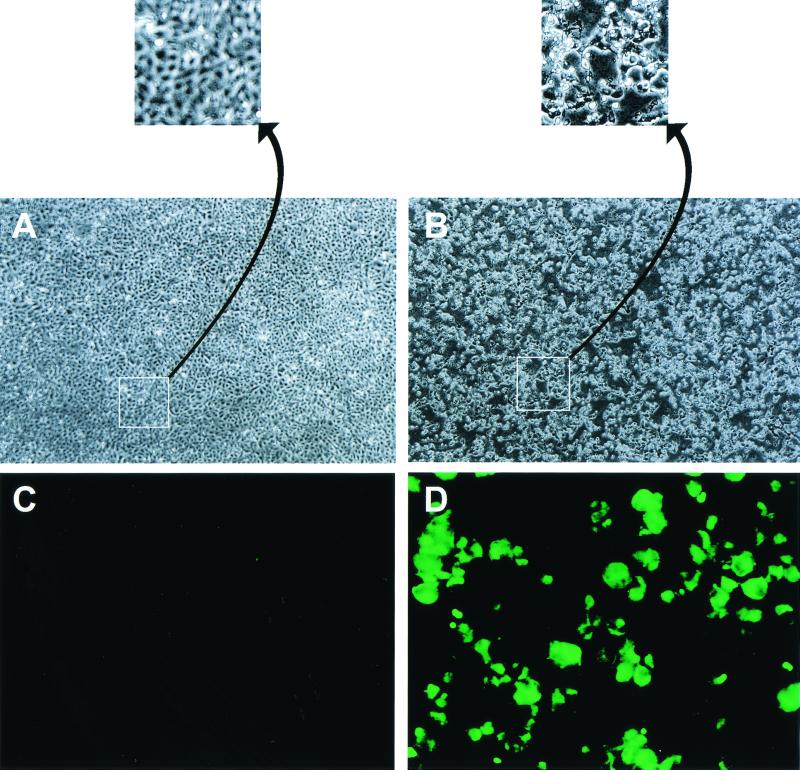
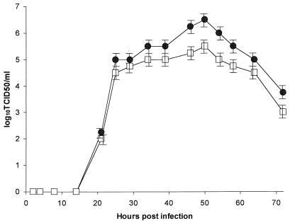
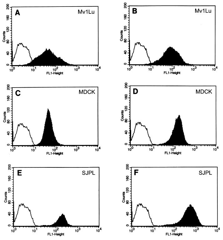
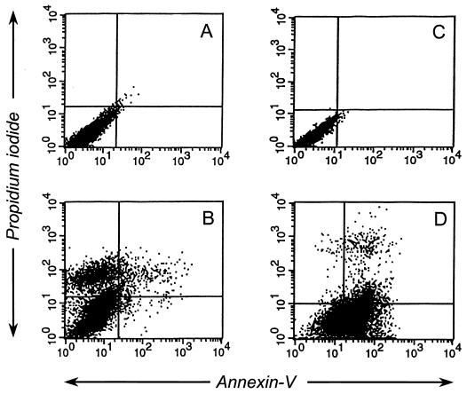
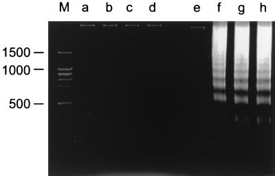
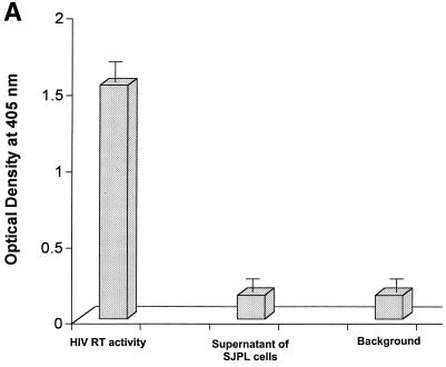
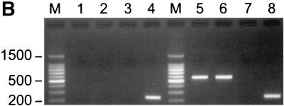
We established a porcine lung epithelial cell line designated St. Jude porcine lung cells (SJPL) and demonstrated that all tested influenza A and B viruses replicated in this cell line. The infectivity titers of most viruses in SJPL cells were comparable to or better than those in MDCK cells. The propagation of influenza viruses from clinical samples in SJPL cells did not lead to antigenic changes in the hemagglutinin molecule. The numbers of both Sia2-3Gal and Sia2-6Gal receptors on SJPL cells were greater than those on MDCK cells. Influenza virus infection of SJPL cells did not lead to apoptosis, as did infection of MDCK cells. No porcine endogenous retrovirus was detected in SJPL cells, and in contrast to MDCK cells, SJPL cells did not cause tumors in nude mice.


Figures

FIG. 1
FIG. 1
CPE and nucleoprotein expression of SJPL cells infected with A/Sydney/5/97 (H3N2). (A and B) Confluent monolayers of SJPL cells in six-well plates were infected with A/Sydney/5/97 (H3N2) (MOI, 2), and 48 h later CPE was evaluated (B). Uninfected cells were used as a control (A). Magnification, ×5 (A and B) and 232% further (insets). (C and D) Confluent monolayers of SJPL cells in two-well Lab-Tek chamber slides were infected with A/Sydney/5/97 (H3N2) (MOI, 5), and 16 h later the cells were incubated with a monoclonal antibody to nucleoprotein and a secondary FITC-labeled goat anti-mouse immunoglobulin antibody (D). Uninfected cells were used as a control (C). Magnification, ×20.
FIG. 2
FIG. 2
Influenza virus growth in SJPL and MDCK cells. Monolayers of SJPL (●) and MDCK (□) cells in a six-well plate were infected with 10 EID50 of B/Memphis/1/84, and aliquots were harvested on different days after infection. The results are from three independent experiments.
FIG. 3
FIG. 3
Influenza virus receptors on SJPL and MDCK cells. SJPL cells, MDCK cells, and Mv1Lu cells (3 × 10⁶ of each) were incubated with the DIG-labeled lectins Maackia amurensis agglutinin (MAA), which binds specifically to Sia2-3Gal, and Sambucus nigra agglutinin (SNA), which binds specifically to Sia2-6Gal. The cells were incubated with a FITC-labeled anti-DIG antibody and then subjected to flow-cytometric analysis. (A) Mv1Lu cells after MAA binding; (B) Mv1Lu cells after SNA binding; (C) MDCK cells after MAA binding; (D) MDCK cells after SNA binding; (E) SJPL cells after MAA binding; (F) SJPL cells after SNA binding. The controls for SJPL, MvILu, and MDCK were populations of cells that were stained only with FITC-labeled anti-DIG antibody (unshaded profiles).
FIG. 4
FIG. 4
Annexin V-Fluos assays and propidium iodide staining of SJPL and MDCK cells infected with A/Sydney/5/97 (H3N2). Ten hours after the cells were infected with A/Sydney/5/97 (H3N2) (MOI, 5), they were incubated with annexin V-Fluos and propidium iodide. (A) Uninfected SJPL cells; (B) infected SJPL cells; (C) uninfected MDCK cells; (D) infected MDCK cells.
FIG. 5
FIG. 5
DNA fragmentation in SJPL and MDCK cells. SJPL cells and MDCK cells were infected with A/Sydney/5/97 (H3N2), A/Chicken/NY/13307-3/95 (H7N2), or A/Swine/IA/17672/88 (H1N1) (MOI, 5), and 18 h later DNA was collected for analysis. The collected DNA was subjected to electrophoresis through a 2% agarose gel. The lanes contained DNA from the following sources: M, molecular marker; a, uninfected SJPL cells; b, SJPL cells infected with A/Sydney/5/97 (H3N2); c, SJPL cells infected with A/Chicken/NY/13307-3/95 (H7N2); d, SJPL cells infected with A/Swine/IA/17672/88 (H1N1); e, uninfected MDCK cells; f, MDCK cells infected with A/Sydney/5/97 (H3N2); g, MDCK cells infected with A/Chicken/NY/13307-3/95 (H7N2); h, MDCK cells infected with A/Swine/IA/17672/88 (H1N1).
FIG. 6
FIG. 6
Reverse transcriptase assay and RT-PCR analysis. (A) Reverse transcriptase activity in the supernatant of SJPL cells was evaluated by using a nonradioactive reverse transcriptase assay. HIV-1 reverse transcriptase (0.125 ng/well in a 96-well plate) was used as a positive control, and ABTS [2′,2-azino-di(3-ethyl-benzthiazoline-6-sulfonic acid)] solution alone was used as a negative control. The sample was tested by the assay four times. (B) Tissue culture supernatants and cellular RNAs were collected from SJPL cells and PK-15 cells. RT-PCR was performed as described in the text. Lanes: M, 100-bp DNA marker; 1, tissue culture supernatant of SJPL cells; 2, cellular RNAs of SJPL cells; 3, cellular RNAs of SJPL cells without reverse transcriptase; 4, cellular RNAs of SJPL cells with β-actin primers; 5, tissue culture supernatant of PK-15 cells; 6, cellular RNAs of PK-15 cells; 7, cellular RNAs of PK-15 cells without reverse transcriptase; 8, cellular RNAs of PK-15 cells with β-actin primers.
FIG. 6
FIG. 6
Reverse transcriptase assay and RT-PCR analysis. (A) Reverse transcriptase activity in the supernatant of SJPL cells was evaluated by using a nonradioactive reverse transcriptase assay. HIV-1 reverse transcriptase (0.125 ng/well in a 96-well plate) was used as a positive control, and ABTS [2′,2-azino-di(3-ethyl-benzthiazoline-6-sulfonic acid)] solution alone was used as a negative control. The sample was tested by the assay four times. (B) Tissue culture supernatants and cellular RNAs were collected from SJPL cells and PK-15 cells. RT-PCR was performed as described in the text. Lanes: M, 100-bp DNA marker; 1, tissue culture supernatant of SJPL cells; 2, cellular RNAs of SJPL cells; 3, cellular RNAs of SJPL cells without reverse transcriptase; 4, cellular RNAs of SJPL cells with β-actin primers; 5, tissue culture supernatant of PK-15 cells; 6, cellular RNAs of PK-15 cells; 7, cellular RNAs of PK-15 cells without reverse transcriptase; 8, cellular RNAs of PK-15 cells with β-actin primers.

References

    1. Armstrong J A, Porterfield J S, Madrid A T D. C-type virus particles in pig kidney cell lines. J Gen Virol. 1971;10:195–198. - PubMed
    1. Ashkenazi A, Dixit V M. Death receptors: signaling and modulation. Science. 1998;281:1305–1308. - PubMed
    1. Carroll S M, Paulson J C. Differential infection of receptor-modified host cells by receptor-specific influenza viruses. Virus Res. 1985;3:165–179. - PubMed
    1. Connor R J, Kawaoka Y, Webster R G, Paulson J C. Receptor specificity in human, avian, and equine H2 and H3 influenza virus isolates. Virology. 1994;205:17–23. - PubMed
    1. Duvall E, Wyllie A H. Death and the cell. Immunol Today. 1986;7:115–119. - PubMed

Publication types