. 2001 Oct;21(19):6470-83.
doi: 10.1128/MCB.21.19.6470-6483.2001.

ETO, a target of t(8;21) in acute leukemia, makes distinct contacts with multiple histone deacetylases and binds mSin3A through its oligomerization domain

Affiliations

ETO, a target of t(8;21) in acute leukemia, makes distinct contacts with multiple histone deacetylases and binds mSin3A through its oligomerization domain

J M Amann et al. Mol Cell Biol. 2001 Oct.

Abstract

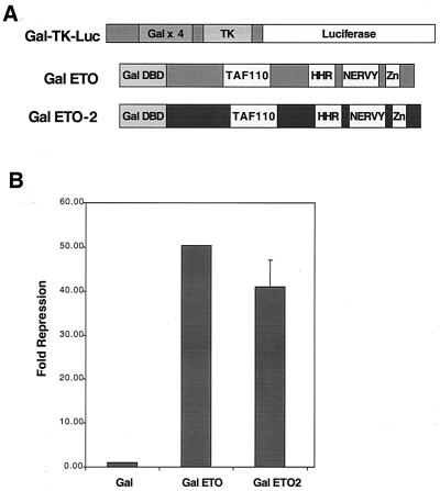
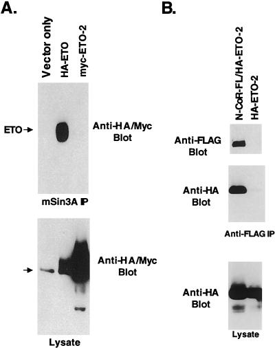
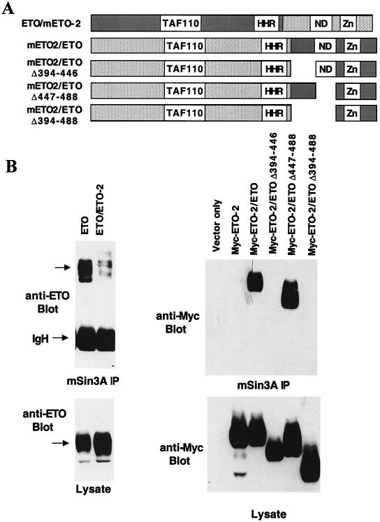
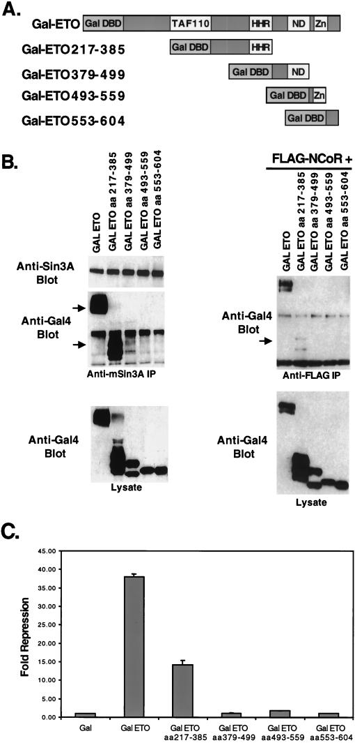
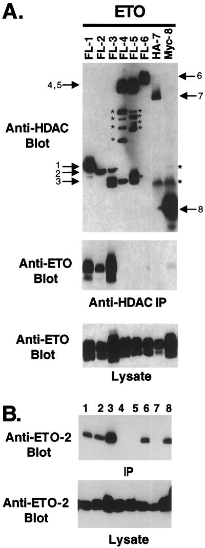
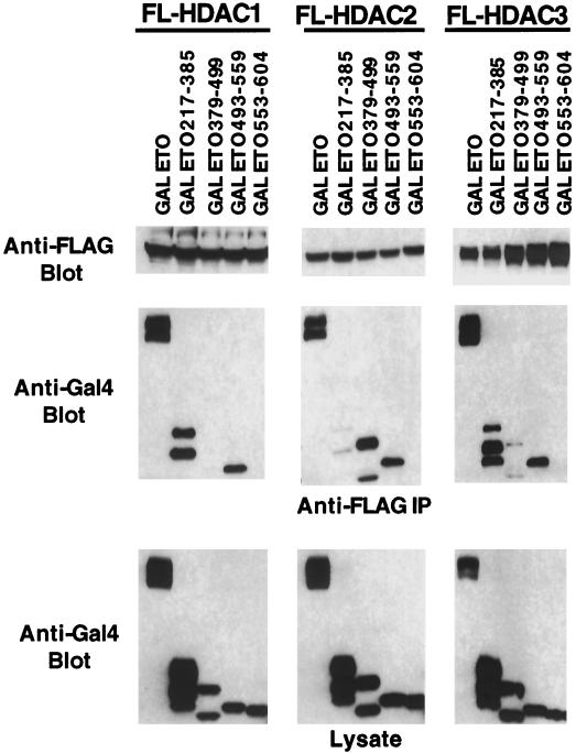
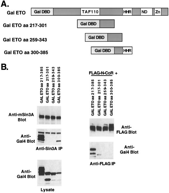
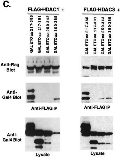
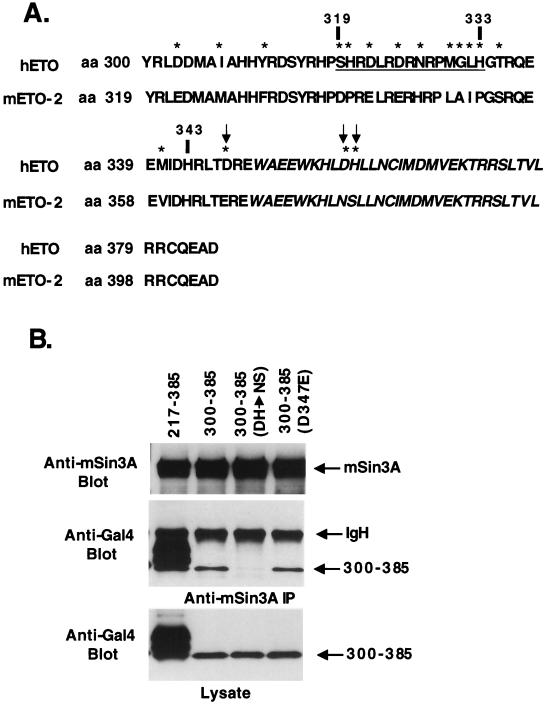
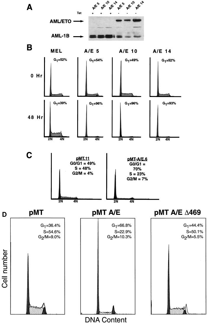
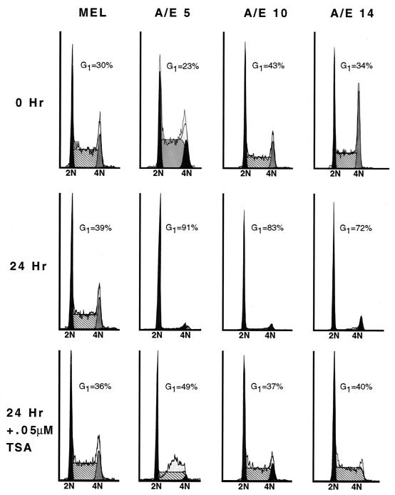
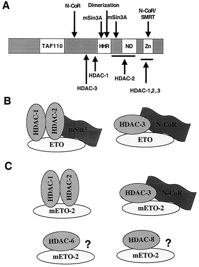
t(8;21) and t(16;21) create two fusion proteins, AML-1-ETO and AML-1-MTG16, respectively, which fuse the AML-1 DNA binding domain to putative transcriptional corepressors, ETO and MTG16. Here, we show that distinct domains of ETO contact the mSin3A and N-CoR corepressors and define two binding sites within ETO for each of these corepressors. In addition, of eight histone deacetylases (HDACs) tested, only the class I HDACs HDAC-1, HDAC-2, and HDAC-3 bind ETO. However, these HDACs bind ETO through different domains. We also show that the murine homologue of MTG16, ETO-2, is also a transcriptional corepressor that works through a similar but distinct mechanism. Like ETO, ETO-2 interacts with N-CoR, but ETO-2 fails to bind mSin3A. Furthermore, ETO-2 binds HDAC-1, HDAC-2, and HDAC-3 but also interacts with HDAC-6 and HDAC-8. In addition, we show that expression of AML-1-ETO causes disruption of the cell cycle in the G(1) phase. Disruption of the cell cycle required the ability of AML-1-ETO to repress transcription because a mutant of AML-1-ETO, Delta469, which removes the majority of the corepressor binding sites, had no phenotype. Moreover, treatment of AML-1-ETO-expressing cells with trichostatin A, an HDAC inhibitor, restored cell cycle control. Thus, AML-1-ETO makes distinct contacts with multiple HDACs and an HDAC inhibitor biologically inactivates this fusion protein.


Figures

FIG. 1
FIG. 1
ETO-2 represses transcription when tethered to a promoter. (A) Schematic diagram of the Gal4-TK-luciferase reporter plasmid and the GAL4-ETO and GAL4–ETO-2 fusion proteins. (B) ETO and ETO-2 repress transcription. GAL4-ETO and GAL4–ETO-2 were transfected with the GAL4 reporter plasmid into NIH 3T3 cells, and relative light units were measured. The data are graphed as the relative fold repression after correction for transfection efficiency.
FIG. 2
FIG. 2
ETO and ETO-2 associate with corepressors. Cos-7 cells were transfected with HA-ETO or Myc–ETO-2, and cell lysates were immunoprecipitated (IP) with anti-mSin3A antibody (A) or were transfected with FLAG-tagged N-CoR and HA–ETO-2 or HA–ETO-2 alone and immunoprecipitated with anti-FLAG antibody (B). Proteins were detected by immunoblot analysis using anti-mSin3A, anti-HA, and anti-Myc antibodies. The bottom panels show the level of expression of ETO and ETO-2. The arrow indicates the presence of a background band.
FIG. 3
FIG. 3
Mapping the C-terminal mSin3A-binding domain. (A) Schematic diagram of ETO/ETO-2 and ETO-2/ETO chimeric proteins. (B) Lysates were made from cells expressing the indicated chimeric proteins and immunoprecipitated with anti-mSin3A antibody. The chimeric proteins were detected using anti-ETO or anti-Myc antibody. Arrows indicate the position of ETO.
FIG. 4
FIG. 4
Mapping the N-terminal mSin3A and N-CoR binding sites. (A) Schematic diagram of the GAL4-ETO chimeric proteins. (B) Lysates were made from cells expressing the indicated chimeric proteins and immunoprecipitated with anti-mSin3A antibody (left). Plasmids expressing FLAG-tagged N-CoR were cotransfected into cells with the GAL4-ETO chimeric proteins (right) and immunoprecipitated with anti-FLAG antibody. Chimeric proteins were detected using anti-GAL4 antibody. Arrows indicate the position of GAL4-ETO or the deletion mutants. (C) GAL4-ETO and GAL4-ETO deletion mutants were transfected with the GAL4 reporter plasmid into NIH 3T3 cells and relative light units were measured. The data are graphed as relative fold repression after correction for transfection efficiency.
FIG. 5
FIG. 5
ETO and ETO-2 bind multiple HDACs. (A) ETO binds HDAC-1, HDAC-2, and HDAC-3. The numbers above the lanes indicate the epitope-tagged form of HDAC-1 to -8 that was coexpressed with ETO. After immunoprecipitation (IP) with anti-FLAG (FL), anti-HA (HA), or anti-Myc (Myc) antibody, copurifying ETO was detected by immunoblot analysis (middle). The bottom panel is an immunoblot of the cell lysates prior to immunoprecipitation. ∗, nonspecific bands and proteolytic fragments. (B) ETO-2 binds HDACs 1, 2, 3, 6, and 8. The experiment was performed exactly as in panel A, but using epitope-tagged mETO-2. The numbers above the lanes indicate the epitope-tagged HDAC that was coexpressed with ETO-2 as described for panel A. Note that anti-HDAC antibody connotes that the experiment was performed with antibodies to the epitope tag for that HDAC.
FIG. 6
FIG. 6
Mapping of the HDAC-binding sites on ETO. FLAG-tagged HDAC-1 to -3 were coexpressed with the GAL4-ETO chimeric proteins from Fig. 4. Copurifying GAL4-ETO proteins were detected using anti-GAL4 immunoglobulin G for immunoblot analysis. Shown are the control anti-FLAG antibody blot showing that each HDAC was expressed and immunoprecipitated (top), copurifying ETO fragments (middle), and the levels of expression of the GAL4-ETO chimeric proteins (bottom).
FIG. 7
FIG. 7
Fine mapping of the N-terminal repression domain of ETO. (A) Schematic diagram of the GAL4-ETO chimeric proteins used. (B) mSin3A and N-CoR bind to distinct ETO domains. (Left) Lysates were made from cells expressing the indicated chimeric proteins and immunoprecipitated (IP) with anti-mSin3A antibody. Immune complexes were analyzed for the presence of mSin3A (top) and GAL4-ETO proteins (anti-GAL4 immunoblot; middle). Shown also are the levels of expression of the GAL4-ETO chimeric proteins (bottom). (Right) GAL4-ETO chimeric proteins were coexpressed with FLAG–N-CoR, and cell lysates containing FLAG-tagged N-CoR were immunoprecipitated with anti-FLAG antibody. Chimeric proteins were detected using anti-GAL4 antibody (below). (C) HDACs bind different domains in ETO. GAL4-ETO chimeric proteins were coexpressed with FLAG–HDAC-1 or FLAG–HDAC-3, and cell lysates containing expressed FLAG-tagged HDAC-1 or HDAC-3 were immunoprecipitated with anti-FLAG antibody. Chimeric proteins copurifying were detected using anti-GAL4 antibody (middle). Shown are the levels of expression of the GAL4-ETO chimeric proteins (bottom) and immunoprecipitated HDAC (top).
FIG. 7
FIG. 7
Fine mapping of the N-terminal repression domain of ETO. (A) Schematic diagram of the GAL4-ETO chimeric proteins used. (B) mSin3A and N-CoR bind to distinct ETO domains. (Left) Lysates were made from cells expressing the indicated chimeric proteins and immunoprecipitated (IP) with anti-mSin3A antibody. Immune complexes were analyzed for the presence of mSin3A (top) and GAL4-ETO proteins (anti-GAL4 immunoblot; middle). Shown also are the levels of expression of the GAL4-ETO chimeric proteins (bottom). (Right) GAL4-ETO chimeric proteins were coexpressed with FLAG–N-CoR, and cell lysates containing FLAG-tagged N-CoR were immunoprecipitated with anti-FLAG antibody. Chimeric proteins were detected using anti-GAL4 antibody (below). (C) HDACs bind different domains in ETO. GAL4-ETO chimeric proteins were coexpressed with FLAG–HDAC-1 or FLAG–HDAC-3, and cell lysates containing expressed FLAG-tagged HDAC-1 or HDAC-3 were immunoprecipitated with anti-FLAG antibody. Chimeric proteins copurifying were detected using anti-GAL4 antibody (middle). Shown are the levels of expression of the GAL4-ETO chimeric proteins (bottom) and immunoprecipitated HDAC (top).
FIG. 8
FIG. 8
The HHR-dimerization motif of ETO contacts mSin3A. (A) Comparison of the sequences of ETO and ETO-2 within the HHR-dimerization domain. Stars highlight the differences between ETO and ETO-2. The HHR is shown in italics. Arrows indicate the amino acids that were changed and analyzed in panel B. The underlined sequence indicates a region of low homology. (B) The HHR is required for binding mSin3A. Lysates were made from cells expressing the indicated GAL4-ETO chimeric proteins and were immunoprecipitated with anti-mSin3A antibody. Shown are immunoprecipitated mSin3A (top), associated GAL4-ETO proteins (middle), and the expression of the GAL4-ETO fusion protiens (bottom). The chimeric proteins were detected using anti-GAL4 antibody.
FIG. 9
FIG. 9
AML-1–ETO-expressing cells accumulate in the G1 phase. (A) Immunoblot analysis of AML-1–ETO in the presence and absence of tetracycline. Cells were cultured in the presence (+) or absence (−) of tetracycline for 48 h prior to the preparation of whole-cell extracts for separation by SDS-PAGE. Proteins were detected using antibodies directed to residues 50 to 177 of AML-1 (the RHD). Arrows indicate the AML-1–ETO and AML-1B bands. Note that the endogenous AML-1B is repressed by AML-1–ETO expression. The numbers following the AML-1–ETO designations indicate individual clonal cell lines. (B) Cell cycle analysis of AML-1–ETO-expressing MEL cells. Cells were cultured in the absence of tetracycline for 48 h before DNA content analysis was determined by flow cytometry after staining the DNA with propidium iodide. (C) Cell cycle analysis of AML-1–ETO-expressing 32D.3 myeloid progenitor cells. These cell lines expressing AML-1–ETO were characterized previously (45). DNA content analysis was determined by flow cytometry after propidium iodide staining. Flow cytometric histograms for the indicated cell lines are shown and the number of cells in each cell cycle phase were determined using the ModFit algorithm. (D) A transcriptionally inactive deletion mutant of AML-1–ETO (Δ469) does not disrupt cell cycle function. Cell cycle analysis of 32D.3 cells expressing AML-1–ETO, the deletion mutant Δ469, or pMT control vector alone were induced with 75 μM ZnSO4 for 16 h, and aliquots were processed for flow cytometric analysis. The names of the 32D.3-derived cell lines are labeled at the top of each panel and the percentages of cells in each phase of the cell cycle, as determined by the ModFit analysis program, are shown within each panel. The apoptotic cells and debris were gated and are not shown in the histograms.
FIG. 10
FIG. 10
TSA ablates the AML-1–ETO-induced G1 phase accumulation of cells. Cell cycle analysis of AML-1–ETO-expressing (A/E) and control (MEL) cell lines by using propidium iodide staining and flow cytometry were used to measure DNA content. The indicated cell lines (lines 5, 10, and 14) were cultured in media containing or lacking tetracycline. After washing the cells to remove tetracycline, half of the cells were cultured in the presence of 0.05 μM TSA. Cell cycle histograms were compiled using the ModFit algorithm.
FIG. 11
FIG. 11
Hypothetical model of ETO and ETO-2 corepression complexes. (A) Schematic diagram of the domains of ETO that bind corepressors. Arrows indicate where the stated proteins contact ETO based on the analysis in this work and work previously described (10, 29, 41). TAF110, a region of ETO with homology to Drosophila TAF110; HHR, hydrophobic heptad repeat; ND, nervy homology domain; Zn, the predicted dual zinc finger motif. (B) Model of ETO-containing complexes. (C) Model of ETO-2-containing complexes. ?, potential corepressor interactions yet to be identified.

References

    1. Ahn M Y, Huang G, Bae S C, Wee H J, Kim W Y, Ito Y. Negative regulation of granulocytic differentiation in the myeloid precursor cell line 32Dcl3 by ear-2, a mammalian homolog of Drosophila seven-up, and a chimeric leukemogenic gene, AML1/ETO. Proc Natl Acad Sci USA. 1998;95:1812–1817. - PMC - PubMed
    1. Cao W, Britos-Bray M, Claxton D F, Kelley C A, Speck N A, Liu P P, Friedman A D. CBF beta-SMMHC, expressed in M4Eo AML, reduced CBF DNA-binding and inhibited the G1 to S cell cycle transition at the restriction point in myeloid and lymphoid cells. Oncogene. 1997;15:1315–1327. - PubMed
    1. Davis J N, Williams B J, Herron J T, Galiano F J, Meyers S. ETO-2: a new member of the ETO-family of nuclear proteins. Oncogene. 1999;18:1375–1383. - PubMed
    1. Erickson P, Gao J, Chang K S, Look T, Whisenant E, Raimondi S, Lasher R, Trujillo J, Rowley J, Drabkin H. Identification of breakpoints in t(8;21) acute myelogenous leukemia and isolation of a fusion transcript, AML1/ETO, with similarity to Drosophila segmentation gene, runt. Blood. 1992;80:1825–1831. - PubMed
    1. Erickson P F, Robinson M, Owens G, Drabkin H A. The ETO portion of acute myeloid leukemia t(8;21) fusion transcript encodes a highly evolutionarily conserved, putative transcription factor. Cancer Res. 1994;54:1782–1786. - PubMed

Publication types

MeSH terms

Substances