. 2001 Oct;21(19):6495-506.
doi: 10.1128/MCB.21.19.6495-6506.2001.

Histone deacetylase activity represses gamma interferon-inducible HLA-DR gene expression following the establishment of a DNase I-hypersensitive chromatin conformation

Affiliations

Histone deacetylase activity represses gamma interferon-inducible HLA-DR gene expression following the establishment of a DNase I-hypersensitive chromatin conformation

A Osborne et al. Mol Cell Biol. 2001 Oct.

Abstract

Expression of the retinoblastoma tumor suppressor protein (Rb) is required for gamma interferon (IFN-gamma)-inducible major histocompatibility complex class II gene expression and transcriptionally productive HLA-DRA promoter occupancy in several human tumor cell lines. Treatment of these Rb-defective tumor cell lines with histone deacetylase (HDAC) inhibitors rescued IFN-gamma-inducible HLA-DRA and -DRB mRNA and cell surface protein expression, demonstrating repression of these genes by endogenous cellular HDAC activity. Additionally, Rb-defective, transcriptionally incompetent tumor cells retained the HLA-DRA promoter DNase I-hypersensitive site. Thus, HDAC-mediated repression of the HLA-DRA promoter occurs following the establishment of an apparent nucleosome-free promoter region and before transcriptionally productive occupancy of the promoter by the required transactivators. Repression of HLA-DRA promoter activation by HDAC activity likely involves a YY1 binding element located in the first exon of the HLA-DRA gene. Chromatin immunoprecipitation experiments localized YY1 to the HLA-DRA gene in Rb-defective tumor cells. Additionally, mutation of the YY1 binding site prevented repression of the promoter by HDAC1 and partially prevented activation of the promoter by trichostatin A. Mutation of the octamer element also significantly reduced the ability of HDAC1 to confer repression of inducible HLA-DRA promoter activation. Treatment of Rb-defective tumor cells with HDAC inhibitors greatly reduced the DNA binding activity of Oct-1, a repressor of inducible HLA-DRA promoter activation. These findings represent the first evidence that HDAC activity can repress IFN-gamma-inducible HLA class II gene expression and also demonstrate that HDAC activity can contribute to promoter repression following the establishment of a DNase I-hypersensitive chromatin conformation.


Figures
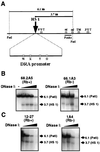
FIG. 1
FIG. 1
Loss of Rb does not result in loss of DNase I-hypersensitive sites in the HLA-DRA promoter. (A) Schematic diagram of HLA-DRA promoter region and coding sequences. The previously described (17) major site of DNase I hypersensitivity in the HLA-DRA promoter is indicated as HS 1. W, W (S/Z) box; X, X1 and X2 boxes; Y, Y box; O, octamer element; α1, α2, and TM, regions of gene coding for α1, α2, and transmembrane domains of DRα, respectively. Digestion of genomic DNA with restriction endonuclease PstI generates an approximately 6.1-kb fragment encompassing the HLA-DRA promoter and most of the coding sequences. If the promoter chromatin is in a generally accessible conformation, further digestion of the genomic DNA with DNase I results in the generation of an approximately 3.7-kb fragment. The 3.7-kb cleavage fragment generated by digestion with PstI and DNase I is detected by hybridization with a radiolabeled DNA probe. (B) Rb-reconstituted (68.2A5) and Rb-defective (66.1A3) subclones of non-small-cell lung carcinoma cell line H2009 were analyzed for their sensitivity to cleavage by DNase I within the HLA-DRA promoter. HS 1 is detected in both Rb-reconstituted and Rb-defective cells and is indicated by an arrow labeled as 3.7 (HS 1). The parental PstI fragment that was not digested with DNase I is indicated by an arrow labeled as 6.1 (PstI). (C) Rb-reconstituted (12-27) and Rb-defective (1A4) subclones of bladder carcinoma cell line 5637 were analyzed exactly as described in panel B for their sensitivity to DNase I cleavage within the HLA-DRA promoter.
FIG. 2
FIG. 2
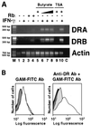
HDAC inhibitors rescue IFN-γ-inducible HLA-DR gene and cell surface expression in Rb-defective bladder carcinoma cells. (A) HDAC inhibitors rescue IFN-γ-inducible HLA-DRA and -DRB mRNA expression in Rb-defective bladder carcinoma cells. RT-PCR was performed using total cytoplasmic RNA from Rb-defective (1A4; lanes 1, 2, and 5 to 10) and Rb-transformed (12-27; lanes 3 and 4) bladder carcinoma cells. Lane M, molecular size marker with 100-bp increments; lane C, negative control for amplification, as no cytoplasmic RNA was added to the RT reaction used as a template for this amplification reaction. Samples were treated with 50 nM TSA or various amounts of sodium butyrate (0.5, 0.25, 0.5, and 1.0 mM for lanes 5 to 8, respectively). Additionally, samples were treated with 400 U of IFN-γ/ml. PCR was performed for 30 cycles for DRA and DRB and 12 cycles for γ-actin to ensure that the reactions were still in the linear range of amplification. The expected band sizes of 325 bp (DRA), 402 bp (DRB), and 730 bp (γ-actin) were obtained for each set of primers. (B) Sodium butyrate rescues IFN-γ-inducible cell surface expression of the HLA-DR heterodimer in Rb-defective bladder carcinoma cells. Rb-defective (5637) bladder carcinoma cells were treated with 400 U of IFN-γ/ml and 1 mM sodium butyrate, and HLA-DR heterodimer expression was detected using primary mouse anti-DR monoclonal antibody (Ab) DA6.147, followed by staining with FITC-conjugated secondary goat anti-mouse (GAM) immunoglobulin G antibody. The plots are as follows: black, untreated; dark gray, IFN-γ; light gray, butyrate; and white, IFN-γ and butyrate.
FIG. 3
FIG. 3
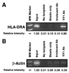
YY1 is present in vivo at the HLA-DRA promoter in Rb-defective 5637 cells. ChIP assays were performed using formaldehyde cross-linked chromatin isolated from 5637 cells. The chromatin was sheared by sonication to an average size of approximately 400 to 600 bp and was immunoprecipitated using only protein G beads, protein G beads and an irrelevant antibody (Ab), or protein G beads and anti-YY1 antibody. Following immunoprecipitation, HLA-DRA promoter fragments were amplified by 35 cycles of PCR using primers spanning the putative HLA-DRA YY1 binding site (A), and β-actin promoter fragments were amplified by 30 cycles of PCR using primers specific for the β-actin promoter (B). The β-actin promoter does not possess a consensus YY1 binding element and is not believed to be regulated by YY1. Input, amplification using 1% the total chromatin introduced into each ChIP reaction as a template; No template, controls in which water was substituted for ChIP samples during PCR amplification of each region of DNA. The relative intensity of each band was determined by densitometry. MW, molecular weight markers.
FIG. 4
FIG. 4
Schematic diagrams of the reporter constructs used in this study. pDRA represents the region, from −176 to +80 of the HLA-DRA promoter, controlling the expression of a luciferase reporter gene. The mutant constructs, pDRA-Octmut and pDRA-YY1mut, are identical to pDRA except for a four-nucleotide substitution mutation (underlined) in the octamer element (5′-ATTTGCAT-3′→5′-CCATGGAT-3′) and a three-nucleotide substitution mutation (underlined) in the YY1 element (5′-AAATGGCCAT-3′→5′-AAATGATTAT-3′), respectively. The double-mutant construct, pDRA-Oct/YY1mut, contains both the four-nucleotide octamer element mutation and the three-nucleotide YY1 element mutation.
FIG. 5
FIG. 5
YY1 overexpression and HDAC activity repress HLA-DRA promoter activation through the +62/+72 YY1 binding element. (A) YY1 requires DNA binding activity for repression of inducible HLA-DRA promoter activation. Different amounts (1, 5, 10, 50, and 100 ng) of expression vector encoding either full-length YY1 protein (pCMV-YY1) (solid line) or a C-terminally truncated mutant YY1 protein which is not capable of binding to DNA [pCEP4F-YY1(1-396)] (broken line) were cotransfected along with 25 ng of promoter-luciferase construct pDRA (closed circles) or pDRA-YY1mut (open circles) into 12-27 cells. The cells were then treated with 400 U of IFN-γ/ml for 24 h prior to assays for luciferase activity. The total amount of transfected DNA in each experiment was normalized by the addition of the empty expression vector pcDNA1/amp or pCEP4F, as appropriate. The data for each reporter construct in the presence of the YY1 expression vector are presented as the percent activation of the same reporter construct in the presence of the appropriate control expression vector (defined as 100%). Specific binding of endogenous and in vitro-translated YY1 to the +62/+72 element and elimination of YY1 binding activity by the GCC→ATT mutation in the pDRA-YY1mut construct or by truncation of the YY1 protein were confirmed by EMSA (see Fig. 7C). (B) HDAC1-mediated repression of inducible HLA-DRA promoter activation requires an intact YY1 binding site. Different amounts (12.5, 25, and 50 ng) of expression vector pCMV-HDAC1 encoding HDAC1 were cotransfected along with 25 ng of construct pDRA (closed circles) or pDRA-YY1mut (open circles) into 12-27 cells. The cells were then treated with 400 U of IFN-γ/ml for 24 h prior to assays for luciferase activity. The total amount of cotransfected DNA was normalized by the addition of empty expression vector pcDNA3. The data for each reporter construct in the presence of the HDAC1 expression vector are presented as the percent activation of the same reporter construct in the presence of control expression vector pcDNA3 (defined as 100%). (C) Activation of the HLA-DRA promoter by TSA requires an intact YY1 binding site. 5637 cells were transfected with 25 ng of promoter-luciferase construct pDRA or pDRA-YY1mut and treated with 400 U of IFN-γ/ml and, where indicated, 100 nM TSA for 24 h prior to assays for luciferase activity.
FIG. 6
FIG. 6
Repression mediated by the +62/+72 YY1 binding element and HDAC activity is relieved by Rb expression. (A) Rb expression prevents repression of the HLA-DRA promoter mediated by the YY1 binding element. Twenty-five nanograms of promoter-luciferase construct pDRA or pDRA-YY1mut was transfected into Rb-defective (5637) or Rb-transformed (12-27) bladder carcinoma cells. The cells were then treated with 400 U of IFN-γ/ml for 24 h prior to assays for luciferase activity. (B) Rb expression prevents activation of the HLA-DRA promoter by TSA. Twenty-five nanograms of promoter-luciferase construct pDRA was transfected into either Rb-defective (5637) or Rb-transformed (12-27) bladder carcinoma cells treated with 400 U of IFN-γ/ml and, where indicated, 100 nM TSA for 24 h prior to assays for luciferase activity.
FIG. 7
FIG. 7
Oct-1 facilitates repression by HDAC. (A) Oct-1 binding to the HLA-DRA octamer element is disrupted by HDAC inhibitors. EMSA were performed using nuclear protein extracts from Rb-defective bladder carcinoma cell line 5637 and its Rb-reconstituted subclone, 12-27. Each lane represents 3 μg of total nuclear protein and 25 fmol of labeled −62/−37 HLA-DRA octamer element probe. Lanes 1 and 2, extracts from 12-27 and 5637 cells, respectively; lanes 3 and 4, extracts from 5637 cells treated with 1 mM sodium butyrate and 200 nM TSA, respectively, for 72 h prior to the isolation of nuclear proteins. The upper band in the autoradiograph represents the Oct-1 protein-DNA complex, as it is supershifted by both anti-Oct-1 antibodies (Ab) (lanes 8 and 9), comigrates with in vitro-translated Oct-1 added to the binding reaction (lane 10), and is competed only by a wild-type (WT) octamer element oligonucleotide (oligo) (lane 6). The band labeled Oct-1T may represent a truncated isoform of Oct-1, as it is supershifted only by an antibody specific for the Oct-1 POU linker domain (lane 9) and not by an antibody specific for the C terminus of Oct-1 (lane 8). Apparently, the DNA binding specificity of this protein is different from that of Oct-1 as well, as it is competed by both wild-type and mutant octamer element oligonucleotides (lanes 6 and 7). (B and C) Binding of NF-Y to the HLA-DRA Y box and YY1 to the +62/+72 HLA-DRA YY1 binding element is not affected by treatment of cells with HDAC inhibitors. EMSA were performed exactly as described above, except that the HLA-DRA octamer element probe was replaced with either a Y box (B) or a YY1 binding site (C) probe.
FIG. 8
FIG. 8
The octamer element represses IFN-γ-inducible HLA-DRA promoter activation and is required for efficient HDAC1-mediated repression of the promoter. (A) The octamer element represses IFN-γ-inducible HLA-DRA promoter activation in Rb-defective cells.Twenty-five nanograms of construct pDRA or pDRA-Octmut was transfected into Rb-defective (5637) and Rb-transformed (12-27) cells treated with 400 U of IFN-γ/ml for 24 h prior to assays for luciferase activity. (B) The octamer element participates in HDAC1-mediated repression of inducible HLA-DRA promoter activation. Twenty-five nanograms of promoter-luciferase construct pDRA, pDRA-YY1mut, pDRA-Octmut, or pDRA-Oct/YY1mut was cotransfected along with 25 ng of expression vector pCMV-HDAC1 or the corresponding empty expression vector, pcDNA3, into 12-27 cells. The cells were then treated with 400 U of IFN-γ/ml for 24 h prior to assays for luciferase activity. The luciferase activity of each reporter construct in the presence of the control vector, pcDNA3, is shown as 100% in each graph, while the luciferase activity of each reporter in the presence of expression vector pCMV-HDAC1 is shown as a percentage relative to the activity obtained with the control vector.
FIG. 9
FIG. 9
Proposed model for HLA-DRA promoter chromatin transitioning through four states of chromatin and culminating in transcriptional activation by CIITA. The HLA-DRA promoter, in the absence of RFX, lacks the hypersensitive site and is repressed by association with nucleosomes (Nuc.) (state I). In the presence of RFX, the HLA-DRA promoter is DNase I hypersensitive (presumably because of a nucleosome-free or topologically altered region at the promoter) and is bound by the required transactivator proteins, such as NF-Y (state III). However, if Rb expression is lost, as in several human tumors, the HLA-DRA promoter retains the DNase I-hypersensitive site yet does not exhibit transcriptionally productive binding by the required transactivator proteins (state II). This situation may be due to prevention of individual factors from accessing their cognate binding sites or transient association of these factors with their binding sites. RFX may establish the hypersensitive site in these Rb-defective cells and then dissociate from the promoter because of its low affinity for nonnucleosomal DNA in the absence of NF-Y. The hypersensitive site is then presumably maintained by other sequence-specific repressor proteins associated with the promoter in these cells, because significant interaction of neither RFX nor NF-Y with the promoter can be detected in Rb-defective cells. HDAC activity tethered to the YY1 element may repress promoter activation by targeting, either directly or indirectly, Oct-1 or by targeting histones that continue to repress the promoter following the formation of the hypersensitive site. Treatment of Rb-defective tumor cells with HDAC inhibitors or reconstitution of these cells with functional Rb leads to decreased Oct-1 DNA binding activity (and possibly subtle alterations in promoter topology) and to complete derepression of the promoter (state III). Treatment of these cells with IFN-γ leads to CIITA expression and subsequent stabilization of transactivator binding at the HLA-DRA promoter by the formation of the MHC class II enhanceosome (state IV). This situation then leads to active transcription from the HLA-DRA promoter.

Similar articles

Cited by

References

    1. Armstrong T D, Clements V K, Martin B K, Ting J P, Ostrand-Rosenberg S. Major histocompatibility complex class II-transfected tumor cells present endogenous antigen and are potent inducers of tumor-specific immunity. Proc Natl Acad Sci USA. 1997;94:6886–6891. - PMC - PubMed
    1. Baskar S, Azarenko V, Garcia Marshall E, Hughes E, Ostrand-Rosenberg S. MHC class II-transfected tumor cells induce long-term tumor-specific immunity in autologous mice. Cell Immunol. 1994;155:123–133. - PubMed
    1. Baskar S, Clements V, Glimcher L, Nabavi N, Ostrand-Rosenberg S. Rejection of MHC class II-transfected tumor cells requires induction of tumor-encoded B7-1 and/or B7-2 costimulatory molecules. J Immunol. 1996;156:3821–3827. - PubMed
    1. Baskar S, Glimcher L, Nabavi N, Jones R T, Ostrand-Rosenberg S. Major histocompatibility complex class II+B7-1+ tumor cells are potent vaccines for stimulating tumor rejection in tumor-bearing mice. J Exp Med. 1995;181:619–629. - PMC - PubMed
    1. Bennett M K, Tawny T N, Jyoti N A, Rosenfeld J M, Osborne T F. Co-stimulation of promoter for low-density lipoprotein receptor gene by sterol regulatory element-binding protein and Sp1 is specifically disrupted by the Yin-yang 1 protein. J Biol Chem. 1999;274:13025–13032. - PubMed

Publication types

MeSH terms

Substances