. 2001 Oct;21(19):6515-28.
doi: 10.1128/MCB.21.19.6515-6528.2001.

Transcriptional coregulation by the cell integrity mitogen-activated protein kinase Slt2 and the cell cycle regulator Swi4

Affiliations

Transcriptional coregulation by the cell integrity mitogen-activated protein kinase Slt2 and the cell cycle regulator Swi4

K Baetz et al. Mol Cell Biol. 2001 Oct.

Abstract

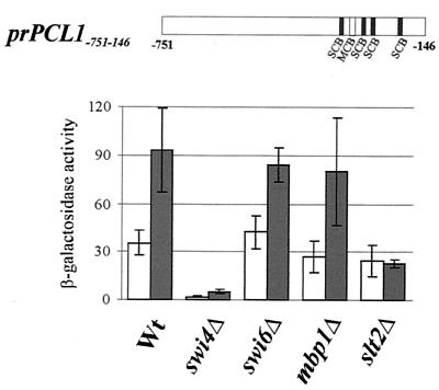
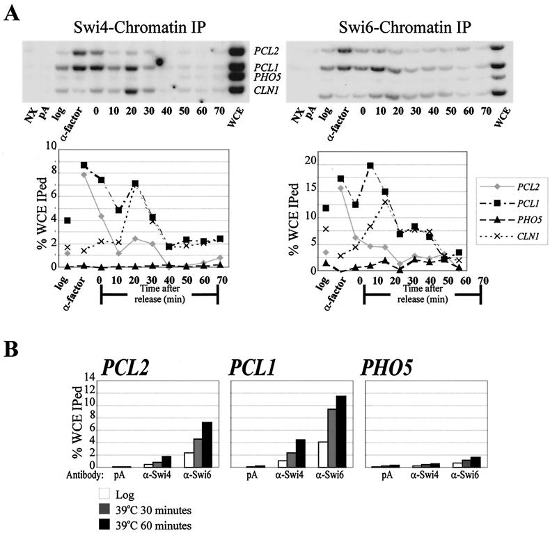
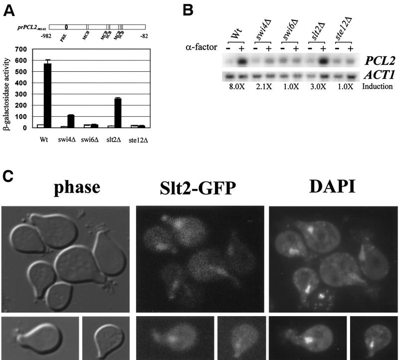
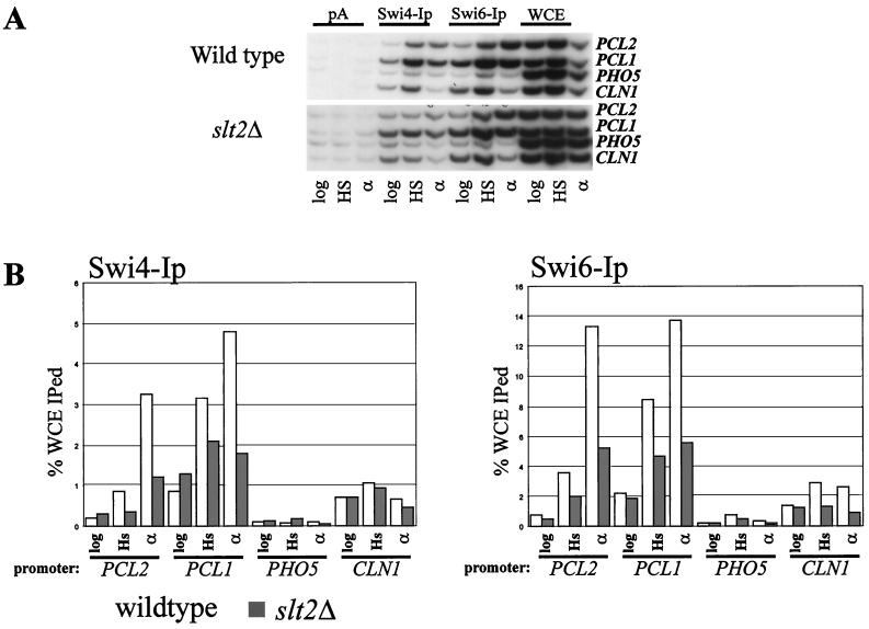
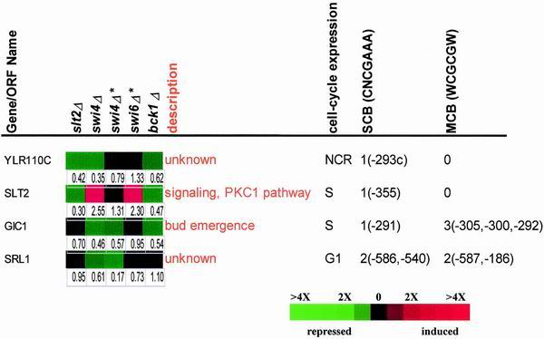
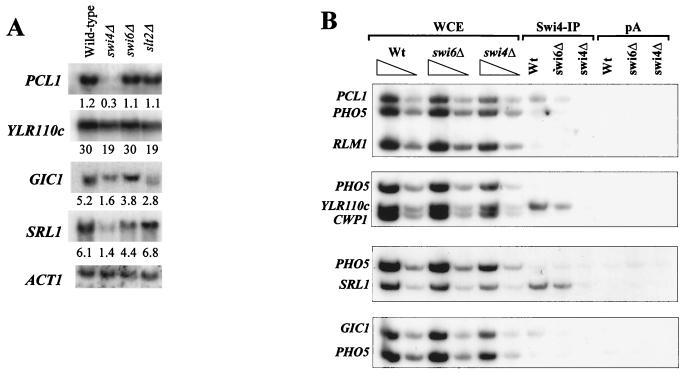
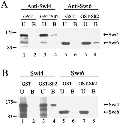
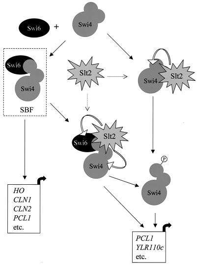
In Saccharomyces cerevisiae, the heterodimeric transcription factor SBF (for SCB binding factor) is composed of Swi4 and Swi6 and activates gene expression at the G(1)/S-phase transition of the mitotic cell cycle. Cell cycle commitment is associated not only with major alterations in gene expression but also with highly polarized cell growth; the mitogen-activated protein kinase (MAPK) Slt2 is required to maintain cell wall integrity during periods of polarized growth and cell wall stress. We describe experiments aimed at defining the regulatory pathway involving the cell cycle transcription factor SBF and Slt2-MAPK. Gene expression assays and chromatin immunoprecipitation experiments revealed Slt2-dependent recruitment of SBF to the promoters of the G(1) cyclins PCL1 and PCL2 after activation of the Slt2-MAPK pathway. We performed DNA microarray analysis and identified other genes whose expression was reduced in both SLT2 and SWI4 deletion strains. Genes that are sensitive to both Slt2 and Swi4 appear to be uniquely regulated and reveal a role for Swi4, the DNA-binding component of SBF, which is independent of the regulatory subunit Swi6. Some of the Swi4- and Slt2-dependent genes do not require Swi6 for either their expression or for Swi4 localization to their promoters. Consistent with these results, we found a direct interaction between Swi4 and Slt2. Our results establish a new Slt2-dependent mode of Swi4 regulation and suggest roles for Swi4 beyond its prominent role in controlling cell cycle transcription.


Figures

FIG. 1
FIG. 1
Slt2/Swi4-dependent and Swi6-independent activation of a PCL1-lacZ reporter gene. Schematic diagram of the PCL1 upstream regulatory region used to create a CYC::lacZ reporter construct. Locations of consensus SCB and MCB motifs are indicated. Wild-type (Wt, BY263), swi4Δ (BY108), swi6Δ (BY107), mbp1Δ (BY551), and slt2Δ (BY1342) strains were transformed with the prPCL1−751-146 reporter plasmid and grown at 30°C to log phase (open bars) or grown at 30°C to log phase and then heat shocked for 30 min at 39°C (filled bars). Cell lysates were made and β-galactosidase activity (Miller units) was determined. Activity depicted is the mean of three experiments; the error bars show the standard deviation for the three experiments.
FIG. 2
FIG. 2
Swi4 and Swi6 localization to the promoters of PCL1 and PCL2 during the cell cycle and after α-factor and heat shock treatment. (A) Cell cycle ChIP using affinity-purified Swi4 antibodies (left panel) and Swi6 antibodies (right panel). A wild-type strain (BY263) was grown to mid- log phase and then blocked in G1 with α-factor. Cells were inoculated into fresh medium lacking α-factor, and samples were taken every 10 min. Samples taken of the unsynchronized log-phase culture (log), the G1 arrested cells (α-factor), and the 10-min time points were cross-linked with formaldehyde. WCEs were made, and ChIP was done using pA alone or pA plus affinity-purified Swi4 or Swi6 antibodies as indicated. In the top panels multiplex PCR was performed to amplify the promoter regions of PCL2, PCL1, PHO5, and CLN1 in each of the ChIP samples, in an immunoprecipitation with extract which was not cross-linked (NX), and in WCEs. The graphs depict the results of phosphorimager analysis of each PCR product. The results are expressed as a percentage of the product in the WCE. The experiment shown is representative of three independent experiments with Swi4 antiserum and two with Swi6 antiserum. (B) Heat shock ChIPs. Cultures of a wild-type strain (BY263) grown at either 30°C (white bars) or 30°C, followed by heat shock at 39°C for 30 min (gray bars) or 60 min (black bars) were cross-linked with formaldehyde. Lysates were made, and ChIP was performed using pA or with affinity-purified Swi4 antibodies (α-Swi4) or affinity-purified Swi6 antibodies (α-Swi6). PCR was performed to amplify the promoters of PCL1, PCL2, and PHO5 in the ChIP samples and in WCE. The bar graphs depict the results of phosphorimager analysis of each PCR product. The results are expressed as a percentage of the product in WCE.
FIG. 3
FIG. 3
Slt2 and SBF activity during α-factor treatment. (A) PCL2 reporter gene assay. Above the graph is a schematic diagram of the PCL2 promoter region used to create the PCL2-lacZ reporter construct prPCL2−-989-82. The locations of the consensus SCB, MCB, and PRE elements are indicated. The graph shows α-factor-induced expression of β-galactosidase from the prPCL2−989-82 reporter plasmid. Wild-type (wt BY263), swi4Δ (BY1321), swi6Δ (BY891), slt2Δ (BY1342), and ste12Δ (BY332) strains were transformed with prPCL2−989-82. The transformants were grown to mid-log phase (open bars) or grown to mid-log phase and treated with 5 μM α-factor for 2 h (filled bars). Cell lysates were made, and the β-galactosidase activity (Miller units) of each strain was determined. Activity depicted is the mean of three experiments; the error bars show the standard deviations for the three experiments. (B) PCL2 Northern blots. The indicated strains were grown to log phase or grown to log phase and treated with 5 μM α-factor for 2 h. Total RNA was isolated and probed with PCL2, followed by the loading control ACT1. Indicated across the bottom is the fold induction of PCL2 expression after α-factor treatment for each strain. Gel shown is representative of three independent experiments. (C) Subcellular localization of Slt2 after α-factor treatment. BY1343 cells (SLT2::GFP) were grown in rich medium to early log phase and then treated with 5 μM α-factor for 2 h. Cells were then washed once with buffer containing DAPI and once with water before examination by fluorescence microscopy at a magnification of ×1,000. Cells were exposed to fluorescent light through an FITC filter for 400 ms in order to visualize Slt2-GFP. Wild-type (BY263) cells treated identically showed no fluorescence. Photographs of the same fields viewed with Nomarski optics (DIC) and stained with DAPI to visualize cell nuclei are shown.
FIG. 4
FIG. 4
Slt2 requirement for SBF localization to the promoters of PCL1 and PCL2. (A) PCR amplification of ChIPs from wild-type and slt2Δ strains. Wild-type (BY263) and slt2Δ (BY1342) cultures grown at 30°C (log), heat shocked for 1 h at 39°C (HS), or treated with α-factor for 2 h (α) were cross-linked with formaldehyde. Lysates were made, and ChIPs were performed using pA alone, Swi4 antibodies (Swi4-Ip), or Swi6 antibodies (Swi6-Ip). Multiplex PCR was used to amplify the promoter regions of PCL2, PCL1, PHO5, and CLN1 from each of the immunoprecipitated chromatin reactions and the WCE. (B) PhosphorImager analysis of each PCR product was performed, and the localization of Swi4 and Swi6 to the promoters of PCL1, PCL2, PHO5, and CLN1 is depicted as percent WCE immunoprecipitated (%WCE Iped). Open bars, wild-type ChIPs; shaded bars, slt2Δ ChIPs. The data shown are representative of two separate experiments.
FIG. 5
FIG. 5
DNA microarray analysis. Experiments are listed along the horizontal axis, and genes are listed along the vertical axis. Three independent DNA microarray datasets were generated (slt2Δ-25°C, swi4Δ-25°C, and bck1Δ-25°C) and compared to two related data sets from previously published data (swi4Δ and swi6Δ [27]). Genes whose expression levels were reduced >1.8-fold in swi4Δ and/or slt2Δ cells are shown. Three control experiments were performed with wild-type RNA (wild-type by wild-type comparison) to validate the cutoff used in the experiments with the mutant strains (see Materials and Methods). Cell cycle expression data were obtained from the Stanford Cell Cycle Expression project and Spellman et al. (52). SCB and MCB elements were found using the SCPD and SGD databases available on the internet. Numbers below the colored squares indicate the intensity ratio between the mutant strain versus the wild-type strain grown under the same conditions and represent the average values from two independent experiments. A value of 1 indicates no change; only genes that were verified by Northern blot analysis are shown.
FIG. 6
FIG. 6
Northern blot analysis and Swi4 localization to the promoters of newly identified Swi4-Slt2-dependent genes. (A) Northern blot analysis of a subset of proposed Swi4- and/or Slt2-dependent genes. Wild-type (Wt, BY263), swi4Δ (BY108), swi6Δ (BY107), and slt2Δ (BY1342) cells were grown to mid-log phase at 30°C, and total RNA was isolated and probed with the indicated open reading frame (ORF). A probe for ACT1 was used as a loading control. Autoradiograms shown are representative of one of three experiments. PhosphorImager analysis was performed, and the signal of each ORF was standardized to the ACT1 loading control (volume ORF/volume ACT1). The average standardized signal of three experiments is indicated beneath each lane. The standard deviation for each triplicate was <10% of the average. (B) Swi4 localization to the promoters of Swi4-Slt2-dependent genes. Wild-type (Wt, BY263), swi6Δ (BY107), and swi4Δ (BY108) cultures grown at 30°C (log) were cross-linked with formaldehyde, WCEs were made, and ChIPs were performed using pA or Swi4 antibodies (Swi4-IP). Multiplex PCR was performed as indicated for the ChIP samples and on fivefold serial dilutions of the WCE to amplify the promoter regions of the genes listed to the left of the panel. RLM1 and PHO5 were included as negative controls. CWP1 was identified as a putative SBF target in a genome-wide ChIP experiment (29) but did not immunoprecipitate with Swi4 antibodies in our experiment.
FIG. 7
FIG. 7
Binding of Slt2 to Swi4 and Swi6 in vitro. (A) Five micrograms of partially purified SBF derived from insect cell extracts was incubated with either GST or GST-Slt2 immobilized on glutathione beads. The unbound (U) and bound (B) fractions were separated by SDS–6% PAGE. The gels were blotted and incubated with Swi4 antiserum (lanes 1 to 4) or Swi6 antiserum (lanes 5 to 8) to identify Swi4 or Swi6 proteins. The migration positions of molecular mass markers are indicated to the left (in kilodaltons). (B) Two microliters of in vitro-translated Swi4 and Swi6 were incubated with either GST or GST-Slt2 immobilized on glutathione beads. The unbound (U) and bound (B) fractions were separately by SDS–6% PAGE. The migration positions of molecular mass markers are indicated to the left (in kilodaltons).
FIG. 8
FIG. 8
Model for Slt2 and Swi4 coordinate regulation of PCL1-like genes. Possible modes of regulation of Swi4 by Slt2 are diagrammed. Swi4 interacts with Swi6 to form SBF (in dashed box) to control expression of G1-specific transcripts (HO, CLN1, CLN2, PCL1, etc.) via binding to SCB elements. In our model, phosphorylation of Swi4 by Slt2, either on its own or complexed with Swi6, may relieve the autoinhibition of Swi4 DNA binding. Regulation of Swi4 by Slt2 may impart a unique Swi6-independent function to Swi4 that directs activation of a subset of Swi4-Slt2-dependent genes (PCL1, SRL1, etc.). Genes controlled by Swi4 and Slt2 include non-cell-cycle-regulated genes and define a novel role for Swi4 beyond its major role in controlling cell-cycle-dependent transcription.

References

    1. Aerne B L, Johnson A L, Toyn J H, Johnston L. Swi5 controls a novel wave of cyclin synthesis in late mitosis. Mol Biol Cell. 1998;9:945–956. - PMC - PubMed
    1. Andrews B, Measday V. The cyclin family of budding yeast: abundant use of a good idea. Trends Genet. 1998;14:66–72. - PubMed
    1. Andrews B J, Moore L A. Interaction of the yeast Swi4 and Swi6 cell cycle regulatory protein in vitro. Proc Natl Acad Sci USA. 1992;89:11852–11856. - PMC - PubMed
    1. Ausubel F M, Brent R, Kingston R E, Moore D D, Seidman J G, Smith J A, Struhl K. Current protocols in molecular biology. Vol. 2. Boston, Mass: John Wiley & Sons, Inc.; 1993.
    1. Baetz K, Andrews B. Regulation of the cell cycle transcription factor Swi4 through auto-inhibition of DNA binding. Mol Cell Biol. 1999;19:6729–6741. - PMC - PubMed

Publication types

MeSH terms

LinkOut - more resources