. 2001 Oct;21(19):6537-48.
doi: 10.1128/MCB.21.19.6537-6548.2001.

Creation of a pluripotent ubiquitin-conjugating enzyme

Affiliations

Creation of a pluripotent ubiquitin-conjugating enzyme

C Ptak et al. Mol Cell Biol. 2001 Oct.

Abstract

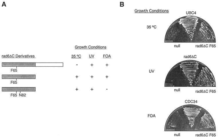
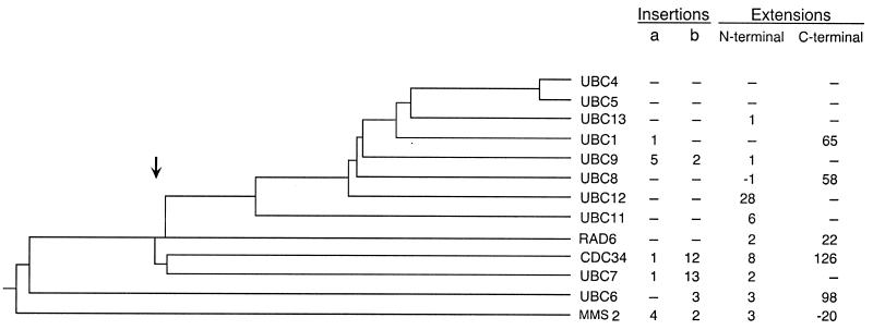
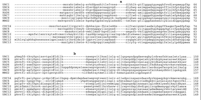
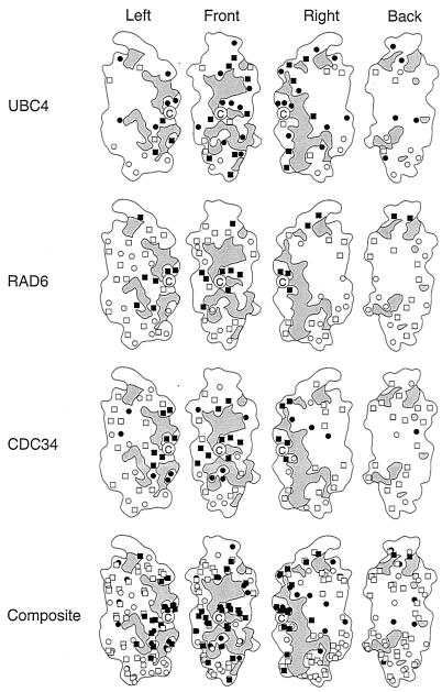
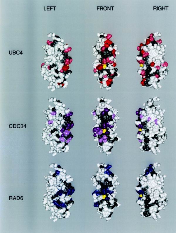
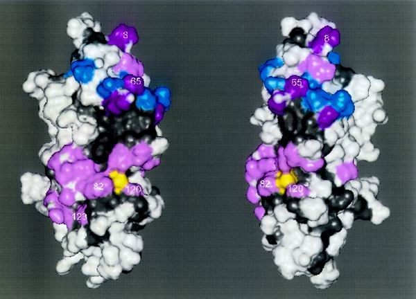
We describe the creation of a pluripotent ubiquitin-conjugating enzyme (E2) generated through a single amino acid substitution within the catalytic domain of RAD6 (UBC2). This RAD6 derivative carries out the stress-related function of UBC4 and the cell cycle function of CDC34 while maintaining its own DNA repair function. Furthermore, it carries out CDC34's function in the absence of the CDC34 carboxy-terminal extension. By using sequence and structural comparisons, the residues that define the unique functions of these three E2s were found on the E2 catalytic face partitioned to either side by a conserved divide. One of these patches corresponds to a binding site for both HECT and RING domain proteins, suggesting that a single substitution in the catalytic domain of RAD6 confers upon it the ability to interact with multiple ubiquitin protein ligases (E3s). Other amino acid substitutions made within the catalytic domain of RAD6 either caused loss of its DNA repair function or modified its ability to carry out multiple E2 functions. These observations suggest that while HECT and RING domain binding may generally be localized to a specific patch on the E2 surface, other regions of the functional E2 face also play a role in specificity. Finally, these data also indicate that RAD6 uses a different functional region than either UBC4 or CDC34, allowing it to acquire the functions of these E2s while maintaining its own. The pluripotent RAD6 derivative, coupled with sequence, structural, and phylogenetic data, suggests that E2s have diverged from a common multifunctional progenitor.


Figures

FIG. 1
FIG. 1
A pluripotent E2. (A) Complementation of defects associated with the ubc4Δ ubc5Δ (35°C), rad6Δ (UV), or cdc34Δ (FOA) mutant strains by various rad6ΔC derivatives. The positions of point substitutions introduced into the rad6ΔC catalytic domain (gray) are indicated, as is the addition of the CDC34 tail domain (white) to the carboxy terminus of rad6ΔC. The ability (+) or inability (−) of each derivative to allow colony formation by each strain under the conditions given was determined. (B) The ability of a plasmid expressing rad6ΔC F65 to complement each disruption strain is compared to that of a negative control plasmid containing no E2 gene (null) or the appropriate positive control plasmid expressing either UBC4, CDC34, or rad6ΔC (see Materials and Methods).
FIG. 2
FIG. 2
Evolutionary relationships among E2s from S. cerevisiae (see Materials and Methods). Also indicated are the lengths of major sequence landmarks for each E2. The letters a and b refer to the insertion positions highlighted in Fig. 3.
FIG. 3
FIG. 3
Structure-based sequence alignment of the E2 catalytic domains from S. cerevisiae (see Materials and Methods). The two positions that accommodate insertions are labeled a and b.
FIG. 4
FIG. 4
Surface map of three E2 catalytic domains based on function. For each E2, the catalytic domain of S. cerevisiae RAD6 has been used as the map template rotated through increments of 90°. Gray areas represent regions that are structurally conserved. The active-site cysteine is marked by the letter C. Amino acid substitutions with no effect on function are indicated with open boxes. Substitutions that affect functions that are specific to each E2 are indicated with filled boxes. Open circles denote residues that are unconserved within each E2 family indicated, while closed circles denote residues that exhibit conservation within an E2 family but variation between families.
FIG. 5
FIG. 5
Three-dimensional map of functionally important surfaces. The three-dimensional surface of S. cerevisiae RAD6 is used as the map template rotated to the left or right by 60°. The amino terminus is found at the top of the structures. The active-site cysteine is yellow. Conserved solvent-accessible regions are gray. Residues of clear specific functional importance for each E2 based on mutation studies are highlighted in dark green (UBC4), dark magenta (CDC34), or blue (RAD6). Residues of potential importance to E2 specificity according to the criteria described in Results are highlighted in light green (UBC4) or pink (CDC34).
FIG. 6
FIG. 6
Surface determinants of E2 function are concentrated in two patches. Mapped onto the S. cerevisiae RAD6 crystal structure are all of the residues which play a role in the functional specificity of either UBC4, CDC34, or RAD6. The two views of RAD6 vary by a rotation of 30°, and the amino terminus is found at the top of the structures. Shown on the surface are (i) residues from UBCH7 which form contacts with the HECT domain of E6-AP (blue), (ii) residues identified as determinants of UBC4, CDC34, or RAD6 function (Fig. 5) that coincide with UBCH7/E6-AP HECT contact points (magenta), and (iii) residues identified as determinants of UBC4, CDC34, or RAD6 function (Fig. 5) that do not coincide with UBCH7/E6-AP HECT contact points (pink). The active-site cysteine is shown (yellow), as are conserved E2 residues (gray). Numbers indicate amino acid positions within rad6ΔC that either play a role in the DNA repair function of RAD6 or allow it to acquire the UBC4 and CDC34 functions.

Similar articles

Cited by

References

    1. Broomfield S, Chow B L, Xiao W. MMS2 encoding a ubiquitin-conjugating-enzyme-like protein is a member of the yeast error-free postreplication repair pathway. Proc Natl Acad Sci USA. 1998;95:5678–5683. - PMC - PubMed
    1. Chen P, Johnson P, Sommer T, Jentsch S, Hochstrasser M. Multiple ubiquitin-conjugating enzymes participate in the in vivo degradation of the yeast MATα2 repressor. Cell. 1993;74:357–369. - PubMed
    1. Cook W J, Jeffery L C, Sullivan M L, Vierstra R D. Three-dimensional structure of a ubiquitin-conjugating enzyme E2. J Biol Chem. 1992;267:15116–15121. - PubMed
    1. Cook W J, Jeffery L C, Xu Y, Chau V. Tertiary structures of class I ubiquitin-conjugating enzymes are highly conserved: crystal structure of yeast UBC4. Biochemistry. 1993;32:13809–13817. - PubMed
    1. Cook W J, Martin P D, Edwards B F, Yamazaki R K, Chau V. Crystal structure of a class I ubiquitin conjugating enzyme (UBC7) from Saccharomyces cerevisiae at 2.9 angstroms resolution. Biochemistry. 1997;36:1621–1627. - PubMed

Publication types

MeSH terms

Substances