. 2001 Oct;21(19):6549-58.
doi: 10.1128/MCB.21.19.6549-6558.2001.

The ornithine decarboxylase gene is essential for cell survival during early murine development

Affiliations

The ornithine decarboxylase gene is essential for cell survival during early murine development

H Pendeville et al. Mol Cell Biol. 2001 Oct.

Abstract

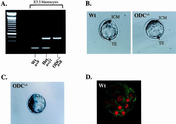
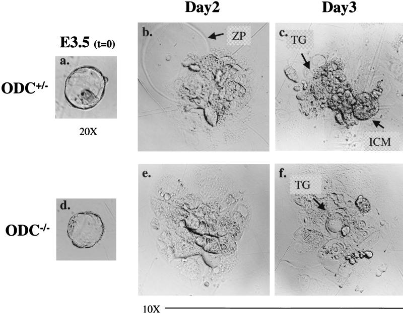
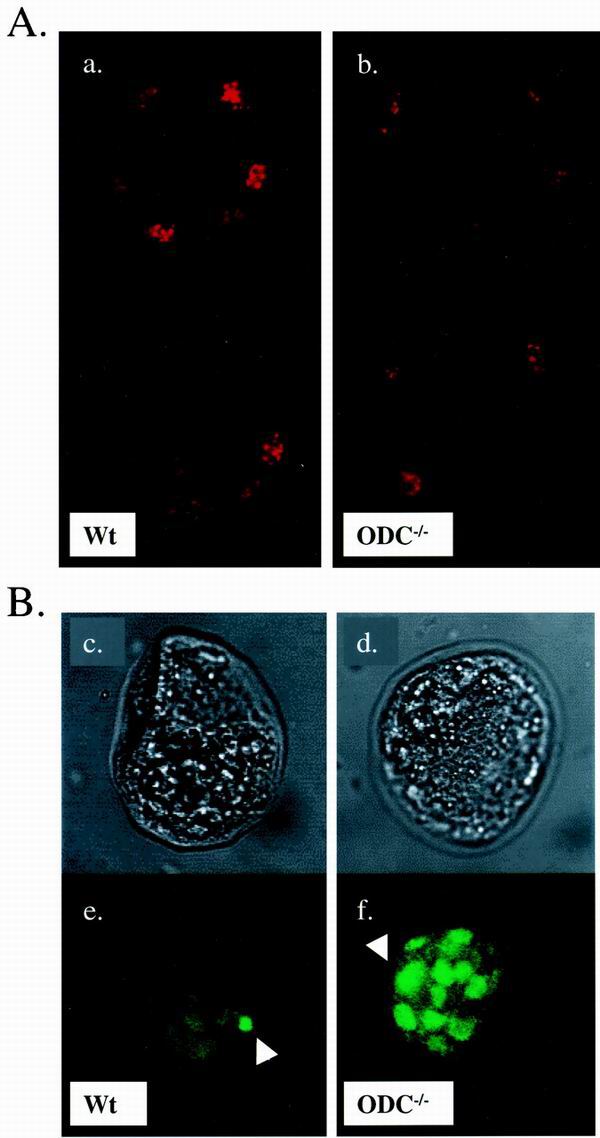
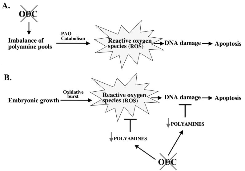
Overexpression and inhibitor studies have suggested that the c-Myc target gene for ornithine decarboxylase (ODC), the enzyme which converts ornithine to putrescine, plays an important role in diverse biological processes, including cell growth, differentiation, transformation, and apoptosis. To explore the physiological function of ODC in mammalian development, we generated mice harboring a disrupted ODC gene. ODC-heterozygous mice were viable, normal, and fertile. Although zygotic ODC is expressed throughout the embryo prior to implantation, loss of ODC did not block normal development to the blastocyst stage. Embryonic day E3.5 ODC-deficient embryos were capable of uterine implantation and induced maternal decidualization yet failed to develop substantially thereafter. Surprisingly, analysis of ODC-deficient blastocysts suggests that loss of ODC does not affect cell growth per se but rather is required for survival of the pluripotent cells of the inner cell mass. Therefore, ODC plays an essential role in murine development, and proper homeostasis of polyamine pools appears to be required for cell survival prior to gastrulation.


Figures

FIG. 1
FIG. 1
(A) Targeting strategy of the ODC genomic locus. Schematics of the wild-type locus (top), targeting vector (middle), and recombined locus (bottom) are shown. Exons are indicated by hatched boxes, and the arrows correspond to the three primers used for PCR genotyping. Abbreviations: St, StuI; H, HindIII; Sc, ScaI; Sl, SalI; Ss, SstI. (B) Southern blot analysis of genomic DNA isolated from ES cell clones. Digestion of the targeted ODC locus with ScaI and hybridization to a 3′ external probe show a 2.8- and an 8.3-kb band specific to the wild-type and targeted alleles, respectively, confirming homologous recombination. (C) Transmission of the ODC mutant allele to the progeny was determined by PCR amplification of tail DNAs using the primers described in Materials and Methods. PCR products were resolved on a 2% agarose gel in Tris-acetate-EDTA buffer. WT and Wt, wild type; mut, mutant; HSV-TK, herpes simplex virus thymidine kinase; IRES, internal ribosome entry site.
FIG. 2
FIG. 2
Histological sections of wild-type (Wt) and ODC−/− mutant embryos in utero. (A) Transverse section through the decidua of a normal embryo at early egg cylinder stage (E5.5). Note the appearance of the proamniotic cavity and the clearly differentiated embryonic and extraembryonic ectoderms. (B) Transverse section through a decidua of a degenerating E5.5 ODC−/− embryo (arrow). No discernible structure can be distinguished. pa, proamniotic cavity; ee, embryonic ectoderm; epc, ectoplacental cone; eee, extraembryonic ectoderm; ve, visceral endoderm.
FIG. 3
FIG. 3
Zygotic ODC is expressed in blastocysts but is dispensable for blastocyst formation. (A) Representative genotypic analysis of E3.5 embryos from ODC+/− breedings. DNA from individual blastocysts was isolated as described in Materials and Methods, and primers were used to amplify fragments from the wild-type and/or ODC mutant allele in each sample. ODC−/− embryos were found at nearly an expected Mendelian ratio (21%). (B) Phase-contrast photographs of wild-type (left panel) and ODC-null (right panel) blastocysts. ODC-deficient blastocysts appear morphologically normal. (C) Expression of zygotic ODC, as detected by LacZ staining. An intense signal is observed in both the ICM and the TE of an ODC-deficient blastocyst. (D) A similar pattern of ODC expression (green) was detected with an ODC-specific antibody in a wild-type blastocyst. Blastocysts were also stained with propidium iodide (red), which detects DNA. Wt, wild type; Het., heterozygous.
FIG. 4
FIG. 4
ODC-deficient E3.5 blastocysts are compromised in their expansion in vitro. Photographs of the same heterozygous E3.5 blastocyst (a) at 2 (b) and 3 (c) days in culture and an ODC-deficient blastocyst (d) at 2 (e) and 3 (f) days in culture are shown. A well-developed ICM is evident in the ODC+/− embryo after 3 days, whereas by this time the ICM of the ODC-deficient blastocyst has already completely degenerated. Only the nondividing trophoblastic giant cells derived from ODC−/− blastocysts remained on the plates. ZP, zona pellucida; TG, trophoblastic giant cells.
FIG. 5
FIG. 5
ODC is required for cell survival in the ICM. (A) Comparison of the proliferation index between wild-type (a) and ODC−/− (b) E3.5 embryos. Blastocysts were isolated, fixed, and stained with an antibody specific to phosphohistone H3 at Ser10. Two serial sections captured by confocal imaging of each blastocyst are shown. Mitotic cells are readily detected in embryos from both genotypes. (B) TUNEL analysis of E3.5 blastocysts. The upper panels show phase-contrast photographs of wild-type (c) and ODC−/− (d) blastocysts, and the lower panels correspond to immunodetection of TUNEL labeling (e and f). The wild-type embryo displays minimal apoptosis (e). In contrast, the ODC-deficient embryo exhibits massive cell death confined to the ICM (f). The arrowheads indicate fluorescent dots corresponding to fragmented DNA. Wt, wild type.
FIG. 6
FIG. 6
Two models of apoptosis caused by loss of ODC. (A) Loss of ODC would be predicted to lead to reductions and an imbalance in polyamine pools. Polyamine catabolism by polyamine oxidase (PAO) would continue and result in the production of ROS, which result in DNA damage and cell death. (B) The rapidly dividing cells of the ICM are at risk of damage due to ROS from the oxidative burst, and these are normally countered by polyamines, which can act as direct scavengers of ROS and/or protect DNA or stimulate DNA repair (21, 47, 48). Loss of ODC would lead to reductions of polyamine pools and thus place these cells at high risk of death from ROS.

References

    1. Ajiro K, Yoda K, Utsumi K, Nishikawa Y. Alteration of cell cycle-dependent histone phosphorylations by okadaic acid. Induction of mitosis-specific H3 phosphorylation and chromatin condensation in mammalian interphase cells. J Biol Chem. 1996;271:13197–13201. - PubMed
    1. Askew D S, Ashmun R A, Simmons B C, Cleveland J L. Constitutive c-myc expression in an IL-3-dependent myeloid cell line suppresses cell cycle arrest and accelerates apoptosis. Oncogene. 1991;6:1915–1922. - PubMed
    1. Auvinen M, Paasinen A, Andersson L C, Holtta E. Ornithine decarboxylase activity is critical for cell transformation. Nature. 1992;360:355–358. - PubMed
    1. Balasundaram D, Tabor C W, Tabor H. Spermidine or spermine is essential for the aerobic growth of Saccharomyces cerevisiae. Proc Natl Acad Sci USA. 1991;88:5872–5876. - PMC - PubMed
    1. Balasundaram D, Tyagi A K. Polyamine-DNA nexus: structural ramifications and biological implications. Mol Cell Biochem. 1991;100:129–140. - PubMed

Publication types

Substances