. 2001 Oct;21(19):6559-73.
doi: 10.1128/MCB.21.19.6559-6573.2001.

Intrachromatid excision of telomeric DNA as a mechanism for telomere size control in Saccharomyces cerevisiae

Affiliations

Intrachromatid excision of telomeric DNA as a mechanism for telomere size control in Saccharomyces cerevisiae

M Bucholc et al. Mol Cell Biol. 2001 Oct.

Abstract

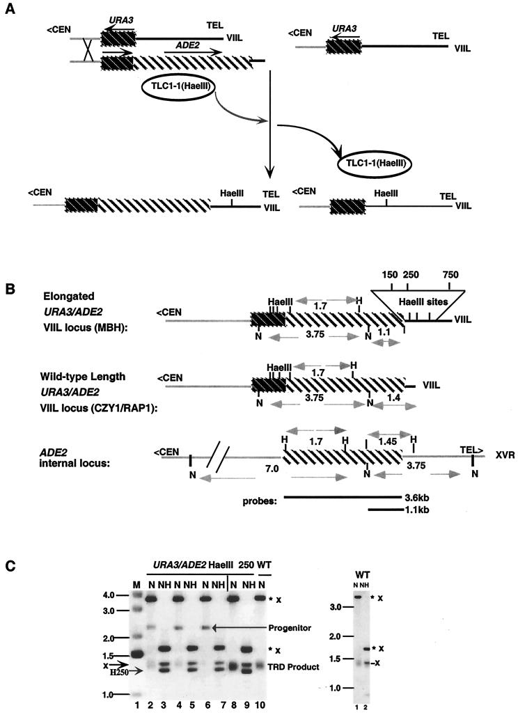
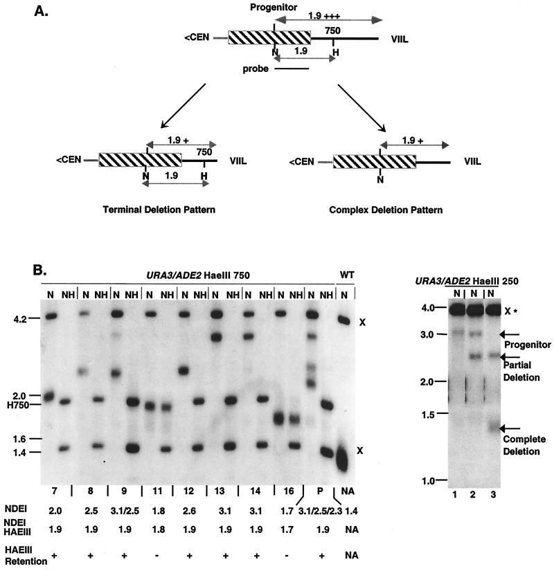
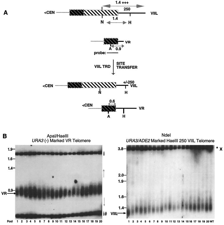
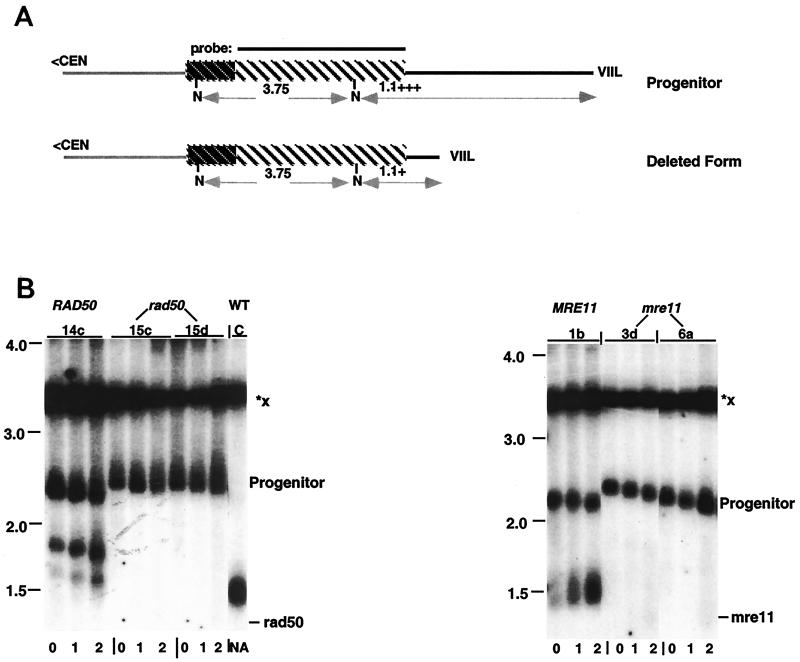
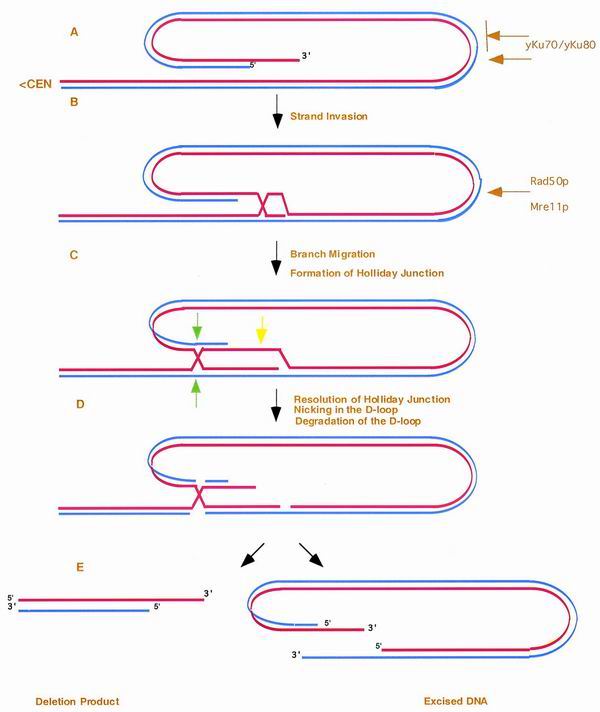
We have previously identified a process in the yeast Saccharomyces cerevisiae that results in the contraction of elongated telomeres to wild-type length within a few generations. We have termed this process telomeric rapid deletion (TRD). In this study, we use a combination of physical and genetic assays to investigate the mechanism of TRD. First, to distinguish among several recombinational and nucleolytic pathways, we developed a novel physical assay in which HaeIII restriction sites are positioned within the telomeric tract. Specific telomeres were subsequently tested for HaeIII site movement between telomeres and for HaeIII site retention during TRD. Second, genetic analyses have demonstrated that mutations in RAD50 and MRE11 inhibit TRD. TRD, however, is independent of the Rap1p C-terminal domain, a central regulator of telomere size control. Our results provide evidence that TRD is an intrachromatid deletion process in which sequences near the extreme terminus invade end-distal sequences and excise the intervening sequences. We propose that the Mre11p-Rad50p-Xrs2p complex prepares the invading telomeric overhang for strand invasion, possibly through end processing or through alterations in chromatin structure.


Figures

FIG. 1
FIG. 1
Incorporation of HaeIII sites into elongated telomeres. (A, left panel) rap1-17 cells containing newly generated URA3/ADE2-marked VIIL telomeres were derived from a URA3(−)-marked VIIL telomere by recombination with a homologous fragment containing the URA3/ADE2 sequences and 80 bp of telomeric seed sequence. Transformation was conducted in the presence of a plasmid borne copy of TLC1-1. The TLC1-1 RNA contains a HaeIII site within its template region. The telomeric seed sequence was elongated to sizes greater than that of the wild type, which is typical of the telomeres present in rap1-17 cells. As a consequence, HaeIII sites were introduced into the URA3/ADE2-marked VIIL telomere, as well as into other unmarked chromosomal telomeres, during the addition of telomeric repeats. After loss of the plasmid borne TLC1-1 gene, the rap1-17 strains containing the elongated URA3/ADE2 HaeIII-marked telomere were mated to an isogenic wild-type strain, and wild-type RAP1 spore colonies containing elongated telomeres were identified. These spore colonies represent the MBH series of strains. (A, left panel) Elongated URA3(−)-marked VIIL telomeres were grown in the presence of TLC1-1 RNA to generate HaeIII sites within both marked and unmarked chromosomal telomeres in rap1-17 cells. Incorporation of HaeIII sites at multiple positions is presumably due to repetitive cycles of deletion and elongation typical of rap1-17 telomeres. After loss of the mutant telomerase RNA, the plasmid-borne copy of rap1-17 was replaced with a wild-type copy of RAP1 through a plasmid shuffle, giving rise to the MUH series of strains (A, right panel). Dark striped regions, the URA3 gene; light striped regions, the ADE2 gene; chromosomal sequences, gray lines; telomeric sequences, black lines; <CEN, direction toward the centromere; arrow, direction of transcription. (B) Restriction maps of the genomic and telomeric ADE2 genes in the strains used in this study (35). WT refers to the strain CZY1/RAP1 carrying a wild-type length URA3/ADE2-marked telomere. Fragment lengths are shown for NdeI (N), NdeI/HaeIII, and HaeIII (H) sites. Each URA3/ADE2-marked VIIL telomere contains one of the three HaeIII sites. The position of a HaeIII site in the telomere was determined by the size of the NdeI/HaeIII fragment after subtracting the length of subtelomeric sequences (1.1 kb). These sites were positioned at 150 bp (HaeIII 150), 250 bp (HaeIII 250), and 750 bp (HaeIII 750) from the subtelomeric junction. The relative positions of HaeIII 150, HaeIII 250, and HaeIII 750 telomeres are shown in the expanded view. The 3.6- and 1.1-kb ADE2 probes used in the study (top and bottom, respectively) are also shown. Designations were described in the Fig. 1A legend. TEL> refers to the direction toward the telomere. (C, left panel) DNA isolated from MBH22-7b colonies, containing the URA3/ADE2 HaeIII 250-marked VIIL telomere, was digested with NdeI (N) or with NdeI and HaeIII (NH) and then subjected to Southern blotting with the 3.6-kb ADE2 probe indicated in panel B. Progenitor refers to the original marked elongated telomere after digestion of DNA isolated from several MBH22-7b subclones with NdeI (lanes 2, 4, and 6). In some MBH22-7b colonies (lane 8), the progenitor had been deleted before subculturing was done for individual colonies. WT refers to NdeI-cleaved DNA derived from CZY1/RAP1. ✽, Restriction fragments derived from the telomeric copy of ADE2; x, restriction fragments derived from the internal ADE2 locus. Size markers (in kilobases) are shown on the left. The positions of the Progenitor, the TRD product, and the NdeI/HaeIII fragment carrying the HaeIII 250 (H250) site are also indicated. M refers to the molecular mass marker. (C, right panel) As a control, DNA was isolated from CZY1/RAP1 and digested with NdeI or with both NdeI and HaeIII. Note that digestion of CZY1/RAP1 generates an internal 1.45-kb NdeI/HaeIII fragment that overlaps with the telomeric fragment. For presentation purposes, the autoradiogram is a composite of two portions of a single gel, with the junction borders indicated by the vertical line on top of the gel. Similar confirmations of HaeIII incorporation were carried out in MBH11-8b and MBH93-6d, carrying the HaeIII 150- and the HaeIII 750-marked telomeres, respectively.
FIG. 2
FIG. 2
Site retention patterns associated with TRD. (A) Site retention was assayed in partial deletions, representing incomplete deletions that are smaller than the distance between the HaeIII site and the terminus. A terminal deletion pattern (left panel) maintains the HaeIII site. A complex deletion TRD pattern (right panel) would lead to both site loss and retention. The shadings of lines and boxes are described in the legend to Fig. 1A. (B, left panel) MBH91-3 (P), which contains both elongated and deleted forms of the URA3/ADE2 HaeIII 750-marked VIIL telomere, was subcultured, and individual colonies were isolated (lanes 7 to 16). DNAs isolated from these colonies were digested with NdeI to determine the extent of the deletion and with NdeI and HaeIII to measure the retention of the site. The resulting Southern blot was probed with the 1.1-kb NdeI/BamHI ADE2 restriction fragment of pL909. Size markers (in kilobases) are shown to the left of the autoradiogram. Below each lane, the telomeric fragment size (NDEI), the NdeI/HaeIII telomeric fragment size (NDEI/HAEIII), and the presence or absence of the HaeIII site (HAEIII RETENTION) are depicted. HaeIII 750 (H750) denotes the position of the terminal NdeI/HaeIII fragment. x, Restriction sites from the genomic ADE2 locus; NA, not applicable due to absence of HaeIII sites. Other designations are described in the legend to Fig. 1. Subclones 11 and 16 have lost the HaeIII site as a consequence of incomplete deletions larger than the distance between the HaeIII site and the extreme terminus. Partial deletions, on the other hand, (subclones 7, 8, 9, and 12) all displayed the HaeIII 750 site. (B, right panel) DNAs isolated from three subclones of MBH21-20b were subjected to digestion with NdeI (N), and subsequent Southern blots were probed with the 3.6-kb BamHI fragment containing the ADE2 gene. Lane 1, the 3.1-kb precursor representing a telomere tract length of 2.0 kb; lane 2, the 2.5-kb partial deletion product representing a 1.4-kb telomeric tract; lane 3, the 1.35-kb complete deletion product representing a tract length of ≈300 bp. All other designations are indicated in the legend to Fig. 1.
FIG. 3
FIG. 3
Relationship between TRD and HaeIII site transfer. (A) Site transfer is assayed by measuring the frequency of HaeIII site movement from the elongated URA3/ADE2 HaeIII 250-marked VIIL telomere to the URA3(−)-marked VR telomere in strain YP17-24c. The shading of lines and boxes has been described in Fig. 1A. (B, left panel) DNA was isolated from 20 batch cultures, each containing 10 independent colonies. DNA was digested with ApaI and HaeIII and probed with a 1.7-kb PstI fragment of pRS316 containing the URA3 gene. The telomeric fragments (VR) were detected in each batch. The position of a unique HaeIII site transferred to the VR telomere (as well as the length of the VR telomere after transfer) is unpredictable and depends on the mechanism leading to the site transfer (gray arrows to the right of the panel). The 1.8-kb fragment represents the left junction of the chromosomal URA3 gene, while the smaller fragments represent two 350-bp HaeIII sites that are common to both telomeric and genomic copies of URA3. The positions of internal URA3 sequences are designated by “i.” Fragments originating from the URA3(−) telomeric fragment are denoted by a hatched mark. Size markers are shown on the left. (B, right panel) DNAs from the same samples were digested with NdeI and Southern blots were probed with the 3.6-kb BamHI fragment containing the ADE2 gene. All samples contained deleted URA3/ADE2-marked VIIL telomeres. WT refers to CZY1/RAP1. Other designations are defined in Fig. 1C.
FIG. 4
FIG. 4
rad50 and mre11 mutations inhibit TRD. (A) Diagram of the restriction maps from the progenitor and deleted ura3/ADE2-marked VIIL telomeres. Line and box shadings are defined in Fig. 1A. (B, left panel) DNA was isolated from a wild-type spore colony (YPR50-14c) and from rad50 spore colonies (YPR50-15c and YPR50-15d) after two rounds of liquid subculturing, corresponding to 20 generations of growth (0 to 2, indicated below each lane). All strains contain the elongated ura3/ADE2-marked VIIL telomere. The DNA was digested with NdeI, and the resulting Southern blots were hybridized with the 3.6-kb BamHI fragment of pL909 carrying the ADE2 gene. The expected mobility of the telomeric fragment in rad50 cells is indicated on the right. Note that an incomplete deletion also accumulates during subculturing of wild-type cells. (B, right panel) DNA was isolated from a wild-type spore colony (YPM11-1b) and from mre11 spore colonies (YPM11-3d and YPM11-6a), containing the elongated ura3/ADE2-marked VIIL telomere, after two rounds of liquid subculturing as described above. DNA was digested and subjected to Southern analysis as described in the text. The expected mobility of the telomeric fragment in mre11 cells is indicated on the right. Size markers (in kilobases) are shown on the left of each panel. Vertical bars above the autoradiograms indicate the positions of lanes from the same gel that were spliced for presentation purposes. C refers to the strain CZY1/RAP1. All other designations were described in the legend to Fig. 1C.
FIG. 5
FIG. 5
Model for telomeric rapid deletion. We propose that the 3′ single strand from the telomeric terminus (A) invades distal telomere tract sequences, leading to the formation of a looped structure (B). (C) After branch migration, the displaced strand forms both a D-loop and Holliday junction. After nicking of the D-loop (yellow arrow), degradation of the D-loop (D), and resolution of the outer strands of the Holliday junction (green arrows), both the TRD product and a linear excision product are produced (E). As shown, we propose that the Ku heterodimer regulates the degree of capping, which is likely to reduce the rate of TRD. Hence, yKu may essentially control the number of telomeres capable of rapid deletion. In this model, the Rad50p and Mre11p components of the MRX complex act to regulate the initial strand invasion either through chromosome condensation or the preparation of ends for recombination. Red line, the leading strand proceeding 5′ to 3′ toward the terminus; blue line, the complementary strand. Dependent upon the direction of resolution and the formation and stability of the D-loop, the intermediate can also give rise to circular forms of the terminal fragment.

References

    1. Aguilera A, Klein H L. Genetic and molecular analysis of recombination events in Saccharomyces cerevisiae occurring in the presence of the hyper-recombination mutation hpr1. Genetics. 1989;122:503–517. - PMC - PubMed
    1. Alani E, Cao L, Kleckner N. A method for gene disruption that allows repeated use of URA3 selection in the construction of multiply disrupted yeast strains. Genetics. 1987;116:541–545. - PMC - PubMed
    1. Bianchi A, de Lange T. Ku binds telomeric DNA in vitro. J Biol Chem. 1999;274:21223–21227. - PubMed
    1. Borts R H, Leung W Y, Kramer W, Kramer B, Williamson M, Fogel S, Haber J E. Mismatch repair-induced meiotic recombination requires the pms1 gene product. Genetics. 1990;124:573–584. - PMC - PubMed
    1. Bosco G, Haber J E. Chromosome break-induced DNA replication leads to nonreciprocal translocations and telomere capture. Genetics. 1998;150:1037–1047. - PMC - PubMed

Publication types

MeSH terms