. 2001 Sep;7(9):1035-40.
doi: 10.1038/nm0901-1035.

Functional small-diameter neovessels created using endothelial progenitor cells expanded ex vivo

Affiliations

Functional small-diameter neovessels created using endothelial progenitor cells expanded ex vivo

S Kaushal et al. Nat Med. 2001 Sep.

Abstract

Arterial conduits are increasingly preferred for surgical bypass because of inherent functional properties conferred by arterial endothelial cells, especially nitric oxide production in response to physiologic stimuli. Here we tested whether endothelial progenitor cells (EPCs) can replace arterial endothelial cells and promote patency in tissue-engineered small-diameter blood vessels (4 mm). We isolated EPCs from peripheral blood of sheep, expanded them ex vivo and then seeded them on decellularized porcine iliac vessels. EPC-seeded grafts remained patent for 130 days as a carotid interposition graft in sheep, whereas non-seeded grafts occluded within 15 days. The EPC-explanted grafts exhibited contractile activity and nitric-oxide-mediated vascular relaxation that were similar to native carotid arteries. These results indicate that EPCs can function similarly to arterial endothelial cells and thereby confer longer vascular-graft survival. Due to their unique properties, EPCs might have other general applications for tissue-engineered structures and in treating vascular diseases.


Figures
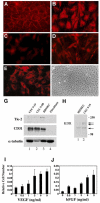
Figure 1
Figure 1. EPCs from the peripheral blood exhibit the morphologic and phenotypic properties of endothelial cells
EPCs exhibit typical cobblestone endothelial morphology (a), CD31 (b), von Willebrand factor (C) and incorporate aLDL (D). Unstimulated EPCs were negative for E-selectin expression (E), but LPS stimulated expression of E-selectin (f). g and h, Whole-cell extracts of two different EPC populations, EPC 9-19 and EPC 9-9B, HDMEC, and human fibroblasts were immunoblotted with antibodies shown on the left of each panel. Human fibroblast extracts were the negative control and HDMEC were the positive control. α-tubulin served as a control for protein loading. The three arrows indicate the fully glycosylated mature form of KDR (230 kDa), the partially glycosylated form of KDR (200 kDa), and unglycosylated form of KDR (150 kDa) . I and j, Proliferation of EPCs in response to increasing concentrations of VEGF and bFGF. Relative cell number was determined after 72 hours of incubation with the growth factors.
Figure 2
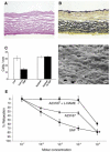
Figure 2. Pre-implantation studies with EPCs seeded on the decellularized matrix
Porcine iliac vessels were harvested and decellularized as described in Methods. a, Histology of the decellularized graft by hematoxylin and eosin stain (magnification 20X).b, Movat stain showed the internal and external elastin layers (dark brown) (magnification 20X).c, EPC density on the luminal surface of the decellularized grafts treated with low (1 dynes/cm2) or gradual (1 to 25 dynes/cm2) shear stress for 48-hours (open bars) and followed by 48 hour treatment with high (25 dynes/cm2) shear stress (black bars). d, Scanning electron microscopy of a EPC-seeded graft treated with gradual increases in shear stress for 48 hours and then an additional 48 hours of high shear stress showed a confluent layer of endothelial cells (magnification 880X, bar = 10 μ). e, The EPC-seeded graft induced NO production. In an organ chamber system, endothelial denuded guinea pig aortic ring was maximally contracted in the presence of U46619 and then relaxation was measured in the presence of increasing concentrations of calcium ionophore A23187 that was perfused directly into the EPC-seeded decellularized graft in the same organ chamber (▲ ). The relaxation effect mediated by A23187 was also measured in the presence of 10−3 M of L-NAME, an endothelial NO-synthase inhibitor (■ ). Relaxation was also recorded in the presence of increasing concentrations of SNP, an endothelial-independent NO donor ( ●). Data represent percent reduction in contraction and represent mean ± SEM derived from 4 grafts. *P<0.001 for dose response of A23187 vs. A23187 and L-NAME.
Figure 3
Figure 3. Long term patency of vascular grafts seeded with EPCs (---) or control vascular grafts with no endothelial cells ()
Patency was defined by Doppler flow studies and confirmed by carotid duplex studies. The EPC-seeded grafts were harvested at 15 days (n=3) or at 130 days (n=4). The non-seeded grafts (controls) were harvested at 15 days (n=4).
Figure 4
Figure 4. Post-implantation studies of the vascular grafts
a, Arteriogram of the EPC-seeded vascular graft 130 days after implantation. The location of the graft on the arteriogram was determined by staples at the sites of the anastomoses and is indicated by the two arrows. b, Seeded graft explanted at 15 days, showing a poorly organized thrombotic deposit overlying acellular graft media (asterisk). c-f, Seeded graft explanted at 130 days. c, Cellular intimal thickening as pannus from the adjacent arterial segments, infiltration of cells into the graft media, and confluent flattened cell monolayer lining the luminal surface of the graft (arrow). d, Section stained for extracellular matrix demonstrates the preservation of the original anatomic features, especially elastin (black). The internal elastic membrane is denoted by a white arrow. e, A macroscopic view of the explanted 130 day EPC-seeded vessel, with 1mm scale below. f, Staining of the cells within the intimal layer (i and inset) for smooth muscle alpha-actin. The medial layer (m) is unstained for alpha-actin. g, Staining of the cells overlying the intimal layer for vWF, denoted by arrow. Magnification in B, C, and F inset = 200X, in D and F= 100X, and in G = 400X. h, The pre-implant graft seeded with endothelial cells labeled with a fluorescent tracer. i, The 15 day and j, 130 explanted grafts showed the presence of CM-DiI-labeled endothelial cells on the luminal surface of the graft. Panels g-i are oriented with the lumen to the left.
Figure 5
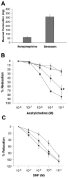
Figure 5. Vasomotor responsiveness of explanted grafts at 130 days
a, Three mid-portion segments of each of the 130-day explant graft were tested for contractility activity in an organ chamber. Increasing doses of norepinephrine and serotonin, ranging from 10−9 M to 10−4 M, contracted the grafts and the maximal responses are shown at 10−4 M concentration. b, Matched segments of EPC-seeded grafts (◆), carotid artery (■), and saphenous vein ( ▲) were contracted with 10−4 M of serotonin and relaxation was assessed in response to increasing concentrations of acetylcholine. In addition, the EPC-seeded graft was relaxed in the presence of of acetylcholine and 10−3 M of L-NAME ( x ). *P<0.001 for EPC-seeded graft vs the L-NAME attenuated response and saphenous vein and P=.088 for EPC-seeded graft vs carotid artery. c, Matched segments similar to (b) were also assessed for relaxation in the presence of SNP. Data represent percent reduction in contraction and represent mean + SEM (n=12).

Comment in

References

    1. Heart and Stroke Facts: Statistical Supplement. American Heart Association; 2000. http://www.americanheart.org/statistics.
    1. Tu JV, et al. Use of cardiac procedures and outcomes in elderly patients with myocardial infarction in the United States and Cannada. N Engl J Med. 1997;336:1500. - PubMed
    1. Weinberg CB, Bell E. A blood vessel model constructed from collagen and cultured vascular cells. Science. 1986;213:397. - PubMed
    1. L’Heureux N, Paquet S, Labbe R, Germain L, Auger FA. A completely biological tissue-engineered human blood vessel. FASEB J. 1998;12:47. - PubMed
    1. Niklason LE, et al. Functional arteries grown in vitro. Science. 1999;284:489. - PubMed

Publication types