. 2001 Sep 3;154(5):925-36.
doi: 10.1083/jcb.200102093.

Checkpoint inhibition of the APC/C in HeLa cells is mediated by a complex of BUBR1, BUB3, CDC20, and MAD2

Affiliations

Checkpoint inhibition of the APC/C in HeLa cells is mediated by a complex of BUBR1, BUB3, CDC20, and MAD2

V Sudakin et al. J Cell Biol. .

Abstract

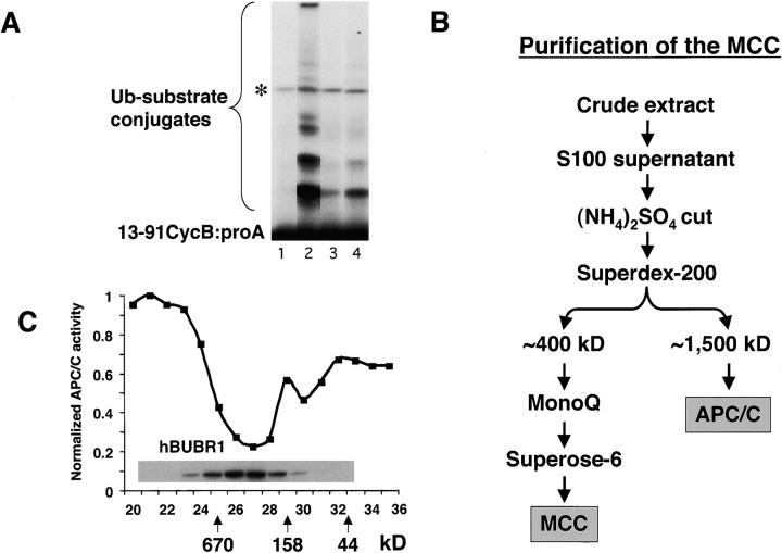
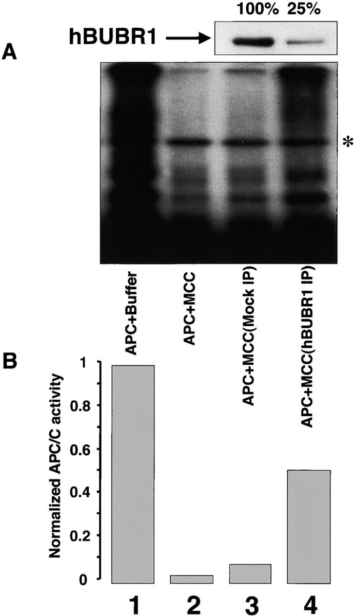
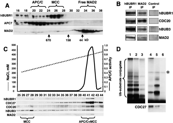
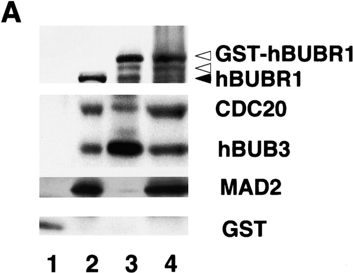
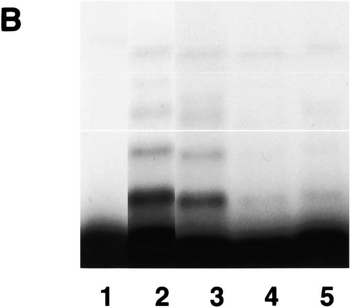
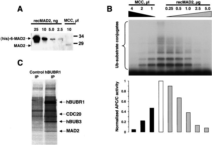
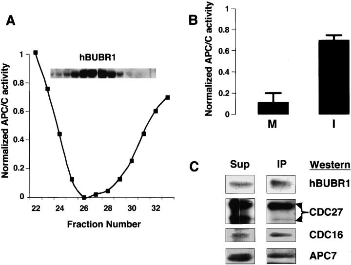
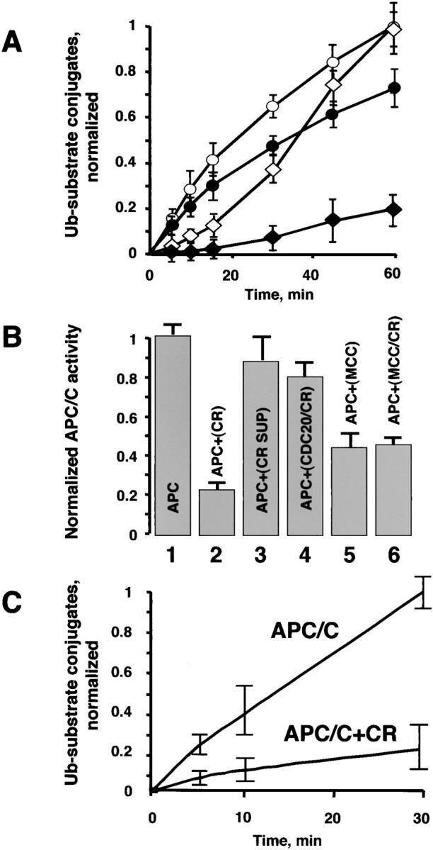
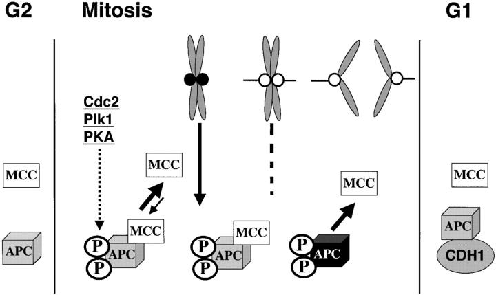
The mitotic checkpoint prevents cells with unaligned chromosomes from prematurely exiting mitosis by inhibiting the anaphase-promoting complex/cyclosome (APC/C) from targeting key proteins for ubiquitin-mediated proteolysis. We have examined the mechanism by which the checkpoint inhibits the APC/C by purifying an APC/C inhibitory factor from HeLa cells. We call this factor the mitotic checkpoint complex (MCC) as it consists of hBUBR1, hBUB3, CDC20, and MAD2 checkpoint proteins in near equal stoichiometry. MCC inhibitory activity is 3,000-fold greater than that of recombinant MAD2, which has also been shown to inhibit APC/C in vitro. Surprisingly, MCC is not generated from kinetochores, as it is also present and active in interphase cells. However, only APC/C isolated from mitotic cells was sensitive to inhibition by MCC. We found that the majority of the APC/C in mitotic lysates is associated with the MCC, and this likely contributes to the lag in ubiquitin ligase activity. Importantly, chromosomes can suppress the reactivation of APC/C. Chromosomes did not affect the inhibitory activity of MCC or the stimulatory activity of CDC20. We propose that the preformed interphase pool of MCC allows for rapid inhibition of APC/C when cells enter mitosis. Unattached kinetochores then target the APC/C for sustained inhibition by the MCC.


Figures

Figure 1.
Figure 1.
Identification of an APC/C inhibitor that copurifies with hBUBR1 kinase. (A) Identification of APC/C inhibitory activity in mitotic lysates. Mitotic lysate (S100) was fractionated through a Superose 6 gel filtration column by FPLC and proteins eluting at approximately the 300–400-kD range were found to inhibit APC/C activity. To maintain some physiological relevance to the APC/C inhibition assays, equal cell equivalents of APC/C and the various column fractions were used. Purified mitotic APC/C was incubated for 30 min with either buffer B alone (lane 2) or fractions from the 300–400-kD range of the Superose 6 column (lane 4) and then assayed for ubiquitin ligase activity. The same column fractions were assayed without addition of APC/C to monitor contaminating APC/C activity (lane 3). A reaction containing only substrate served as a negative control (lane 1). Asterisk denotes an iodinated contaminant bacterial protein that is not a substrate for APC/C and was excluded from the quantitation. (B) Flow chart of the purification of the APC/C inhibitory complex from HeLa cells. (C) Inhibitor of APC/C cofractionates with the hBUBR1 kinase complex. The elution profiles of hBUBR1 (inset) and APC/C inhibitory activity from the final Superose 6 column is shown. Arrows point to thyroglobulin (670 kD), γ-globulin (158 kD), and ovalbumin (44 kD), which served as migration standards.
Figure 2.
Figure 2.
MCC inhibition of the APC/C requires hBUBR1. Purified mitotic APC/C was incubated in the presence of buffer (lane 1), MCC (lane 2), MCC that was mock depleted with nonimmune IgG (lane 3) or immunodepleted with hBUBR1 antibodies (lane 4), and then assayed for ubiquitin ligase activity. (A) Ubiquitination assay. Mock- and hBUBR1-immunodepleted supernatants were probed for hBUBR1 (top panel). Asterisk denotes the contaminant iodinated protein. (B) Quantification of the ubiquitination assay on phosphorimager.
Figure 3.
Figure 3.
APC/C inhibitor is a complex of mitotic checkpoint proteins. (A) MAD2 comigrates with the hBUBR1 kinase complex. Mitotic HeLa extracts were separated through a Superose 6 column and the column fractions were probed for MAD2, hBUBR1, and the APC7. Fractions exhibiting APC/C inhibitory activity are denoted as MCC. (B) MCC consists of hBUBR1, hBUB3, MAD2, and CDC20. Fractions eluting from the 300–400-kD region of the Superose 6 column in A that exhibited APC/C inhibitory activity were pooled and immunoprecipitated with nonimmune IgG, anti-hBUBR1, and anti-MAD2 antibodies, washed, and probed with for hBUBR1, hBUB3, MAD2, and CDC20. (C) MCC can exist independently of APC/C. Fractions 19–28 from the Superose 6 column shown in A were combined and rechromatographed through a MonoQ anion exchange column by FPLC. Fractions were assayed for APC/C activity (solid line) and probed for hBUBR1, hBUB3, CDC20, MAD2, and CDC27 by Western blots. (D) Separation of active and inactive APC/C. Fractions 40–42 from the MonoQ column shown in C that exhibited APC/C activity and MCC were immunodepleted successively with anti-hBUBR1 and anti-CDC20 antibodies. The ubiquitin ligase activity was tested in the supernatants and immunoprecipitates. (Lane 1) Input APC/C activity; (lanes 2 and 3, respectively) APC/C activity in the supernatants after depletion of hBUBR1 and CDC20; (lanes 4 and 5, respectively) APC/C activity associated with corresponding hBUBR1 and CDC20 immunoprecipitates; and (lane 6) immunoprecipitate performed with nonimmune IgG. hBUBR1, CDC20, and nonimmune IgG immunoprecipitates were probed for CDC27 to estimate relative amount of APC/C (bottom panel). Asterisk denotes the contaminant iodinated protein.
Figure 4.
Figure 4.
Affinity-purified GST–hBUBR1:MCC inhibits APC/C. (A) GST–hBUBR1 is associated with the MCC. A GST–hBUBR1 construct was transfected into cells and the protein was affinity purified using glutathione beads. Affinity-purified GST–hBUBR1 (lane 3) and the remaining supernatant (lane 4) were probed for hBUBR1, hBUB3, CDC20, and MAD2. In parallel, a GST construct was transfected and the affinity-purified GST (lane 1) and the remaining supernatant (lane 2) was probed with the same antibodies and a GST antibody to confirm expression of GST in transfected cells. The black arrowhead points to endogenous hBUBR1 and the open arrowheads point to GST–hBUBR1 and its degradation product (as confirmed by GST Western blot, unpublished data). (B) Affinity-purified GST–hBUBR1:MCC inhibits APC/C. Mitotic APC/C (lane 2) was incubated with GST (lane 3), conventionally purified MCC (lane 4), and GST-MCC (lane 5) and assayed for ubiquitin ligase activity. A control reaction lacking APC/C (lane 1) provided the level of input substrate that was used for calculating APC/C activity in the various reactions.
Figure 4.
Figure 4.
Affinity-purified GST–hBUBR1:MCC inhibits APC/C. (A) GST–hBUBR1 is associated with the MCC. A GST–hBUBR1 construct was transfected into cells and the protein was affinity purified using glutathione beads. Affinity-purified GST–hBUBR1 (lane 3) and the remaining supernatant (lane 4) were probed for hBUBR1, hBUB3, CDC20, and MAD2. In parallel, a GST construct was transfected and the affinity-purified GST (lane 1) and the remaining supernatant (lane 2) was probed with the same antibodies and a GST antibody to confirm expression of GST in transfected cells. The black arrowhead points to endogenous hBUBR1 and the open arrowheads point to GST–hBUBR1 and its degradation product (as confirmed by GST Western blot, unpublished data). (B) Affinity-purified GST–hBUBR1:MCC inhibits APC/C. Mitotic APC/C (lane 2) was incubated with GST (lane 3), conventionally purified MCC (lane 4), and GST-MCC (lane 5) and assayed for ubiquitin ligase activity. A control reaction lacking APC/C (lane 1) provided the level of input substrate that was used for calculating APC/C activity in the various reactions.
Figure 5.
Figure 5.
Characterization of the MCC. (A) Estimation of MAD2 content in MCC. Quantitative immunoblot of purified MCC with known amounts of recombinant MAD2. Recombinant (His) 6-tagged MAD2 migrated slower than native MAD2. (B) Comparison of APC/C inhibitory activity between the recombinant oligomeric MAD2 and purified MCC. Increasing amounts of oligomeric MAD2 and purified MCC were added to the APC/C assay to compare inhibitory activities. Phosphorimage of the APC/C reactions (upper panel) was quantified to determine the amount of recombinant MAD2 and MCC required to achieve equivalent levels of inhibition (lower panel). (C) MCC subunits exist in near equal stoichiometry. HeLa cells were labeled in 35S-Trans label for 6 h and mitotic and interphase cells were separated and then incubated with nonimmune IgG or hBUBR1 antibodies. Phosphorimage of the immunoprecipitate obtained from mitotic cells shows the radiolabeled hBUBR1, hBUB3, MAD2, and CDC20. The counts from each subunit were normalized to their cysteine and methionine contents (without initiating Met) to estimate their stoichiometry.
Figure 6.
Figure 6.
Regulation of the MCC. (A) MCC is present and active in interphase HeLa cells. hBUBR1 complex was purified from HeLa cells that were arrested at the G1/S boundary by a double thymidine block. The hBUBR1 and APC/C inhibitor profiles (tested by APC/C partially purified on gel filtration column) from the final Superose 6 column are shown. (B) Only APC/C from mitotic cells is sensitive to inhibition by MCC. APC/C purified from mitotic (M) and interphase (I) cells were incubated in the absence and presence of the MCC that was purified from interphase HeLa cells. Relative APC/C activity denotes the ratio of the ubiquitin ligase activity in reactions performed in the presence and absence of MCC. (C) hBUBR1 binds preferentially APC/C that contains hyperphosphorylated CDC27. Mitotic HeLa lysates were fractionated through a Superose 6 column as described and the portion containing intact APC was immunoprecipitated with hBUBR1 antibodies. The immunoprecipitate (IP) and remaining supernatant (Sup) were separated on SDS-PAGE and probed with hBUBR1, CDC27, CDC16, and APC7 antibodies.
Figure 7.
Figure 7.
Chromosomes inhibit APC/C. (A) Chromosomes prolong the inhibition of the APC/C activity in mitotic lysates. APC/C activity in lysates prepared from interphase (○) and mitotic (⋄) cells were monitored for up to 60 min. The same lysates were assayed for ACP/C activity in the presence of chromosomes (• and ♦). (B) Chromosomes inhibit APC/C fraction directly. Partially purified mitotic APC/C (gel filtration form of the APC/C was prepared as described above), MCC and in vitro–translated CDC20 were preincubated, respectively, in the presence or absence of chromosomes at 300°C. Upon completion of the preincubation, the APC/C inhibitory assays were assembled as described below. After 30 min, E1, E2-C, and radio- labeled substrate were added to initiate the ubiquitin ligase reaction and APC/C activity was determined over the course of 30 min. (1) APC. APC/C was preincubated alone; (2) APC+CR. APC/C was preincubated with chromosomes; (3) APC+CR SUP. Chromosomes were preincubated, the mix was centrifuged for 10 min at 14,000 rpm to remove chromosomes, and the supernatant was tested for the APC/C inhibitory activity; (4) APC+CDC20/CR. In vitro–translated CDC20 was preincubated with chromosomes, the mix was centrifuged for 10 min at 14.000 rpm to remove chromosomes, and the supernatant was tested for the APC/C inhibitory activity; (5) APC+MCC. MCC was preincubated alone; (6) APC+MCC/CR. MCC was preincubated with chromosomes, the mix was centrifuged for 10 min at 14,000 rpm to remove chromosomes, and the supernatant was tested for the APC/C inhibitory activity. All reactions were supplemented with CDC20 to maintain the same concentration with the samples where preincubation of CDC20 with chromosomes has been tested. (C) Chromosomes inhibit highly purified APC/C. Gel filtration fraction of the APC/C was further purified on MonoQ anion exchange FPLC column as described above. The APC/C was preincubated with or without chromosomes and the ubiquitination reaction was initiated upon addition of all necessary ingredients as described above. 0, 5, 10, and 30 min time points were taken to determine the APC/C activity. In all experiments presented in the figure the Ub–substrate conjugates were visualized and quantified as described above.
Figure 8.
Figure 8.
Model for MCC function and regulation. MCC is present throughout the cell cycle but is incapable of inhibiting APC/C until it has undergone mitotic modifications. APC/C is normally rapidly activated in mitosis by phosphorylations and association with CDC20. These modifications may also be recognized by the MCC so that APC/C is prevented from ubiquitinating target proteins until all chromosomes are aligned. We speculate that the interaction between MCC and APC/C is not stable so APC/C is not permanently inhibited. In the presence of unattached kinetochores (•), signals initiated at kinetochores modify the APC/C or APC-bound MCC to enhance their interactions and thus prolong its inhibition. The signal from the kinetochore can either be MAD2 as proposed or a kinase cascade that is initiated at the kinetochore by hBUBR1, hBUB1, or other kinases. After chromosomes align, the signal from kinetochores decays along with the modifications. The MCC then dissociates and APC/C becomes active to drive cells out of mitosis. We believe that MCC may not inhibit APC/C during interphase because it either lacks the appropriate phosphorylations or the APC/C is bound to a different substrate specificity factor, CDH1.

Comment in

Similar articles

Cited by

References

    1. Abrieu, A., J.A. Kahana, K.W. Wood, and D.W. Cleveland. 2000. CENP-E as an essential component of the mitotic checkpoint in vitro. Cell. 102:817–826. - PubMed
    1. Campbell, M.S., G.K.T. Chan, and T.J. Yen. 2001. Mitotic checkpoint proteins HsMAD1 and HsMAD2 are associated with nuclear pore complexes in interphase. J. Cell Sci. 114:953–963. - PubMed
    1. Chan, G.K.T., B.T. Schaar, and T.J. Yen. 1998. Characterization of the kinetochore binding domain of CENP-E reveals interactions with the kinetochore proteins CENP-F and hBUBR1. J. Cell Biol. 143:49–63. - PMC - PubMed
    1. Chan, G.K.T., S.A. Jablonski, V. Sudakin, J.C. Hittle, and T.J. Yen. 1999. Human BUBR1 is a mitotic checkpoint kinase that monitors CENP-E functions at kinetochores and binds the cyclosome/APC. J. Cell Biol. 146:941–954. - PMC - PubMed
    1. Charles, J.F., S.L. Jaspersen, R.L. Tinker-Kulberg, L. Hwang, A. Szidon, and D.O. Morgan. 1998. The Polo-related kinase Cdc5 activates and is destroyed by the mitotic cyclin destruction machinery in S. cerevisiae. Curr. Biol. 8:497–507. - PubMed

Publication types

MeSH terms

Substances