. 2001 Sep 3;154(5):1045-57.
doi: 10.1083/jcb.200104016.

Kettin, a major source of myofibrillar stiffness in Drosophila indirect flight muscle

Affiliations

Kettin, a major source of myofibrillar stiffness in Drosophila indirect flight muscle

M Kulke et al. J Cell Biol. .

Abstract

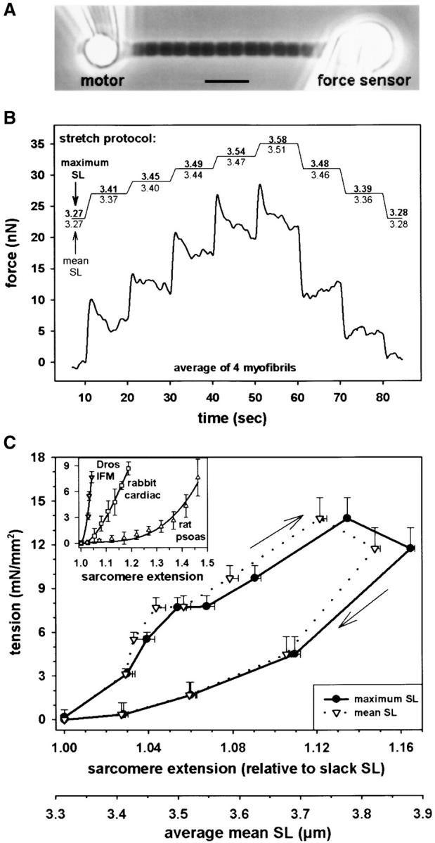
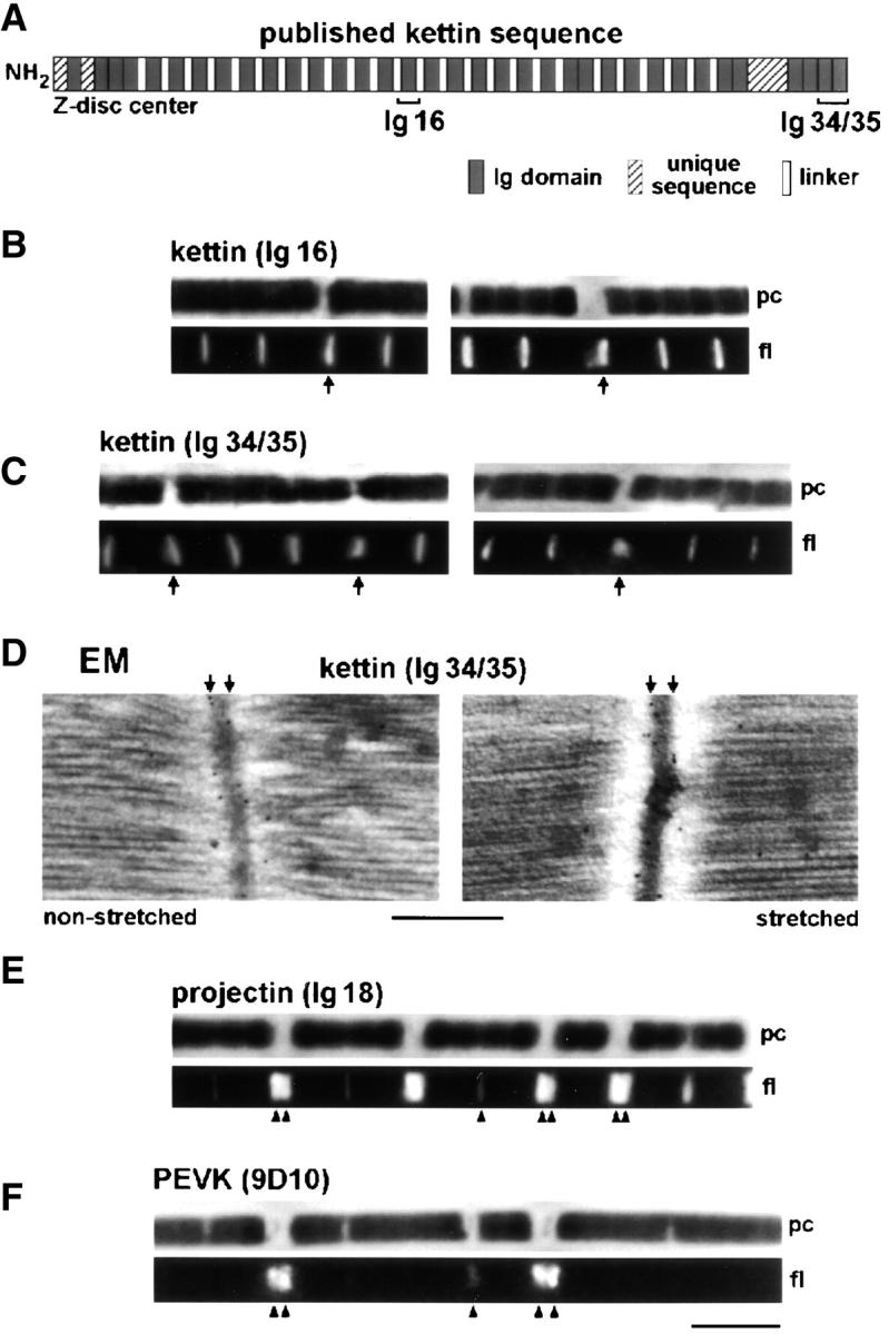
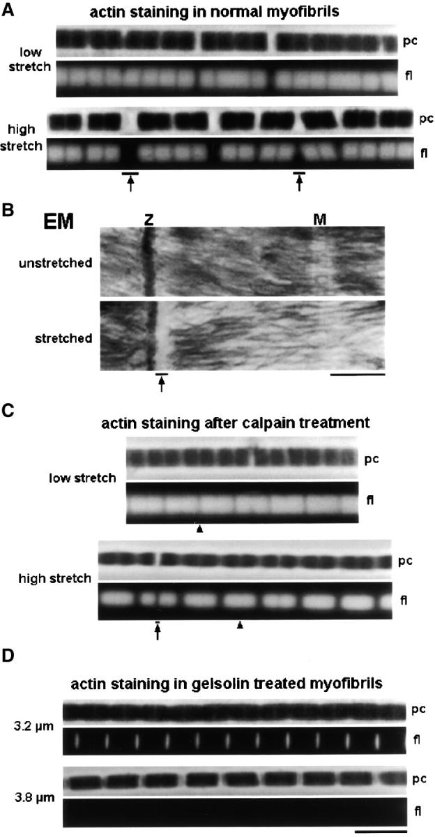
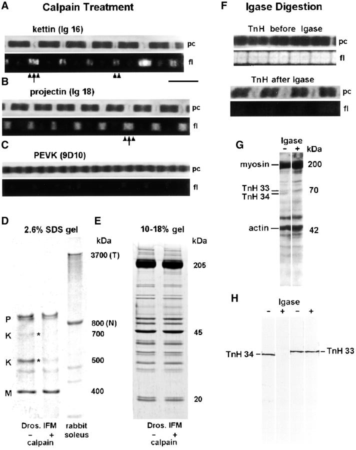
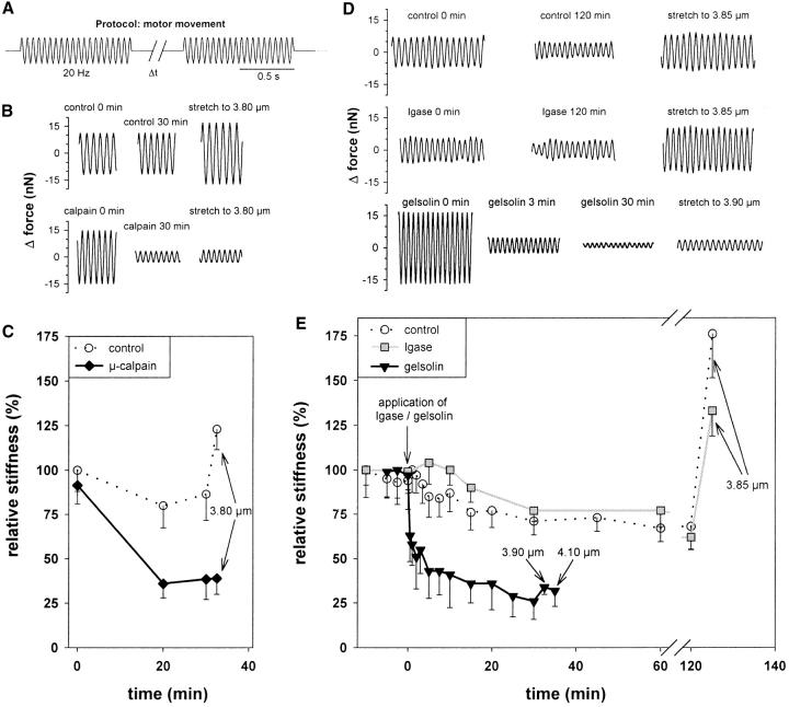
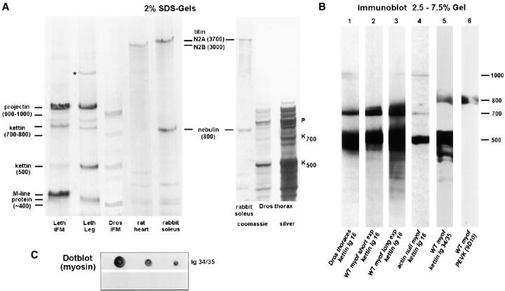
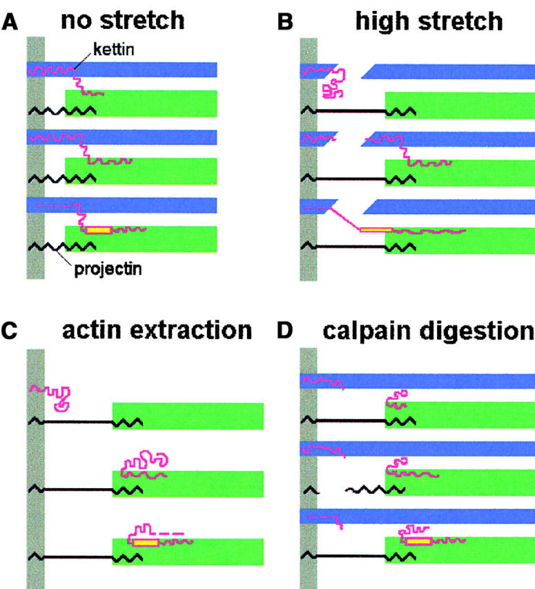
Kettin is a high molecular mass protein of insect muscle that in the sarcomeres binds to actin and alpha-actinin. To investigate kettin's functional role, we combined immunolabeling experiments with mechanical and biochemical studies on indirect flight muscle (IFM) myofibrils of Drosophila melanogaster. Micrographs of stretched IFM sarcomeres labeled with kettin antibodies revealed staining of the Z-disc periphery. After extraction of the kettin-associated actin, the A-band edges were also stained. In contrast, the staining pattern of projectin, another IFM-I-band protein, was not altered by actin removal. Force measurements were performed on single IFM myofibrils to establish the passive length-tension relationship and record passive stiffness. Stiffness decreased within seconds during gelsolin incubation and to a similar degree upon kettin digestion with mu-calpain. Immunoblotting demonstrated the presence of kettin isoforms in normal Drosophila IFM myofibrils and in myofibrils from an actin-null mutant. Dotblot analysis revealed binding of COOH-terminal kettin domains to myosin. We conclude that kettin is attached not only to actin but also to the end of the thick filament. Kettin along with projectin may constitute the elastic filament system of insect IFM and determine the muscle's high stiffness necessary for stretch activation. Possibly, the two proteins modulate myofibrillar stiffness by expressing different size isoforms.


Figures

Figure 1.
Figure 1.
Passive force measurements on single Drosophila IFM myofibrils. (A) Phase–contrast image of myofibril suspended between glue-coated needles. (B) Stretch protocol and averaged force response of four myofibrils. Maximum SL refers to the length of the longest sarcomere, and mean SL refers to an average of all sarcomeres. (C) Passive tension-SL relationship. Data represent quasi steady-state tension (mean ± SEM; n = 24 myofibrils). Average slack SL was 3.32 μm. (Inset) Comparison of stress-strain curves of Drosophila IFM and mammalian myofibrils (Linke, 2000). Bar, 5 μm.
Figure 2.
Figure 2.
Immunolabeling of relaxed Drosophila IFM sarcomeres with various antibodies. (A) Domain structure of kettin (Kolmerer et al., 2000) and position of kettin antibodies used. (B and C) IF microscopy of stretched single myofibrils stained with kettin antibodies and Cy-3–conjugated IgG. (D) IEM of sarcomeres at rest length and after small stretch, stained with α-kettin Ig34/35. (E and F) IF of stretched single myofibrils stained with α-projectin (E) and α-PEVK antibodies (F). pc, phase–contrast image; fl, fluorescence image. Bars: (D) 0.5 μm; (F, IF images) 5 μm.
Figure 3.
Figure 3.
Actin filaments in stretched Drosophila IFM sarcomeres. (A) Single myofibrils stained with rhodamine-phalloidin. Note actin filament breakage at Z-discs (arrows). (B) Electron micrographs of IFM sarcomeres. Myofilament breakage at the Z-disc (Z) was observed at modest stretch (arrow). M, M-line; (c) Actin staining of stretched single myofibrils after digestion with μ-calpain. Actin filaments of opposing half sarcomeres usually remained connected at the Z-disc (arrowheads); actin was seen rarely to be broken (arrow). (D) Actin staining of sarcomeres after extraction with 0.2–0.3 mg/ml (top) and >0.3 mg/ml (bottom) gelsolin fragment. Bars: (B) 0.5 μm; (all IF images) 5 μm.
Figure 4.
Figure 4.
Actin-extracted, stretched, Drosophila IFM myofibrils. (A) Images of single myofibrils at different SLs stained with α-kettin Ig16. Both Z-discs (arrow) and A-band edges (arrowheads) are labeled. (B) Examples of intensity profiles and graph showing the SL-dependent spacing (mean ± SD; n = 22) of kettin-Ig16 epitopes at the A-band edge measured across the M-line (•) or Z-disc (▴). For comparison, the distance between A-band edges is also shown (○ and ▵). (C) Sequence of fluorescence images of a single myofibril at ∼4.0 μm SL stained with α-Ig34/35. Upon exposure of the myofibril to 20 Hz sinusoidal length oscillations, the intensity of Z-disc epitopes gradually decreased (arrows), whereas that of epitopes at the A-band edge increased (arrowheads). (D) IEM of actin-extracted fibers stained with α-Ig34/35. A larger area is shown in the top left image. Images at bottom depict sarcomeres at two different degrees of stretch: ∼3.8 and ∼4.2 μm SL. Nanogold particles indicate the position of kettin epitopes at the Z-disc periphery (arrows) and A-band edge (arrowheads). The histogram shows the nanogold particle distance from the center of the Z-disc measured in ∼3.8-μm-long sarcomeres. A major peak is at ∼60 nm, a minor peak at 360–400 nm, out from the Z-disc center. (E) IF of single myofibril stained with α-projectin antibody. The A-band edge is stained strongly (arrowheads) and the Z-disc faintly (arrow). (F) α-PEVK antibody did not stain actin-extracted myofibrils. Bars: (D) 0.5 μm; (all IF images) 5 μm.
Figure 5.
Figure 5.
Effect of treatment with μ-calpain (A–E) and Igase (F–H) on Drosophila IFM myofibrils. (A) IF of single myofibril stained with α-kettin Ig16 after calpain treatment and stretch; the A-band edge is labeled (arrowheads), and the Z-disc is labeled also in some sarcomeres (arrow). (B) α-Projectin staining of stretched sarcomeres showed a fuzzy epitope at the A-band edge (arrowheads) and also Z-disc labeling (arrow). (C) PEVK sequence was not stained by 9D10 antibody in calpain-treated, stretched, myofibrils. (D) Low percentage SDS-gel to show the effect of μ-calpain (3 μg/ml, 25°C, 45 min) on high molecular weight proteins. Only kettin (K) isoforms are substantially digested, whereas projectin (P) and M-line protein (M) remain largely intact. Nebulin (N) and titin (T) from rabbit soleus muscle are used as standards. (E) Lower molecular weight proteins are not affected by μ-calpain treatment as detectable on 10–18% SDS-gradient gels. (F) Igase-mediated digestion of troponin H effectively decreased the staining intensity of α-TnH34 antibodies (MAC 143) on myofibrils. (G) 12% SDS-gel electrophoresis of washed IFM myofibrils to separate TnH33 and TnH34 isoforms. Igase treatment eliminated the TnH34 isoform. (H) Western blot with antibodies to either TnH33 or TnH34 confirms that Igase treatment removed TnH34 but left TnH33 intact.
Figure 6.
Figure 6.
Stiffness measurements on single Drosophila IFM myofibrils. (A) Protocol: 20 Hz sinusoidal oscillations were applied for 1–2 s; the rest interval (Δt) was 1–20 min. (B) Examples of oscillatory force response of control specimens and calpain-treated myofibrils. (C) Stiffness (mean ± SD; n = 3) versus time after calpain treatment (solid line) in comparison with control stiffness (dotted line). SL was 3.7 μm except after 30 min when myofibrils were stretched to 3.8 μm. (D) Examples of force response of control specimens, igase-digested, and gelsolin-treated single myofibrils. (E) Stiffness (mean ± SD; n = 3) versus time in control myofibrils (dotted curve), during igase digestion (gray solid curve), and during actin extraction (black solid curve). SL before the stretch at the end of the experimental protocol was 3.7 μm.
Figure 7.
Figure 7.
Isoforms of large insect muscle proteins studied by SDS-PAGE and immunoblotting and dotblot to probe kettin-myosin binding. (A) Coomassie-stained 2% SDS-gels to separate projectin and kettin of Lethocerus IFM/leg muscle or Drosophila IFM myofibrils. Drosophila thoraces frozen in liquid nitrogen immediately after dissection were also included in the analysis (right two lanes; right lane is silver stain). For size comparison and to construct a calibration curve for high molecular weight proteins, large rat cardiac and rabbit psoas proteins were used. P, projectin; K, kettin; *unidentified band. (B) Immunoblots. Proteins in Drosophila thoraces (lane 1) or washed IFM myofibrils (lanes 2–6) were separated on 2.5–7.5 SDS-gradient gels. Lane 4 was run with IFM myofibrils from the Drosophila actin-null mutant, KM88. Immunoblots in lanes 1–4 were incubated with α-kettin Ig16; lane 3 is a longer exposure of lane 2, whereas lanes 3 and 4 were exposed for the same time to compare relative amounts of 500-kD kettin in wild-type and actin-null. Lane 5 was incubated with α-kettin Ig34/35 and lane 6 with 9D10 antibody to PEVK. Molecular masses are estimated relative to kettin (500 kD) and projectin (900 kD). (C) Dotblot to show binding of expressed kettin Ig34/35 domains to myosin. Upper strip, dots of 2.0, 1.0, and 0.5 μg of Lethocerus myosin incubated in Ig34/35 followed by anti-His and second antibody; lower strip, myosin dots incubated with anti-His and second antibody only.
Figure 8.
Figure 8.
Model of the arrangement of kettin (red), projectin (black), actin (blue), and myosin (green) in Drosophila IFM sarcomeres. The model attempts to explain this study's results obtained under the respective experimental conditions indicated above each panel. Kettin is drawn in different length variants to take into account the presence of 500-, 700-, and 800-kD isoforms (from top to bottom in each panel). Yellow color indicates PEVK domain (position of PEVK domain was tentatively induced from the genome sequence of D-titin; Machado and Andrew, 2000; Zhang et al., 2000). Gray bars indicate Z-disk. For further explanation, see text.

References

    1. Ayme-Southgate, A., P. Lasko, C. French, and M.L. Pardue. 1991. Drosophila has a twitchin/titin-related gene that appears to encode projectin. Proc. Natl. Acad. Sci. USA. 88:7973–7977. - PMC - PubMed
    1. Benian, G.M., A. Ayme-Southgate, and T.L. Tinley. 1999. The genetics and molecular biology of the titin/connectin-like proteins of invertebrates. Rev. Physiol. Biochem. Pharmacol. 138:235–268. - PubMed
    1. Bullard, B., K.S. Hammond, and B.M. Luke. 1977. The site of paramyosin in insect flight muscle and the presence of an unidentified protein between myosin filaments and Z-line. J. Mol. Biol. 115:417–440. - PubMed
    1. Bullard, B., K. Leonard, A. Larkins, G. Butcher, C. Karlik, and E. Fyrberg. 1988. Troponin of asynchronous flight muscle. J. Mol. Biol. 294:621–637. - PubMed
    1. Bullard, B., D. Goulding, C. Ferguson, and K. Leonard. 2000. Links in the chain: the contribution of kettin to the elasticity of insect muscles. Adv. Exp. Med. Biol. 481:207–218. - PubMed

Publication types