. 2001 Sep 3;154(5):1069-79.
doi: 10.1083/jcb.200103111.

Proteolytic exposure of a cryptic site within collagen type IV is required for angiogenesis and tumor growth in vivo

Affiliations

Proteolytic exposure of a cryptic site within collagen type IV is required for angiogenesis and tumor growth in vivo

J Xu et al. J Cell Biol. .

Erratum in

  • J Cell Biol 2001 Nov 26;155(5):859. Yuen SM [corrected to Moon YS]

Abstract

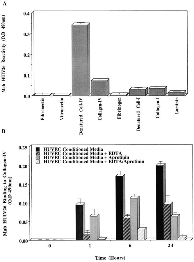
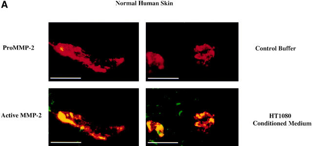
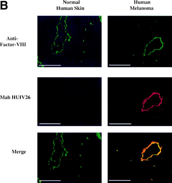
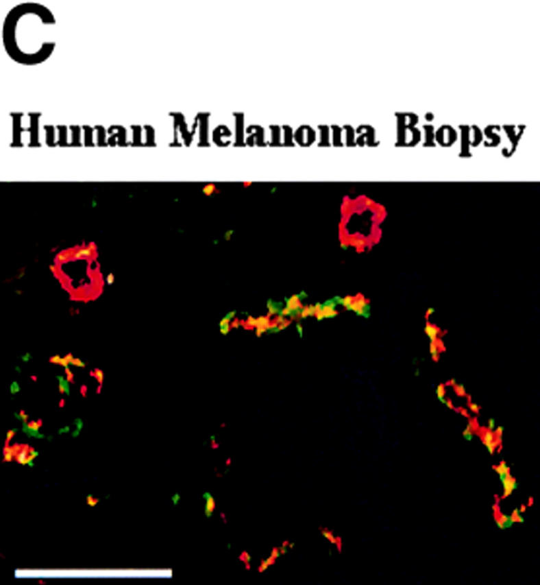
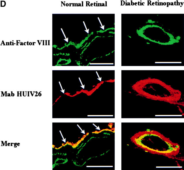
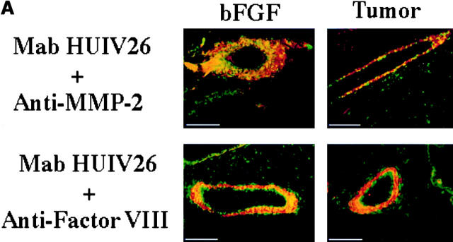
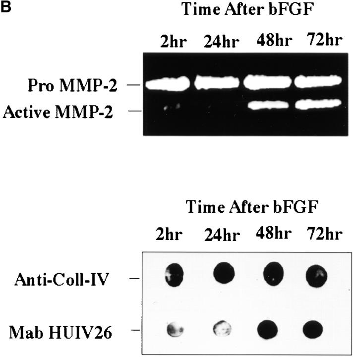
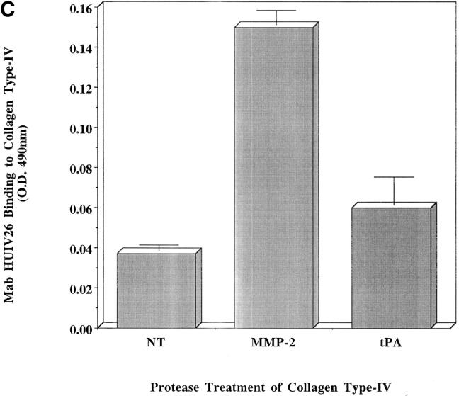
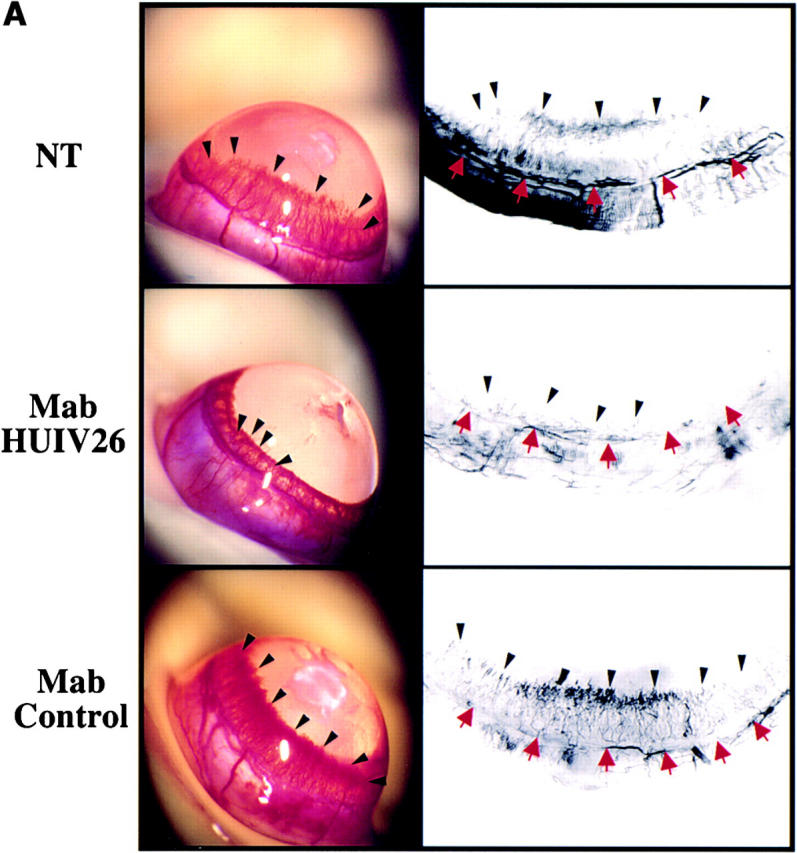
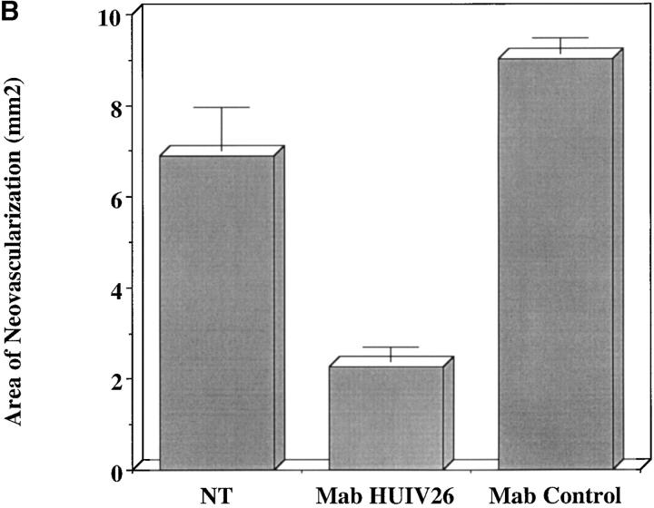
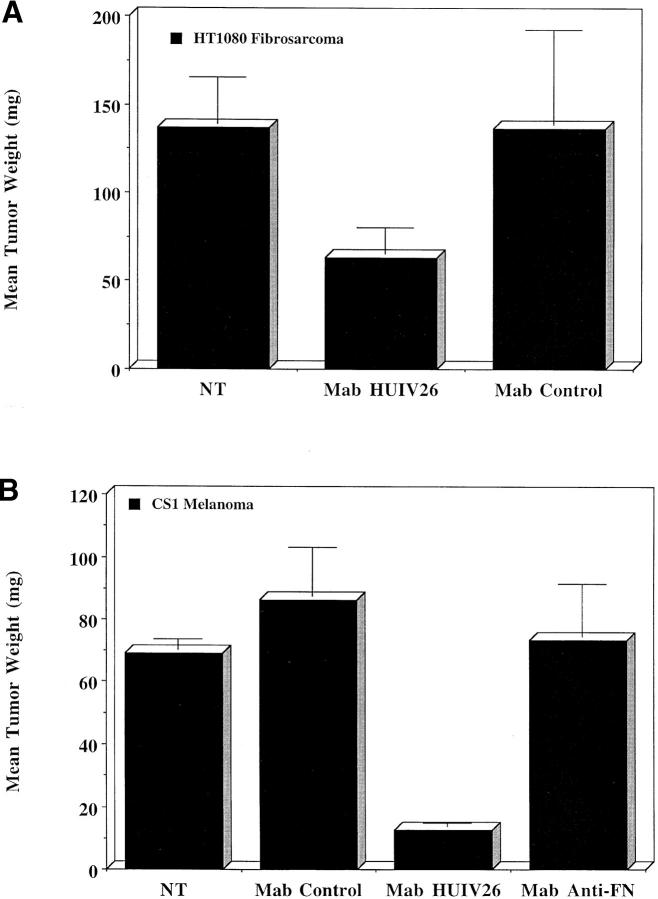
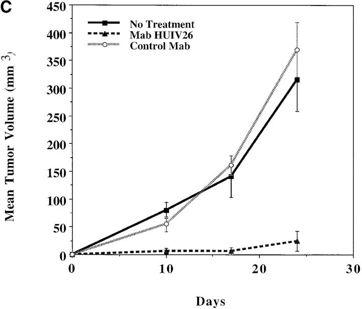
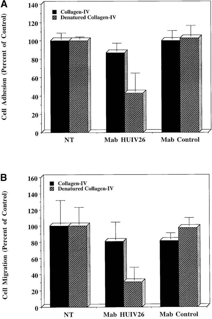
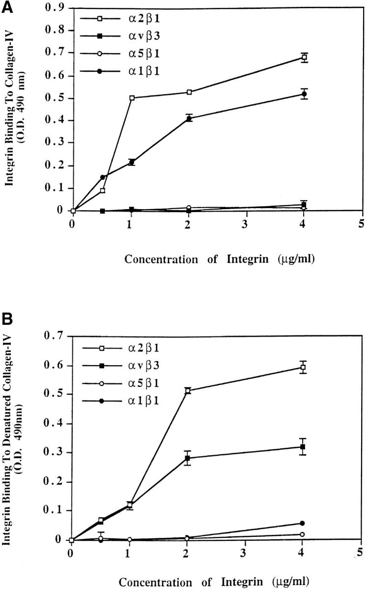
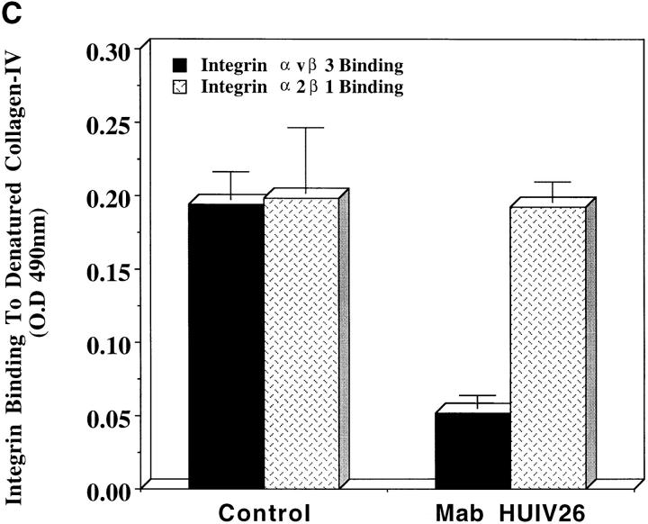
Evidence is provided that proteolytic cleavage of collagen type IV results in the exposure of a functionally important cryptic site hidden within its triple helical structure. Exposure of this cryptic site was associated with angiogenic, but not quiescent, blood vessels and was required for angiogenesis in vivo. Exposure of the HUIV26 epitope was associated with a loss of alpha1beta1 integrin binding and the gain of alphavbeta3 binding. A monoclonal antibody (HUIV26) directed to this site disrupts integrin-dependent endothelial cell interactions and potently inhibits angiogenesis and tumor growth. Together, these studies suggest a novel mechanism by which proteolysis contributes to angiogenesis by exposing hidden regulatory elements within matrix-immobilized collagen type IV.


Figures

Figure 1.
Figure 1.
Mab HUIV26 reactivity with denatured/proteolyzed collagen IV in solid phase ELISA. Microtiter plates were coated with ECM components at a concentration of 25 μg/ml. (A) Mab HUIV26 was added (1 μg/ml), followed 1 h later with goat anti–mouse peroxidase–labeled IgG. All data was corrected for nonspecific binding of secondary antibody. Data bars represent the mean OD ± standard deviations from triplicate wells. (B) Microtiter wells were coated with triple helical collagen IV at 25 μg/ml. Concentrated (20×) HUVEC serum–free–conditioned media was added to the wells in the presence or absence of EDTA, aprotinin, or both and allowed to incubate for 1, 6, and 24 h. The plates were next washed, blocked, and incubated with Mab HUIV26 or control antibody. All data was corrected for nonspecific secondary antibody binding. Data bars represent the mean OD ± standard deviations from triplicate wells.
Figure 2.
Figure 2.
Exposure of the HUIV26 cryptic site within human tissue by proteolysis. Frozen sections of human tissue were mounted on glass slides. (A) Nonfixed normal human skin was incubated for 2 h with either control buffer, pro or activated MMP-2 (1.0 μg/ml), or HT1080 tumor–conditioned medium. The sections were costained with Mab HUIV26 and a polyclonal antibody directed to factor VIII–related antigen. Red indicates factor VIII staining of blood vessels, green indicates exposure of the HUIV26 cryptic epitope, and yellow indicates colocalization. Photo were taken at 630× under oil immersion (B) Normal human skin or malignant melanoma were costained with Mab HUIV26 and a polyclonal antibody directed to factor VIII–related antigen. Red indicates exposure of the HUIV26 cryptic site, green indicates human blood vessels, and yellow indicates colocalization. Photographs were taken at 200× magnification. (C) Representative example of human melanoma tumor tissue costained with Mab HUIV26 (green) and polyclonal antibody directed to factor VIII–related antigen (red), indicating that not all tumor vessels stain positive for HUIV26 epitope. Photos were taken at low power (200×). (D) Normal human retinal tissue or retina from subjects with diabetic retinopathy were costained with Mab HUIV26 and a polyclonal antibody to factor VIII–related antigen. Green indicates human blood vessels and red indicates HUIV26 cryptic sites. Arrows indicate nonspecific fluorescence of retinal pigmented epithelium due to auto-fluorescence of lipofusion. Photomicrographs were taken at 200× magnification. Bars, 50.0 μm.
Figure 2.
Figure 2.
Exposure of the HUIV26 cryptic site within human tissue by proteolysis. Frozen sections of human tissue were mounted on glass slides. (A) Nonfixed normal human skin was incubated for 2 h with either control buffer, pro or activated MMP-2 (1.0 μg/ml), or HT1080 tumor–conditioned medium. The sections were costained with Mab HUIV26 and a polyclonal antibody directed to factor VIII–related antigen. Red indicates factor VIII staining of blood vessels, green indicates exposure of the HUIV26 cryptic epitope, and yellow indicates colocalization. Photo were taken at 630× under oil immersion (B) Normal human skin or malignant melanoma were costained with Mab HUIV26 and a polyclonal antibody directed to factor VIII–related antigen. Red indicates exposure of the HUIV26 cryptic site, green indicates human blood vessels, and yellow indicates colocalization. Photographs were taken at 200× magnification. (C) Representative example of human melanoma tumor tissue costained with Mab HUIV26 (green) and polyclonal antibody directed to factor VIII–related antigen (red), indicating that not all tumor vessels stain positive for HUIV26 epitope. Photos were taken at low power (200×). (D) Normal human retinal tissue or retina from subjects with diabetic retinopathy were costained with Mab HUIV26 and a polyclonal antibody to factor VIII–related antigen. Green indicates human blood vessels and red indicates HUIV26 cryptic sites. Arrows indicate nonspecific fluorescence of retinal pigmented epithelium due to auto-fluorescence of lipofusion. Photomicrographs were taken at 200× magnification. Bars, 50.0 μm.
Figure 3.
Figure 3.
Exposure of the HUIV26 cryptic epitope is associated with the expression and activation of MMP-2 in vivo. bFGF-treated CAMs or CAMs containing CS1 melanoma tumors were costained with either Mab HUIV26 and polyclonal anti–MMP-2 (top), or Mab HUIV26 and polyclonal antifactor VIII–related antigen (bottom). (A) Tissues were visualized by incubation with rhodamine- and FITC-conjugated secondary antibodies. Top, red indicates MMP-2 and green indicates HUIV26 cryptic epitope. Bottom, red indicates factor VIII staining of blood vessels, green indicates HUIV26 cryptic epitope, and yellow indicates colocalization. Photographs were taken at 200× magnification. (B) CAMs of 10-d-old embryos were stimulated with bFGF and total CAM lysates were prepared at 2, 24, 48, and 72 h. Top, gelatin zymogram of total CAM lysates after stimulation with bFGF. Bottom, dot blot of total CAM lysates. Total collagen IV (triple helical and denatured) was detected with a polyclonal antibody to both native and denatured collagen IV. Denatured collagen IV was detected with Mab HUIV26. (C) Microtiter plates were coated with triple helical collagen type IV (25 μg/ml). MMP-2 (500 ng/ml), tPA (6 U/ml, specific activity 700 μg/mg protein), or NT (control buffer) were incubated for 18 h. The wells were washed, blocked with BSA, and the HUIV26 cryptic sites were detected with Mab HUIV26 (1.0 μg/ml). Data bars represent the mean OD ± standard deviations from triplicate wells. Bars, 50.0 μm.
Figure 3.
Figure 3.
Exposure of the HUIV26 cryptic epitope is associated with the expression and activation of MMP-2 in vivo. bFGF-treated CAMs or CAMs containing CS1 melanoma tumors were costained with either Mab HUIV26 and polyclonal anti–MMP-2 (top), or Mab HUIV26 and polyclonal antifactor VIII–related antigen (bottom). (A) Tissues were visualized by incubation with rhodamine- and FITC-conjugated secondary antibodies. Top, red indicates MMP-2 and green indicates HUIV26 cryptic epitope. Bottom, red indicates factor VIII staining of blood vessels, green indicates HUIV26 cryptic epitope, and yellow indicates colocalization. Photographs were taken at 200× magnification. (B) CAMs of 10-d-old embryos were stimulated with bFGF and total CAM lysates were prepared at 2, 24, 48, and 72 h. Top, gelatin zymogram of total CAM lysates after stimulation with bFGF. Bottom, dot blot of total CAM lysates. Total collagen IV (triple helical and denatured) was detected with a polyclonal antibody to both native and denatured collagen IV. Denatured collagen IV was detected with Mab HUIV26. (C) Microtiter plates were coated with triple helical collagen type IV (25 μg/ml). MMP-2 (500 ng/ml), tPA (6 U/ml, specific activity 700 μg/mg protein), or NT (control buffer) were incubated for 18 h. The wells were washed, blocked with BSA, and the HUIV26 cryptic sites were detected with Mab HUIV26 (1.0 μg/ml). Data bars represent the mean OD ± standard deviations from triplicate wells. Bars, 50.0 μm.
Figure 4.
Figure 4.
Effects of purified Mab HUIV26 on angiogenesis in vivo. Rat corneal micropocket assays were performed to assess the effects of Mab HUIV26 on angiogenesis. (A) Representative corneas from rats implanted with hydron pellets containing bFGF (top), bFGF + Mab HUIV26 (middle) or bFGF + control Mab (bottom). Black arrows indicate angiogenic neovessels. Red arrows indicate preexisting limbal vessels. (B) Quantification of the area of neovascularization within rat corneas. Data bars represent the mean area of neovascularization from the limbus to hydron pellet. Experiments were performed at least twice with five to seven eyes per condition.
Figure 4.
Figure 4.
Effects of purified Mab HUIV26 on angiogenesis in vivo. Rat corneal micropocket assays were performed to assess the effects of Mab HUIV26 on angiogenesis. (A) Representative corneas from rats implanted with hydron pellets containing bFGF (top), bFGF + Mab HUIV26 (middle) or bFGF + control Mab (bottom). Black arrows indicate angiogenic neovessels. Red arrows indicate preexisting limbal vessels. (B) Quantification of the area of neovascularization within rat corneas. Data bars represent the mean area of neovascularization from the limbus to hydron pellet. Experiments were performed at least twice with five to seven eyes per condition.
Figure 5.
Figure 5.
Effects of systemic administration of purified Mab HUIV26 on tumor growth in vivo. The effects of Mab HUIV26 on tumor growth was assessed in two independent models, including the chick embryo (A and B) and the SCID Mouse (C). HT1080 human fibrosarcoma cells (4 × 10⁵) or CS-1 melanoma tumor cells (5 × 10⁶) were inoculated on the CAMs of 10-d-old chick embryos. 24 h later, the embryos received a single intravenous injection of 100 μg of Mab HUIV26 or isotype-matched control. (A) Quantitation of HT1080 tumor growth in the chick embryo. (B) Quantitation of CS-1 tumor growth within the chick embryo. Data bars represent the mean tumor weights ± the standard errors from 5 to 10 embryos per condition. (C) SCID mice were injected subcutaneously with 2 × 10⁶ M21 human melanoma cells. 3 d later mice were treated i.p. daily for 24 d with 100 μg of either Mab HUIV26 or an isotype-matched control antibody. Tumor size was monitored with calipers and tumor volumes were determined. Data represents the mean ± standard errors of the tumor volumes. All experiments were conducted 3 to 4 times with 5 to 10 animals per condition.
Figure 5.
Figure 5.
Effects of systemic administration of purified Mab HUIV26 on tumor growth in vivo. The effects of Mab HUIV26 on tumor growth was assessed in two independent models, including the chick embryo (A and B) and the SCID Mouse (C). HT1080 human fibrosarcoma cells (4 × 10⁵) or CS-1 melanoma tumor cells (5 × 10⁶) were inoculated on the CAMs of 10-d-old chick embryos. 24 h later, the embryos received a single intravenous injection of 100 μg of Mab HUIV26 or isotype-matched control. (A) Quantitation of HT1080 tumor growth in the chick embryo. (B) Quantitation of CS-1 tumor growth within the chick embryo. Data bars represent the mean tumor weights ± the standard errors from 5 to 10 embryos per condition. (C) SCID mice were injected subcutaneously with 2 × 10⁶ M21 human melanoma cells. 3 d later mice were treated i.p. daily for 24 d with 100 μg of either Mab HUIV26 or an isotype-matched control antibody. Tumor size was monitored with calipers and tumor volumes were determined. Data represents the mean ± standard errors of the tumor volumes. All experiments were conducted 3 to 4 times with 5 to 10 animals per condition.
Figure 6.
Figure 6.
Effects of Mab HUIV26 on human endothelial cell adhesion and migration. Microtiter plates (96 wells) and Transwell membranes were coated with triple helical or denatured colla- gen type IV (25 μg/ml). (A) Subconfluent HUVECs (105) were resuspended in adhesion buffer and allowed to attach in the presence or absence of Mab HUIV26 or an isotype-matched control antibody for 30 min. Nonattached cells were removed by washing and the attached cells were stained with crystal violet. (B) Subconfluent HUVECs (105) were resuspended in migration buffer and allowed to migrate in the presence or absence of Mab HUIV26 or an isotype-matched control antibody. Cells remaining on the top side of the membrane were removed and cells that had migrated to the under side were stained with crystal violet. Cell adhesion and migration was quantified by measuring the OD of eluted dye at 600 nm. Data bars represent the mean OD ± standard deviation from triplicate wells expressed as a percentage of control.
Figure 7.
Figure 7.
Integrin binding to triple helical and denatured human collagen IV. Microtiter wells were coated with either triple helical or denatured human collagen IV (25 μg/ml). Purified human integrins α1β1, α2β1, α5β1, or αvβ3 (0.5–4 μg/ml) were allowed to bind to triple helical collagen IV (A) or denatured collagen IV (B) for 1 h at 37°C. Integrin binding was detected with antiintegrin antibodies. (C) Purified human integrins α2β1 and αvβ3 (1.0 μg/ml) were allowed to bind to denatured collagen IV–coated plates for 1 h at 37°C in the presence or absence of Mab HUIV26 or an isotype-matched control antibody. Integrin binding was detected by incubation with either polyclonal antibody directed to α2 or α3 integrins. Data bars represent the mean OD ± standard deviations from triplicate wells.
Figure 7.
Figure 7.
Integrin binding to triple helical and denatured human collagen IV. Microtiter wells were coated with either triple helical or denatured human collagen IV (25 μg/ml). Purified human integrins α1β1, α2β1, α5β1, or αvβ3 (0.5–4 μg/ml) were allowed to bind to triple helical collagen IV (A) or denatured collagen IV (B) for 1 h at 37°C. Integrin binding was detected with antiintegrin antibodies. (C) Purified human integrins α2β1 and αvβ3 (1.0 μg/ml) were allowed to bind to denatured collagen IV–coated plates for 1 h at 37°C in the presence or absence of Mab HUIV26 or an isotype-matched control antibody. Integrin binding was detected by incubation with either polyclonal antibody directed to α2 or α3 integrins. Data bars represent the mean OD ± standard deviations from triplicate wells.

References

    1. Blood, C.H., and B.R. Zetter. 1990. Functional interactions with the vasculature: angiogenesis and tumor metastasis. Biochim. Biophys. Acta. 1032:89–118. - PubMed
    1. Bowersox, J.C., and N. Sorgenete. 1982. Chemotaxis of aortic endothelial cells in response to fibronectin. Cancer Res. 42:2547–2551. - PubMed
    1. Brooks, P.C., A.M.P. Montgomery, M. Rosenfeld, R.A. Reisfeld, T. Hu, G. Klier, and D.A. Cheresh. 1994. Integrin αvβ3 antagonists promote tumor regression by inducing apoptosis of angiogenic blood vessels. Cell. 79:1157–1164. - PubMed
    1. Brooks, P.C., S. Stromblad, L.C. Sanders, T.L. von Schalscha, R.T. Aimes, W.G. Stetler-Stevenson, J.P. Quigley, and D.A. Cheresh. 1996. Localization of matrix metalloproteinase MMP-2 to the surface of invasive cells by interaction with integrin. Cell. 85:683–693. - PubMed
    1. Brooks, P.C., A.M.P. Montgomery, and D.A. Cheresh. 1998. Use of the 10 day old chick embryo model for studying angiogenesis. A.R. Howlett, editor. Humana Press Inc., Totowa, NJ. 257–269. - PubMed

Publication types

MeSH terms