. 2001 Sep 3;194(5):591-9.
doi: 10.1084/jem.194.5.591.

T cell homeostasis: thymus regeneration and peripheral T cell restoration in mice with a reduced fraction of competent precursors

Affiliations

T cell homeostasis: thymus regeneration and peripheral T cell restoration in mice with a reduced fraction of competent precursors

A R Almeida et al. J Exp Med. .

Abstract

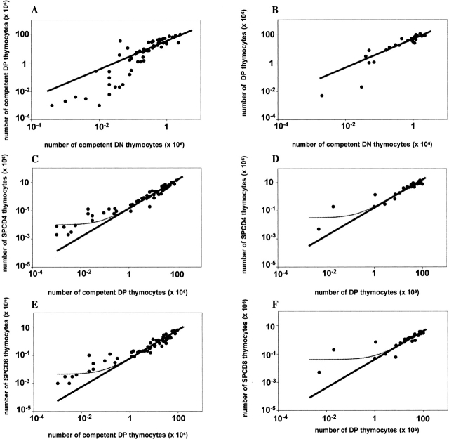
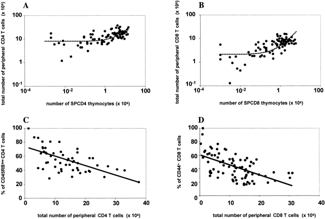
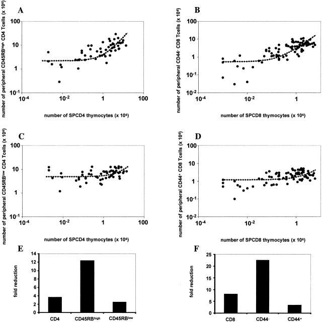
We developed a novel experimental strategy to study T cell regeneration after bone marrow transplantation. We assessed the fraction of competent precursors required to repopulate the thymus and quantified the relationship between the size of the different T cell compartments during T cell maturation in the thymus. The contribution of the thymus to the establishment and maintenance of the peripheral T cell pools was also quantified. We found that the degree of thymus restoration is determined by the availability of competent precursors and that the number of double-positive thymus cells is not under homeostatic control. In contrast, the sizes of the peripheral CD4 and CD8 T cell pools are largely independent of the number of precursors and of the number of thymus cells. Peripheral "homeostatic" proliferation and increased export and/or survival of recent thymus emigrants compensate for reduced T cell production in the thymus. In spite of these reparatory processes, mice with a reduced number of mature T cells in the thymus have an increased probability of peripheral T cell deficiency, mainly in the naive compartment.


Figures

Figure 1
Figure 1
Thymus regeneration. Lethally irradiated Rag2−/− mice were reconstituted with BM cells from normal B6.Ly5a alone or diluted among incompetent BM cells from either B6.Ly5bTCRα−/− (A, C, and E) or B6.Ly5bCD3ε−/− (B, D, and F) donors. 8 to 20 wk after reconstitution the chimeras were killed and the number of competent Ly5a cells was evaluated in the different thymus cell compartments. For each chimera (•), the relationship between the number of competent cells in the CD3CD4CD8 (DN), CD4+CD8+ (DP) compartments is shown in A and B, between the DP and the SPCD4 compartments in C and D, and in the DP and the SPCD8 compartments in E and F. The curves show the relationships between DN, DP, SPCD4, and SPCD8 cells as predicted by the mathematical model (see Mathematical Analysis). All datasets were fitted twice: once including all data points (thin lines), and once excluding the mice with very low DN (<105) or DP (<106) cell numbers (thick lines). In A and B, both of the fits could not be significantly improved by adding a homeostatic term. Moreover, both fits predicted a too high number of competent DP cells in mice with very few competent DN cells, suggesting that an additional mechanism (not included in the model) is involved. The model we used was sufficient, however, to conclude that in mice with at least 5% of the normal number of competent DN cells, the number of competent cells in the DP compartment was proportional to the size of the DN compartment (see thick lines). Likewise, in C–F we found a proportionality between the numbers of competent DP cells and SP cells (see thick lines) in all mice except the ones with very low numbers of competent DP cells (<106). The addition of a small homeostatic term only helped to describe the relatively high SP cells numbers in the latter mice (see thin lines), while it did not improve the fits between the model and the data from all other mice. Except in very poorly reconstituted mice, the size of each thymus compartment is thus proportional to the size of the compartment that precedes it. The parameter values (see Mathematical Analysis) that gave the best fits to the data are: s/m = (A) 35, (B) 47, (C) 0.14, (D) 0.17, (E) 0.06, (F) 0.04 (thick lines), and p/m = (C) 0.01, (D) 0.03, (E) 0.005, and (F) 0.04 (thin lines).
Figure 2
Figure 2
Restoration of the total peripheral T cell pools. Panel A shows the relationship between the number of peripheral Ly5a CD4 T cells (SPL+LN) and the number of competent Ly5a SPCD4 cells in the thymus of each individual B6.Ly5a/B6.Ly5bTCRα−/− or B6.Ly5a/B6.CD3ε−/−Ly5b chimera. (B) The same but for CD8 T cells. The data were fitted to the steady-state level corresponding to including the homeostatic term (dashed lines), as this significantly improved the fits to the data, even if mice with very low SP numbers (<105) were not taken into account (not shown). Parameter results are: s/m = (A) 1.5, (B) 2.8, and p / m = (A) 8.0, (B) 2.1. Panel C shows the percentage of activated/memory CD45RBlowCD4+ T cells as a function of the total number of peripheral CD4 T cells, while D shows the percentage of activated/memory CD44+CD8+ cells as a function of the total number of peripheral CD8 T cells. The lines in C and D are linear regression lines with r = −0.6 in both cases.
Figure 3
Figure 3
Restoration of the naive and activated/memory T cell pools. Panel A shows the relationship between the number of SPCD4 cells and the number of peripheral naive CD45RBhighCD4+ T cells. (B) The relationship between the number of SPCD8 and the number of peripheral naive CD44CD8+ T cells. (C) The relationship between the number of SPCD4 and the number of peripheral activated/memory CD45RBlowCD4+ T cells. (D) The relationship between the number of SPCD8 and the number of peripheral activated/memory CD44+CD8+ T cells in all B6.Ly5a/B6.Ly5bTCRα−/− and B6.Ly5a/B6.Ly5bCD3ε−/− chimeras. The data were fitted to the steady-state level corresponding to including the homeostatic term, as this significantly improved the fits to the data, even if mice with very low SP numbers (<105) were excluded (not shown). Parameter results are: s/m = (A) 2.0, (B) 2.6, (C) 0.5, (D) 0.5, and p / m = (A) 2.2, (B) 0.5, (C) 5.0, (D) 1.2. E and F show the fold reductions in the total, naive (CD45RBhighCD44) and activated/memory (CD45RBlowCD44+) CD4 (E) and CD8 (F) peripheral compartments resulting from a 100-fold reduction (compared with fully reconstituted mice) in the thymus SPCD4 and SPCD8 compartments, respectively.
Figure 4
Figure 4
TCR repertoires in chimeras with normal and low T cell numbers. Representation of the different Vβ TCR families by the spleen T cells of different BM chimeras. Mice were reconstituted with either 100% BM cells from normal B6.Ly5a mice (A) or with a mixture of 1% BM cells from normal B6.Ly5a mice and 99% BM cells from T cell–deficient B6.Ly5a/B6.Ly5bTCRα−/− donors (B). Each bar represents the percentage of CD3+CD4+ T cells expressing each Vβ family in individual mice as assessed by flow cytometry. Similar results were obtained with CD3+CD8+ spleen T cells. Note that although the representation of each Vβ family is identical in all mice reconstituted with 100% BM cells from normal donors, it shows individual variations in mice reconstituted with a limited fraction of competent BM cells.

References

    1. Mackall C.L., Hakim F.T., Gress R.E. Restoration of T-cell homeostasis after T-cell depletion. Semin. Immunol. 1997;9:339–346. - PubMed
    1. Douek D.C., Vescio R.A., Betts M.R., Brenchley J.M., Hill B.J., Zhang L., Berenson J.R., Collins R.H., Koup R.A. Assessment of thymic output in adults after haematopoietic stem-cell transplantation and prediction of T-cell reconstitution. Lancet. 2000;355:1875–1881. - PubMed
    1. Roux E., Dumont-Girard F., Starobinski M., Siegrist C.A., Helg C., Chapuis B., Roosnek E. Recovery of immune reactivity after T-cell-depleted bone marrow transplantation depends on thymic activity. Blood. 2000;96:2299–2303. - PubMed
    1. Cavazzana-Calvo M., Hacein-Bey S., de Saint Basile G., Gross F., Yvon E., Nusbaum P., Selz F., Hue C., Certain S., Casanova J.L. Gene therapy of human severe combined immunodeficiency (SCID)-X1 disease. Science. 2000;288:669–672. - PubMed
    1. Mackall C.L., Gress R.E. Thymic aging and T-cell regeneration. Immunol. Rev. 1997;160:91–102. - PubMed

Publication types

MeSH terms