. 2001 Sep 3;194(5):645-56.
doi: 10.1084/jem.194.5.645.

Localized gene-specific induction of accessibility to V(D)J recombination induced by E2A and early B cell factor in nonlymphoid cells

Affiliations

Localized gene-specific induction of accessibility to V(D)J recombination induced by E2A and early B cell factor in nonlymphoid cells

P Goebel et al. J Exp Med. .

Abstract

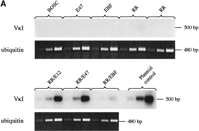
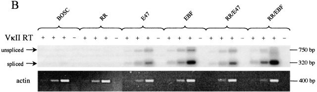
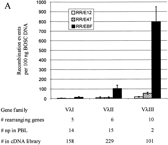
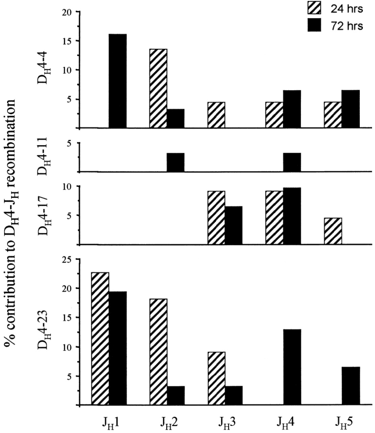
Accessibility of immunoglobulin (Ig) gene segments to V(D)J recombination is highly regulated and is normally only achieved in B cell precursors. We previously showed that ectopic expression of E2A or early B cell factor (EBF) with recombination activating gene (RAG) induces rearrangement of IgH and IgL genes in nonlymphoid cells. VkappaI genes throughout the locus were induced to rearrange after transfection with E2A, suggesting that the entire Vkappa locus was accessible. However, here we show that Ig loci are not opened globally but that recombination is localized. Gene families are interspersed in the D(H), Vkappa, and Vlambda loci, and we show that certain families and individual genes undergo high levels of recombination after ectopic expression of E2A or EBF, while other families within the same locus are not induced to rearrange. Furthermore, in some families, induction of germline transcription correlates with the level of induced recombination, while in others there is no correlation, suggesting that recombination is not simply initiated by induction of germline transcription. The induced repertoire seen at 24 hours does not change significantly over time indicating the absence of many secondary rearrangements and also suggesting a direct targeting mechanism. We propose that accessibility occurs in a local manner, and that binding sites for factors facilitating accessibility are therefore likely to be associated with individual gene segments.


Figures

Figure 1
Figure 1
Southern blot analysis and quantitation of PCR amplified Vκ recombination products. (A) A representative blot for the amplification of the VκI family is shown. Aliquots were taken after 28, 31, and 34 cycles of amplification. The top panel shows samples from cells transfected with E47, EBF, or with RAG1 and RAG2 (RR) alone compared with mock-transfected BOSC23 cells. The bottom panel shows DNA from cells transfected with each transcription factor together with the RAG proteins. The bottom panel also shows amplification obtained from 10 fg of a plasmid control sample used for quantitation. The equality of genomic DNA used in each sample was confirmed by amplification of the same genomic DNA samples with primers for the ubiquitin-conjugating enzyme, shown under each lane of the blot, taken at 25, 28, and 31 cycles. (B) Levels of induced Vκ–Jκ rearrangements. Hybridization signals from PCR products in the linear amplification range were quantitated by PhosphorImager and normalized to the plasmid standards. Samples from cells that had only been transfected with the transcription factors alone did not result in measurable PCR products and were excluded from quantitation. Normalized values are expressed as the number of recombination events detected per 100 ng of transfected BOSC genomic DNA. The bar graph shows the average ±SEM of the normalized signals. The numbers under each family represent the number of recombining genes in the proximal half of the Vκ locus seen in large databases. The second and third row of numbers represents nonproductive (estimates of recombination frequency) and productive (expressed repertoire) sequences in vivo (references 28 and 30).
Figure 1
Figure 1
Southern blot analysis and quantitation of PCR amplified Vκ recombination products. (A) A representative blot for the amplification of the VκI family is shown. Aliquots were taken after 28, 31, and 34 cycles of amplification. The top panel shows samples from cells transfected with E47, EBF, or with RAG1 and RAG2 (RR) alone compared with mock-transfected BOSC23 cells. The bottom panel shows DNA from cells transfected with each transcription factor together with the RAG proteins. The bottom panel also shows amplification obtained from 10 fg of a plasmid control sample used for quantitation. The equality of genomic DNA used in each sample was confirmed by amplification of the same genomic DNA samples with primers for the ubiquitin-conjugating enzyme, shown under each lane of the blot, taken at 25, 28, and 31 cycles. (B) Levels of induced Vκ–Jκ rearrangements. Hybridization signals from PCR products in the linear amplification range were quantitated by PhosphorImager and normalized to the plasmid standards. Samples from cells that had only been transfected with the transcription factors alone did not result in measurable PCR products and were excluded from quantitation. Normalized values are expressed as the number of recombination events detected per 100 ng of transfected BOSC genomic DNA. The bar graph shows the average ±SEM of the normalized signals. The numbers under each family represent the number of recombining genes in the proximal half of the Vκ locus seen in large databases. The second and third row of numbers represents nonproductive (estimates of recombination frequency) and productive (expressed repertoire) sequences in vivo (references 28 and 30).
Figure 2
Figure 2
Induction of VκI and VκII germline transcription after transient transfection of BOSC23 cells. (A) RNA was isolated from cells transfected with the indicated expression vectors or from mock-transfected BOSC cells. cDNA was prepared with a VκI family-specific primer located downstream of the RSS and amplified with primers in the leader and FR3. This results in amplification of unspliced and spliced transcripts as indicated on the blot. Aliquots taken from each PCR at consecutive intervals (25, 28, and 31 cycles) are indicated with RT+. One aliquot from each RT control PCR taken at 31 cycles was also included to test for possible genomic contamination in the RNA preparation. The equal input amounts of RNA was confirmed by actin PCR (25, 28, and 31 cycles), shown for each set beneath the blot. (B) RNA from the same transfected cells was also analyzed for the presence of VκII germline transcripts. The analysis was performed as described in A, using primer combinations specific for VκII germline transcripts but aliquots were taken at 34, 37, and 40 cycles. As for VκI, the unspliced and spliced transcripts were detected and are indicated on the blot. Actin PCRs confirmed equal input amounts of RNA.
Figure 2
Figure 2
Induction of VκI and VκII germline transcription after transient transfection of BOSC23 cells. (A) RNA was isolated from cells transfected with the indicated expression vectors or from mock-transfected BOSC cells. cDNA was prepared with a VκI family-specific primer located downstream of the RSS and amplified with primers in the leader and FR3. This results in amplification of unspliced and spliced transcripts as indicated on the blot. Aliquots taken from each PCR at consecutive intervals (25, 28, and 31 cycles) are indicated with RT+. One aliquot from each RT control PCR taken at 31 cycles was also included to test for possible genomic contamination in the RNA preparation. The equal input amounts of RNA was confirmed by actin PCR (25, 28, and 31 cycles), shown for each set beneath the blot. (B) RNA from the same transfected cells was also analyzed for the presence of VκII germline transcripts. The analysis was performed as described in A, using primer combinations specific for VκII germline transcripts but aliquots were taken at 34, 37, and 40 cycles. As for VκI, the unspliced and spliced transcripts were detected and are indicated on the blot. Actin PCRs confirmed equal input amounts of RNA.
Figure 3
Figure 3
Levels of induced Vλ–Jλ and DH–JH recombination events after transient transfection Induced recombination events for the members of several Vλ and DH gene families were analyzed as outlined in the legend to Fig. 1. Hybridization signals were quantitated, normalized to plasmid standards, and are represented as recombination events per 100 ng of transfected BOSC genomic DNA. (A) Levels of induced Vλ–Jλ rearrangements. The number of rearranging genes within each family are indicated. As an estimate of the contribution of each family to the peripheral repertoire, the number of nonproductive sequences identified in PBL 34 and their contribution within a large cDNA library (reference 35) are shown. (B) Levels of induced DH3 and DH4 rearrangements. The number of rearranging genes within each family and the contribution of each family to complete VDJ rearrangements isolated from peripheral blood (reference 23) are shown.
Figure 3
Figure 3
Levels of induced Vλ–Jλ and DH–JH recombination events after transient transfection Induced recombination events for the members of several Vλ and DH gene families were analyzed as outlined in the legend to Fig. 1. Hybridization signals were quantitated, normalized to plasmid standards, and are represented as recombination events per 100 ng of transfected BOSC genomic DNA. (A) Levels of induced Vλ–Jλ rearrangements. The number of rearranging genes within each family are indicated. As an estimate of the contribution of each family to the peripheral repertoire, the number of nonproductive sequences identified in PBL 34 and their contribution within a large cDNA library (reference 35) are shown. (B) Levels of induced DH3 and DH4 rearrangements. The number of rearranging genes within each family and the contribution of each family to complete VDJ rearrangements isolated from peripheral blood (reference 23) are shown.
Figure 4
Figure 4
Frequency of EBF-induced DH–JH recombination events 24 and 72 h after transfection. Genomic DNA from transfected BOSC cells was harvested either 24 or 72 h after transfection with RAG1, RAG2, and EBF expression vectors. DH–JH recombination products were amplified as outlined in the legend to Fig. 1 and the resulting PCR products were cloned and sequenced. Data were derived from 31 d 3 sequences and 21 d 1 sequences. The bar graphs represent the percent contribution of individually detected DH–JH recombination events to all the sequences for that particular time point.

Similar articles

Cited by

References

    1. Yancopoulos G.D., Alt F.W. Regulation of the assembly and expression of variable-region genes. Annu. Rev. Immunol. 1986;4:339–368. - PubMed
    1. Sleckman B.P., Gorman J.R., Alt F.W. Accessibility control of antigen-receptor variable-region gene assemblyrole of cis-acting elements. Annu. Rev. Immunol. 1996;14:459–481. - PubMed
    1. Sleckman B.P., Bassing C.H., Bardon C.G., Okada A., Khor B., Bories J.C., Monroe R., Alt F.W. Accessibility control of variable region gene assembly during T-cell development. Immunol. Rev. 1998;165:121–130. - PubMed
    1. Havran W.L., Allison J.P. Developmentally ordered appearance of thymocytes expressing different T-cell antigen receptors. Nature. 1988;335:443–445. - PubMed
    1. Goldman J.P., Spencer D.M., Raulet D.H. Ordered rearrangement of variable region genes of the T cell receptor γ locus correlates with transcription of the unrearranged genes. J. Exp. Med. 1993;177:729–739. - PMC - PubMed

Publication types

MeSH terms