. 2001 Sep 3;194(5):677-84.
doi: 10.1084/jem.194.5.677.

Development of spontaneous autoimmune peripheral polyneuropathy in B7-2-deficient NOD mice

Affiliations

Development of spontaneous autoimmune peripheral polyneuropathy in B7-2-deficient NOD mice

B Salomon et al. J Exp Med. .

Erratum in

  • J Exp Med 2001 Nov 5;194(9):1393

Abstract

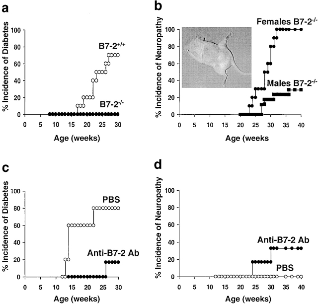
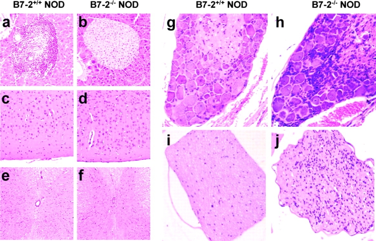
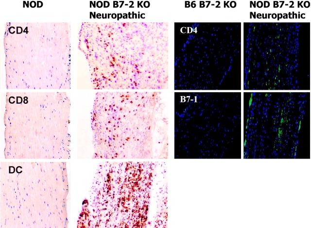
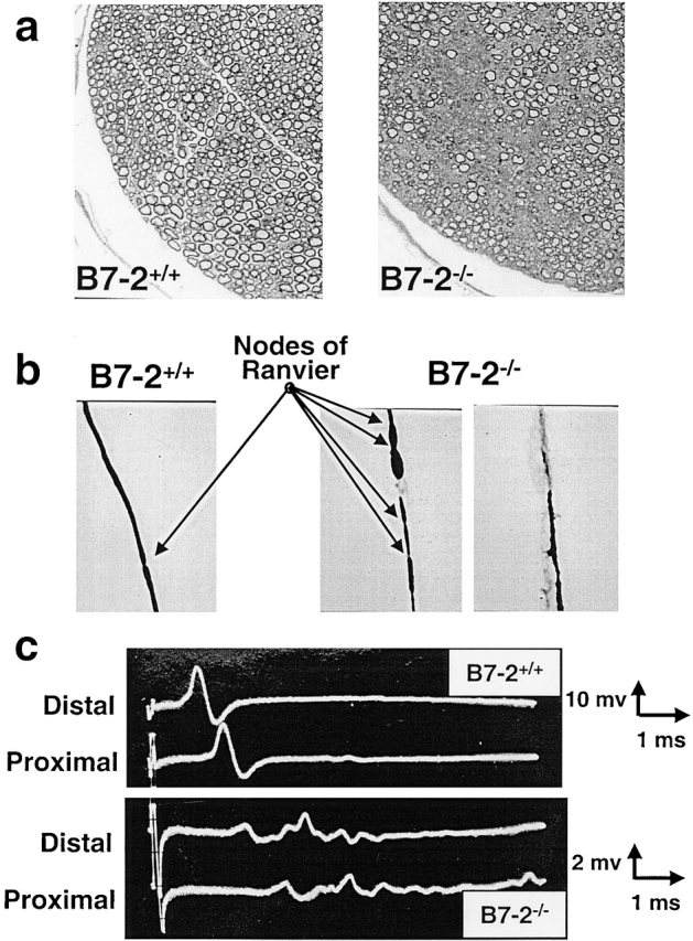
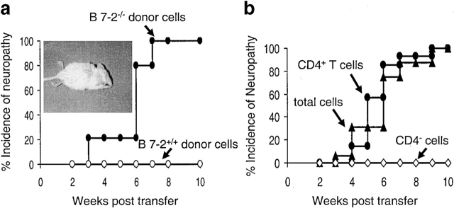
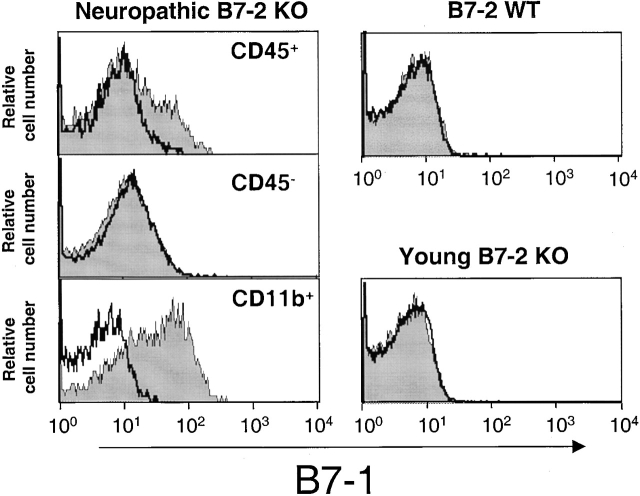
An increasing number of studies have documented the central role of T cell costimulation in autoimmunity. Here we show that the autoimmune diabetes-prone nonobese diabetic (NOD) mouse strain, deficient in B7-2 costimulation, is protected from diabetes but develops a spontaneous autoimmune peripheral polyneuropathy. All the female and one third of the male mice exhibited limb paralysis with histologic and electrophysiologic evidence of severe demyelination in the peripheral nerves beginning at 20 wk of age. No central nervous system lesions were apparent. The peripheral nerve tissue was infiltrated with dendritic cells, CD4(+), and CD8(+) T cells. Finally, CD4(+) T cells isolated from affected animals induced the disease in NOD.SCID mice. Thus, the B7-2-deficient NOD mouse constitutes the first model of a spontaneous autoimmune disease of the peripheral nervous system, which has many similarities to the human disease, chronic inflammatory demyelinating polyneuropathy (CIDP). This model demonstrates that NOD mice have "cryptic" autoimmune defects that can polarize toward the nervous tissue after the selective disruption of CD28/B7-2 costimulatory pathway.


Figures

Figure 1
Figure 1
B7-2 blockade in NOD mice prevents diabetes but induces a peripheral neuropathy (a and b). B7-2–deficient mice (reference 14) were backcrossed to NOD mice for nine generations and then intercrossed to generate B7-2 WT (B7-2+/+) and B7-2 KO (B7-2−/−) NOD mice. B7-2+/+ (n = 10) and B7-2−/− (n = 10) females were checked weekly for blood glucose levels (a) and B7-2−/− females and males were checked weekly for clinical signs of neuropathy (hind leg weakness and inability to grasp wire bar lid of cages) (b). Inset: neuropathic mouse displaying deterioration of hind limb control and coordination. (c and d) WT NOD females were treated with 50 μg of anti–B7-2 blocking mAb (GL-1) every other day between 2 to 4 wk of age plus at weeks 5, 6, and 7 (n = 6) or with PBS (n = 5). Incidence of diabetes (c) and neuropathy (d) were evaluated at weekly intervals.
Figure 2
Figure 2
Inflammation in the peripheral nervous system in neuropathic B7-2−/− NOD mice. Hematoxylin and eosin staining on sections of pancreas (a and b), brain cortex (c and d), spinal cord (e and f), dorsal root ganglion (g and h), and sciatic nerve (i and j) in 25–30-wk-old B7-2−/− neuropathic females (b, d, f, h, and j) and age-matched B7-2+/+ control females (a, c, e, g, and i). (a–d and f–j) Original magnification: ×500. (e and f) Original magnification: ×250. B7-2+/+ mice, but not B7-2−/− mice, exhibit severe insulitis (a) whereas B7-2−/− mice, but not B7-2+/+ mice, exhibit inflammation in dorsal root ganglions (h) and nerves (j).
Figure 4
Figure 4
Nerves of B7-2−/− neuropathic NOD mice are highly infiltrated with CD4+, CD8+ T cells and dendritic cells. (Left panel) Frozen sections of sciatic nerve of a 30-wk-old B7-2−/− neuropathic female and an age match control mouse were immunostained for the presence of CD4+ and CD8+ T cells and dendritic cells (DC). No infiltrate was detected in B7-2+/+ mice (dark oblong structures are nuclei of Schwann cells) whereas B7-2−/− mice presented abundant infiltration (dark round structures are stained cells). (Right panel) Frozen sections of sciatic nerve of a 28-wk-old B7-2−/− neuropathic female and an age-matched B7-2−/− B6 male mouse were immunostained for the presence of CD4 (green, top panel) and B7-1 (green, bottom panel). No infiltrate or B7-1 staining was detected in a B7-2−/− B6 mouse whereas B7-2−/− NOD mice presented abundant infiltration and B7-1 expression.
Figure 3
Figure 3
B7-2−/− neuropathic NOD mice have a demyelinating disease. (a) Toluidine blue staining on cross section of sciatic nerves showing destruction of myelin sheets in B7-2−/− neuropathic mice evidenced by lower sheet density (right panel). (b) Individual nervous fibers released by tease preparation of sciatic nerves showing clear evidence of demyelination in B7-2−/− mice (right panels). In addition some fibers have increased number of nodes of Ranvier (arrows) due to myelin sheet regeneration by newly dividing Schwann cells (middle panel). (c) Compound muscle action potentials were obtained upon distal (ankle) and proximal (thigh) stimulation of sciatic nerve. Examples of nerve conduction studies showing prolonged latencies, slowed conduction velocity, and dispersion of compound muscle action potentials in B7-2−/− neuropathic mice compared with controls (bottom panel).
Figure 5
Figure 5
Essential role of CD4+ T cells in the neuropathy of B7-2−/− NOD mice. (a) Total spleen and lymph node cells from B7-2−/− neuropathic NOD mice or age match NOD controls were stimulated in vitro with anti-CD3 and anti-CD28 mAbs and injected into five NOD.SCID mice. Symptoms of neuropathy were then checked weekly. Inset: neuropathic NOD.SCID mouse displaying complete paralysis of hind limbs. (b) CD4+ T cells, CD4-depleted cells, and whole cells, purified from spleen and lymph nodes of B7-2−/− neuropathic NOD mice, were stimulated in vitro with anti-CD3 and anti-CD28 mAbs and injected into 14, 9, and 16 NOD.SCID mice, respectively. Data are from three independent experiments.
Figure 6
Figure 6
Expression of B7-1 in B7-2KO NOD mice. Sciatic nerve tissues from 28-wk-old neuropathic B7-2 KO or 12–14-wk-old B7-2 KO and B7-2 WT females were digested with collagenase. Indicated cell subpopulations (CD45 cell population in B7-2 WT and young B7-2 KO mice) were then analyzed for B7-1 expression (shaded histograms) by flow cytometry. White histograms show the staining with the isotypic control Ab.

Comment in

References

    1. Jenkins M.K., Schwartz R.H. Antigen presentation by chemically modified splenocytes induces antigen-specific T cell unresponsiveness in vitro and in vivo. J. Exp. Med. 1987;165:302–319. - PMC - PubMed
    1. Linsley P.S., Brady W., Grosmaire L., Aruffo A., Damle N.K., Ledbetter J.A. Binding of the B cell activation antigen B7 to CD28 costimulates T cell proliferation and interleukin 2 mRNA accumulation. J. Exp. Med. 1991;173:721–730. - PMC - PubMed
    1. Harding F.A., McArthur J.G., Gross J.A., Raulet D.H., Allison J.P. CD28-mediated signalling co-stimulates murine T cells and prevents induction of anergy in T-cell clones. Nature. 1992;356:607–609. - PubMed
    1. Salomon B., Bluestone J.A. Complexities of CD28/B7CTLA-4 costimulatory pathways in autoimmunity and transplantation. Annu. Rev. Immunol. 2001;19:225–252. - PubMed
    1. Finck B.K., Linsley P.S., Wofsy D. Treatment of murine lupus with CTLA4Ig. Science. 1994;265:1225–1227. - PubMed

Publication types

MeSH terms