. 2001 Sep 11;98(19):10658-63.
doi: 10.1073/pnas.181029198. Epub 2001 Sep 4.

Compaction of single DNA molecules induced by binding of integration host factor (IHF)

Affiliations

Compaction of single DNA molecules induced by binding of integration host factor (IHF)

B M Ali et al. Proc Natl Acad Sci U S A. .

Abstract

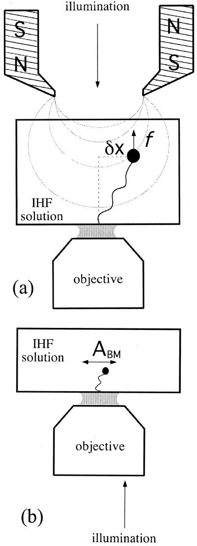
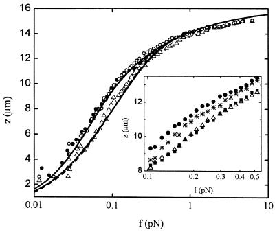
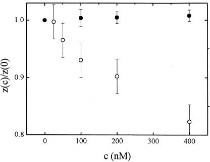
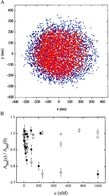
We studied the interaction between the integration host factor (IHF), a major nucleoid-associated protein in bacteria, and single DNA molecules. Force-extension measurements of lambda DNA and an analysis of the Brownian motion of small beads tethered to a surface by single short DNA molecules, in equilibrium with an IHF solution, indicate that: (i) the DNA-IHF complex retains a random, although more compact, coiled configuration for zero or small values of the tension, (ii) IHF induces DNA compaction by binding to multiple DNA sites with low specificity, and (iii) with increasing tension on the DNA, the elastic properties of bare DNA are recovered. This behavior is consistent with the predictions of a statistical mechanical model describing how proteins bending DNA are driven off by an applied tension on the DNA molecule. Estimates of the amount of bound IHF in DNA-IHF complexes obtained from the model agree very well with independent measurements of this quantity obtained from the analysis of DNA-IHF crosslinking. Our findings support the long-held view that IHF and other histone-like proteins play an important role in shaping the long-scale structure of the bacterial nucleoid.


Figures

Figure 1
Figure 1
(a) Experimental setup for force–extension measurements of λ-phage DNA–IHF complexes. A force is exerted on the DNA-tethered paramagnetic bead by a pair of magnets, whose height can be controlled to change the force's magnitude. (b) Experimental setup for measurements of the amplitude of transversal Brownian motion ABM of small beads tethered by short DNA molecules to a glass slide in the presence of IHF.
Figure 2
Figure 2
Extension z versus force f curves for a λ-phage DNA molecule in protein solutions of different concentration: 0 nM IHF (full circles), 1,250 nM IHF (empty triangles), and 1,250 nM of mutant βR46C (empty circles). The data for 0 nM data were fitted with Eq. 2 (full line), assuming fixed persistence and molecular lengths A0 and L0, respectively. The 1,250 nM data were fitted with the full model (see text and ref. 22), assuming either IHF sliding along the DNA molecule (full line) or fixed IHF positions (dashed line). (Inset) Magnification of the region of maximal difference between experimental force–distance curves for different IHF concentrations, showing behavior at an intermediate concentration, 250 nM IHF (stars), as well as the absence of hysteresis at a typical large concentration, 1,250 nM. Extension (empty triangles) of the nucleoprotein complex is followed by retraction (full squares).
Figure 3
Figure 3
Height z(c) of a 2.8-μm bead tethered by a λ-phage DNA molecule as a function of IHF concentration c, normalized by the height at c = 0 in the presence of 0.02 mg/ml poly(dI-dC) (full circles) and in its absence (empty circles). The molecule is stretched with a constant force of 150 fN.
Figure 4
Figure 4
Brownian motion of beads tethered by short DNA fragments. (A) x,y coordinates of a bead undergoing Brownian motion without IHF (blue) and with 1.0 μM IHF (red). The bead was tethered by a 1,288-bp DNA molecule with a specific IHF site. (B) Normalized amplitude of Brownian motion ABM(c)/ABM(0) of tethered beads as a function of wild-type IHF concentration c: 1,288-bp DNA with a specific IHF site (full circles), 850-bp DNA without a specific site (empty circles), 1,288-bp DNA with a specific site in the presence of 0.02 mg/ml of poly(dI-dC) (full squares). ABM(c)/ABM(0) as a function of mutant βR46C concentration c in the case of a bead tethered by a 1,200-bp DNA molecule (empty squares).
Figure 5
Figure 5
Buoyant densities of DNA–IHF complexes. Radioactive λ DNA alone (full circles) incubated with native IHF (full squares) or with mutant IHF (empty circles) was fixed with formaldehyde and fractionated by equilibrium density gradient centrifugation in CsCl. The radioactivity in each fraction is shown as a function of the density, as calculated from the refractive index. Unlabeled λ DNA added to all gradients as an internal standard banded at the position marked by the arrow.

References

    1. Pettijohn D E. J Biol Chem. 1988;263:12793–12798. - PubMed
    1. Trun N J, Marko J F. Am Soc Microbiol News. 1998;64:276–283.
    1. Drlica K, Rouviere-Yaniv J. Microbiol Rev. 1987;51:301–319. - PMC - PubMed
    1. Sinden R R, Pettijohn D E. Proc Natl Acad Sci USA. 1981;78:224–228. - PMC - PubMed
    1. Zimmerman S B, Minton A P. Rev Biophys Biomol Struct. 1993;22:27–65. - PubMed

Publication types

LinkOut - more resources