. 2001 Sep 11;98(19):10722-7.
doi: 10.1073/pnas.191067898. Epub 2001 Sep 4.

Suppression of Ras-mediated tumorigenicity and metastasis through inhibition of the Met receptor tyrosine kinase

Affiliations

Suppression of Ras-mediated tumorigenicity and metastasis through inhibition of the Met receptor tyrosine kinase

K A Furge et al. Proc Natl Acad Sci U S A. .

Abstract

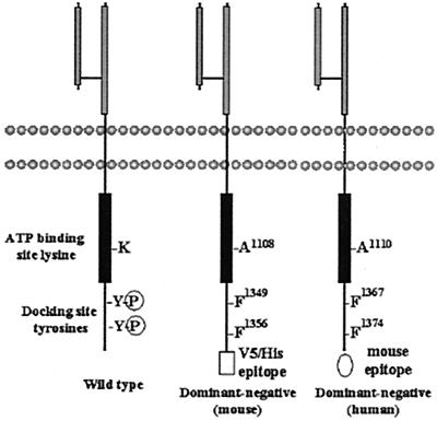
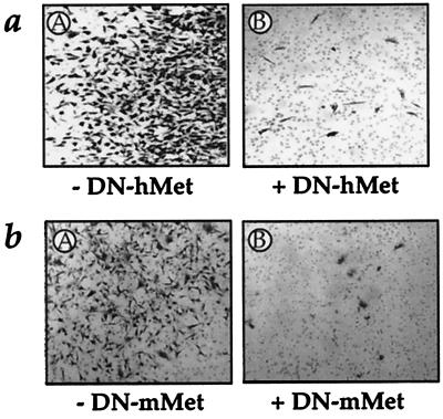
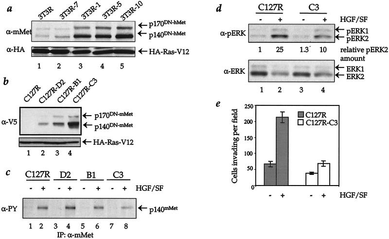
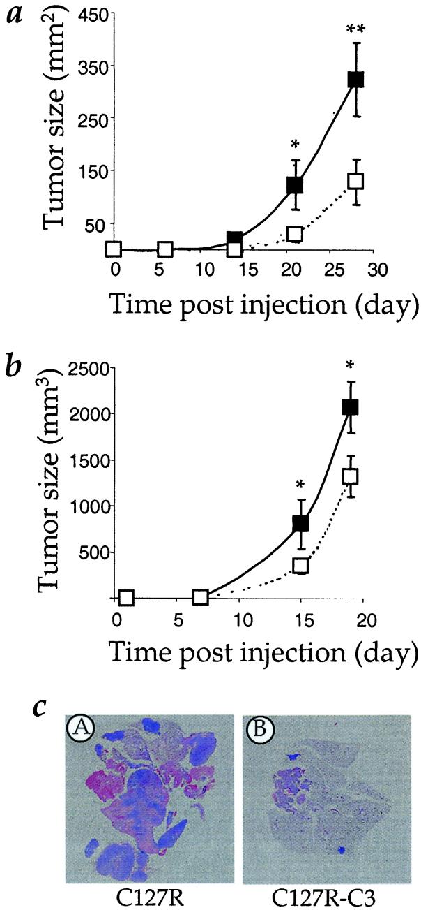
Mutations in the Ras family of GTP binding proteins represent one of the most frequently observed genetic alterations in human cancers. We and others have recently demonstrated that expression of Met, the tyrosine kinase receptor for hepatocyte growth factor/scatter factor (HGF/SF), is significantly up-regulated in Ras-transformed cells. Because HGF/SF-Met signaling is proposed to play a prominent role in tumor development and progression, we assessed the possible requirement for Met during Ras-mediated tumor growth and metastasis. To disrupt endogenous Met signaling, we constructed dominant-negative mutants of both human and murine Met and showed that these can inhibit HGF/SF-mediated Met signaling and cell invasion of ras-transformed cells in vitro. Moreover, ectopic expression of dominant-negative Met mutants reduced the s.c. tumor growth of ras-transformed cells and dramatically suppressed their ability to form lung metastases in vivo. Our data demonstrate that Met plays a prominent role during Ras-mediated tumor growth and metastasis, and further suggest that agents that inhibit HGF/SF-Met signaling may represent an important therapeutic avenue for the treatment of a variety of malignant tumors.


Figures

Figure 1
Figure 1
Schematic of dominant-negative Met mutant receptors. Light and dark gray boxes designate the Met extracellular domain and the intracellular tyrosine kinase domain, respectively. A lysine critical for kinase activity was mutated to alanine and two tyrosines in the C-terminal region of the protein that are required for interactions with a number of effector proteins were mutated to phenylalanine. The mouse dominant-negative Met contains a C-terminal V5 epitope tag. The C-terminal 21 aa of the human dominant-negative Met were replaced by the C-terminal 12 aa of mouse Met.
Figure 2
Figure 2
Expression of DN-Met inhibits HGF/SF-mediated invasion of B16-F1 melanoma cells. (a) B16-F1 cells stably transfected with control vector (pRK5, A) or human dominant-negative Met (DN-hMet/pRK5, B) were placed onto the upper chamber of Matrigel filters and lowered into wells containing 50 ng/ml HGF/SF for 96 h. Invading cells adhering to the underside of the filter were stained and photographed. (b) B16-F1 cells stably transfected with control vector (pcDNA6, A) or mouse dominant-negative Met (DN-mMet/pcDNA6, B) and assayed as in a.
Figure 3
Figure 3
Expression of DN-Met suppresses HGF/SF-mediated Met signaling in ras-transformed cells. (a) Cell extracts from NIH 3T3 cells stably transfected with V12 H-Ras and either control plasmid (lane 1, 3T3R) or a plasmid encoding dominant-negative human Met (lanes 2–5) were analyzed by Western blotting with anti-mouse Met (SP260) and anti-HA (Y-11) antibodies. Clone designations are shown above. (b) Cell extracts from ras-transformed C127 cells transfected with either control plasmid (lane 1, C127R) or a plasmid encoding dominant-negative mouse Met (lanes 2–4) were analyzed by Western blotting with anti-HA (Y-11) and anti-V5 antibodies. Clone designations are shown above. (c) The indicated C127 clones were mock treated (lanes 1, 3, 5, and 7) or treated with 50 ng/ml HGF/SF for 5 min (lanes 2, 4, 6, and 8). Met was precipitated from cell extracts by using anti-mouse Met antiserum (SP260), and the immunoprecipitates were assayed by Western blotting using antiphosphotyrosine antibodies (4G10). (d) The indicated C127 clones were mock treated (lanes 1 and 3) or treated with HGF/SF as in c (lanes 2 and 4). Cell extracts were assayed by Western blotting using anti-phospho-ERK1/ERK2 (E10) and anti-ERK1/ERK2 (SC-94/SC-154) antibodies. The degree of ERK2 phosphorylation was determined relative to total ERK2 levels by densitometry of Western blots using QUANTISCAN software. (e) Indicated ras-transformed C127 cells were placed onto the upper chamber of Matrigel filters and lowered into wells containing media with or without 50 ng/ml HGF/SF for 22 h. Invading cells adhering to the underside of the filter were stained and quantified by counting. Data represent mean cell number from triplicate experiments (±SE).
Figure 4
Figure 4
Effect of dominant-negative Met expression on the growth and metastasis of ras-transformed cells. (a) ras-transformed NIH 3T3 control cells (clone 3T3R, solid line) or cells expressing dominant-negative human Met (clone 3T3R-10, hashed line) were injected s.c. into the back of athymic nude female mice and the size of the resulting tumors measured at the indicated times. Data represent mean tumor area (±SE) from four mice. A one-tailed t test was used to determine the statistical difference between the tumors with * designating P ≤ 0.09 and ** designating P ≤ 0.02. (b) ras-transformed C127 control cells (clone C127R, solid line) or cells expressing dominant-negative murine Met (clone C127R-C3, hashed line) were injected and analyzed as described in a. Data represent mean tumor volume (±SE) from five mice with * designating P ≤ 0.09 computed as in a. (c) Photographs of representative hematoxylin and eosin stained lung sections obtained 4 weeks post tail vein injection of ras-transformed C127 control cells (C127R) or cells expressing dominant-negative mouse Met (C127R-C3). Tumor lesions appear as intense blue staining.

Similar articles

Cited by

References

    1. Hanahan D, Weinberg R A. Cell. 2000;100:57–70. - PubMed
    1. Greenlee R, Murray T, Bolden S, Wingo P. CA Cancer J Clin. 2000;50:7–33. - PubMed
    1. Collard J G, Roos E, La, R. e G, Habets G G. Cancer Surv. 1988;7:691–710. - PubMed
    1. Bos J L. Cancer Res. 1989;49:4682–4689. - PubMed
    1. Chambers A F, Tuck A B. Crit Rev Oncog. 1993;4:95–114. - PubMed

Publication types

Substances