. 2001 Sep 11;98(19):10799-804.
doi: 10.1073/pnas.191124098. Epub 2001 Sep 4.

Crystal structure of the human CD4 N-terminal two-domain fragment complexed to a class II MHC molecule

Affiliations

Crystal structure of the human CD4 N-terminal two-domain fragment complexed to a class II MHC molecule

J H Wang et al. Proc Natl Acad Sci U S A. .

Abstract

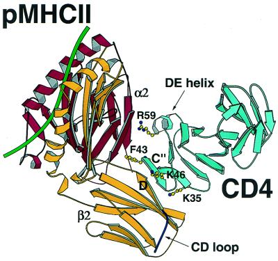
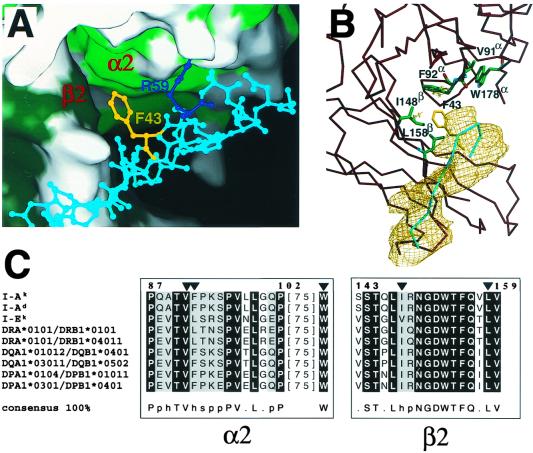
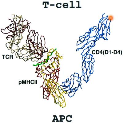
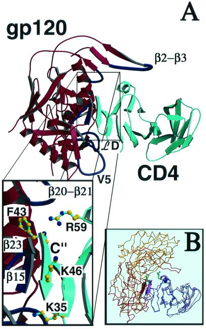
The structural basis of the interaction between the CD4 coreceptor and a class II major histocompatibility complex (MHC) is described. The crystal structure of a complex containing the human CD4 N-terminal two-domain fragment and the murine I-A(k) class II MHC molecule with associated peptide (pMHCII) shows that only the "top corner" of the CD4 molecule directly contacts pMHCII. The CD4 Phe-43 side chain extends into a hydrophobic concavity formed by MHC residues from both alpha 2 and beta 2 domains. A ternary model of the CD4-pMHCII-T-cell receptor (TCR) reveals that the complex appears V-shaped with the membrane-proximal pMHCII at the apex. This configuration excludes a direct TCR-CD4 interaction and suggests how TCR and CD4 signaling is coordinated around the antigenic pMHCII complex. Human CD4 binds to HIV gp120 in a manner strikingly similar to the way in which CD4 interacts with pMHCII. Additional contacts between gp120 and CD4 give the CD4-gp120 complex a greater affinity. Thus, ligation of the viral envelope glycoprotein to CD4 occludes the pMHCII-binding site on CD4, contributing to immunodeficiency.


Figures

Figure 1
Figure 1
Ribbon diagram of the CD4–pMHCII complex. The murine I-Ak MHC class II molecule with a CA peptide (green) bound to the antigen-presenting platform interacts with hCD4 (cyan) through both the α2 (red) and the β2 (yellow) domains of pMHCII and domain 1 (D1) of hCD4. Residues Lys-35, Phe-43, Lys-46, and Arg-59 on CD4 D1 essential for binding are highlighted. The CD loop (delineated with an arrow) on the β2 domain of the I-Ak molecule is shown to have no direct interactions with CD4. Note also how CD4 D2 (unlabeled, cyan) makes no contact to pMHCII. All the figures were prepared with molscript (46) and RASTER 3D (47).
Figure 2
Figure 2
The conserved hydrophobic pocket formed by class II MHC α2 and β2 domain residues into which CD4 Phe-43 inserts. (A) Class II MHC is depicted in surface representation form and adjacent CD4 residues in ball-and-stick format. Hydrophobic residues from domain α2 (light green) and domain β2 (dark green) of the I-Ak molecule are shown. The positions of the side chains of Phe-43 (yellow) and Arg-59 (blue) have been optimized by molecular dynamics by using cns (24). The surface representation was prepared with spock. (B) The same region is presented with residues that contribute to the hydrophobic pocket shown, including Val-91, Phe-92, and Trp-178 from the α2 domain and Ile-148 and Leu-158 from the β2 domain of the MHC class II molecule in green. The Cα trace of the MHC class II molecule in this region is depicted in dark red. The Cα trace of the C" strand of the CD4 molecule is depicted in cyan, and the Phe-43 residue is shown in yellow. A portion of an omit map at a contour level of 1σ is shown in gold for the C" strand of CD4, which was obtained by rigid body refinement with this segment excluded from the calculation. (C) Sequence alignment of the highly conserved contact regions in MHC class II for different murine (I-Ak, I-Ad, and I-Ek) and human (DRA*0101/DRB1*0101, DRA*0101/DRB1*04011, DQA1*01012/DQB1*0401, DQA1*03011/DQB1*0502, DPA1*0104/DPB1*01011, DPA1*0301/DPB1*0401) alleles by using clustalx (48). Residues Val-91, Phe-92, and Trp-178 of the α2 domain and Ile-148 and Leu-158 of the β2 domain are indicated by a triangle. The consensus is defined so that entirely conserved residues are depicted by their amino acid code, whereas p indicates conservation of a polar residue, h conservation of a hydrophobic residue, and s conservation of a small side-chain-containing residue.
Figure 3
Figure 3
Model of the ternary CD4–pMHCII–TCR complex. The assembly of several crystal structures reveals a ternary complex composed of the hCD4 D1-D4, pMHCII, and TCR having a V-shaped configuration. The D10 TCR–CA/I-Ak complex consists of the α (bright red) and β (yellow) subunits of the I-Ak molecule, the CA peptide (green), and the two TCR subunits α (dark red) and β (beige). The hCD4 D1-D4 is shown in blue, with the approximate position of the D4 glycan given as an orange ball. The assembly is oriented with the T-cell membrane on top and the antigen-presenting cell membrane on the bottom.
Figure 4
Figure 4
Molecular mimicry of gp120 binding to CD4 relative to MHC class II. (A) The CD4 D1D2–gp120 complex (ref. , with Fab not included for clarity) is shown in similar orientation to the complex of CD4 D1D2/I-Ak in Fig. 1. The interactions that are in common are displayed in the box, which shows the Phe-43 inserted in a hydrophobic pocket involving gp120 β15 and β23, the formation of an antiparallel mini-β-sheet between the C" strand of CD4 and β15 from gp120 as well as interactions between CD4 Arg-59 and the loop connecting β20 to β21 in gp120. Besides these core interactions, gp120 has additional contacts through the V1/V2 loop connecting the β2 and β3 strands, the ℒD loop, and the V5 loop. (B) Overlay of CD4–CA/I-Ak and CD4–gp120 (42) complexes based on hCD4 D1 (blue and pink, respectively) with I-Ak in yellow and gp120 in red. Phe-43 and Arg-59 side chains are shown.

Similar articles

Cited by

References

    1. Wang J-H, Reinherz E L. Curr Opin Struct Biol. 2000;10:656–661. - PubMed
    1. Janeway C A., Jr Annu Rev Immunol. 1992;10:645–674. - PubMed
    1. Kruisbeek A M, Mond J J, Fowlkes B J, Carmen J A, Bridges S, Longo D L. J Exp Med. 1985;161:1029–1047. - PMC - PubMed
    1. Marrack P, Kappler J. Adv Immunol. 1986;38:1–30. - PubMed
    1. Gao G F, Tormo J, Gerth U C, Wyer J R, McMichael A J, Stuart D I, Bell J I, Jones E Y, Jakobsen B K. Nature (London) 1997;387:630–634. - PubMed

Associated data