

Replication of enhancer-deficient amphotropic murine leukemia virus in human cells
- PMID: 11535815
- PMCID: PMC58571
- DOI: 10.1073/pnas.191182098
Replication of enhancer-deficient amphotropic murine leukemia virus in human cells
Abstract
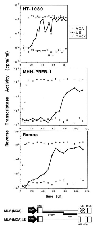
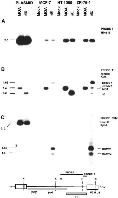
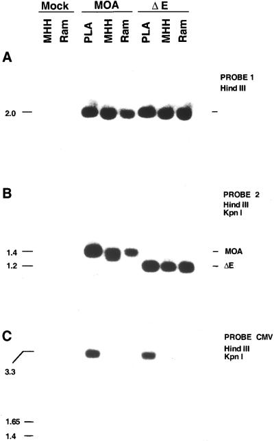
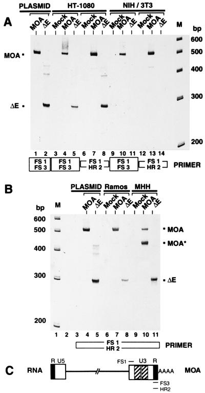
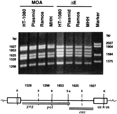
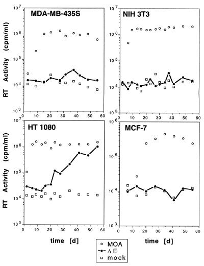
Amphotropic murine leukemia virus (MLV) replicates in cells from various mammalian species, including humans, and is a potential contaminant in MLV vector preparations for human gene transfer studies. The generation of replication-competent virus is considered less likely with vectors that delete the viral transcription elements. This conclusion is based on data obtained in rodents, where MLV replication depends on the expression of viral genes under the control of 75-bp enhancer elements in the long terminal repeat. We demonstrate here that in some human cells replication of amphotropic MLV is possible in the absence of these enhancer elements. Replication of the enhancer-deficient virus MLV-(MOA)Delta E is observed in selected human sarcoma and B lymphoma lines and proceeds at a lower rate than that of the intact virus. No insertion of a foreign promoter or enhancer into the long terminal repeat was detected. Our data suggest the presence of a secondary enhancer element within the MLV provirus that can in selected human cells mediate virus transcription and replication in the absence of the 75-bp U3 enhancers.
Figures
Similar articles
-
Transcriptional inactivation of amphotropic murine leukemia virus replication in human cells.J Med Virol. 2003 Feb;69(2):267-72. doi: 10.1002/jmv.10274. J Med Virol. 2003. PMID: 12683417
-
Replication of enhancer-deficient amphotropic murine leukemia virus in human fibrosarcoma but not in primary human fibroblasts.J Med Virol. 2002 Oct;68(2):278-84. doi: 10.1002/jmv.10202. J Med Virol. 2002. PMID: 12210420
-
Amphotropic murine leukemia virus replication in human mammary epithelial cells and the formation of cytomegalovirus-promoter recombinants.Virology. 2001 Dec 5;291(1):91-100. doi: 10.1006/viro.2001.1199. Virology. 2001. PMID: 11878879
-
A genetic model of the host range restriction of murine leukemia viruses: a review and a hypothesis.Jpn J Exp Med. 1973 Feb;43(1):1-7. Jpn J Exp Med. 1973. PMID: 4349499 Review. No abstract available.
-
Viral and cellular transcription enhancers.Oxf Surv Eukaryot Genes. 1985;2:24-48. Oxf Surv Eukaryot Genes. 1985. PMID: 3916927 Review. No abstract available.
Cited by
-
Sequence analysis of porcine endogenous retrovirus long terminal repeats and identification of transcriptional regulatory regions.J Virol. 2003 Jan;77(1):142-9. doi: 10.1128/jvi.77.1.142-149.2003. J Virol. 2003. PMID: 12477819 Free PMC article.
-
Multiple modifications allow high-titer production of retroviral vectors carrying heterologous regulatory elements.J Virol. 2004 Feb;78(3):1384-92. doi: 10.1128/jvi.78.3.1384-1392.2004. J Virol. 2004. PMID: 14722293 Free PMC article.
References
-
- Marcel T, Grausz J D. Hum Gene Ther. 1996;7:2025–2046. - PubMed
Publication types
MeSH terms
Substances
LinkOut - more resources
Full Text Sources
