. 2001 Sep 11;98(19):10572-7.
doi: 10.1073/pnas.191375098. Epub 2001 Sep 4.

Cloning and characterization of a histone deacetylase, HDAC9

Affiliations

Cloning and characterization of a histone deacetylase, HDAC9

X Zhou et al. Proc Natl Acad Sci U S A. .

Abstract

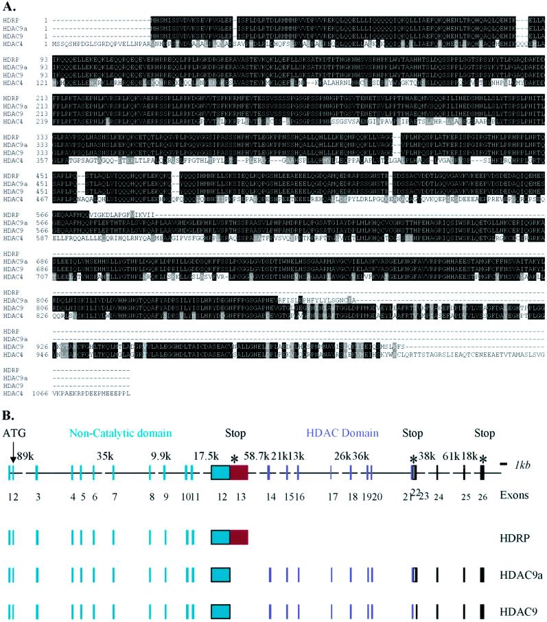
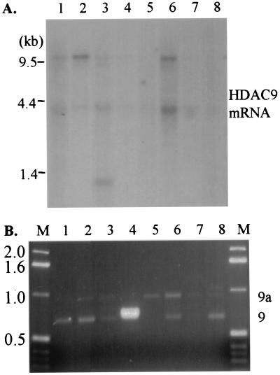
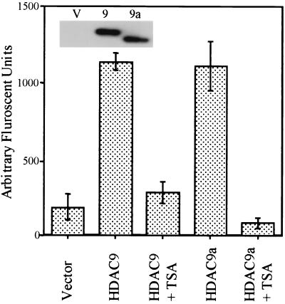
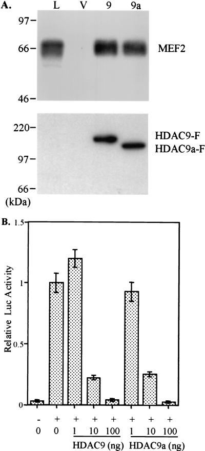
Histone deacetylase (HDAC) catalyzes the removal of the acetyl group from the lysine residues in the N-terminal tails of nucleosomal core histones. Eight human HDACs have been identified so far. Here, we report the identification of a ninth member of the HDAC family, designated HDAC9. HDAC9 is a class II HDAC and its gene resides on human chromosome 7. HDAC9 has several alternatively spliced isoforms. One of these isoforms is histone deacetylase-related protein or myocyte enhancer-binding factor 2-interacting transcriptional repressor that we and others have previously reported and which does not possess an HDAC catalytic domain. The longest of the HDAC9 isoforms contains 1,011 aa. The isoform, designated HDAC9a, is 132 aa shorter at the C terminus than HDAC9. Also, we have identified isoforms of HDAC9 that lack the nuclear localization signal. Similar to histone deacetylase-related protein, HDAC9 transcripts are expressed at high levels in brain and skeletal muscle. The ratio of HDAC9 and HDAC9a transcripts differs among the tissues examined. HDAC9 and HDAC9a contain the HDAC catalytic domain, and Flag-tagged HDAC9 and HDAC9a possess deacetylase activity. HDAC9 and HDAC9a also repress myocyte enhancer-binding factor 2-mediated transcription. In the present study, we have identified HDAC9 and a number of alternatively spliced isoforms of HDAC9 with potentially different biological activities.


Figures

Figure 1
Figure 1
Amino acid sequence alignment and HDAC9 gene structure. (A) Amino acid sequence alignment of HDAC9, HDAC9a, HDRP, and HDAC4. Deduced amino acid sequences of HDAC9 (GenBank accession no. AY032737) and HDAC9a (accession no. AY032738) are aligned with HDRP (accession no. BAA34464) and HDAC4 (accession no. NP_006028). Identical residues in all proteins are shown in reverse type on black background; similar residues are shaded in gray. (B) Human HDAC9 gene structure. The HDAC9 gene is schematically depicted. Color-coded boxes represent exons present in different isoforms; lines represent introns; broken lines represent larger introns (with size in bp at top). The 5′ untranslated region cDNA and coding region cDNA are represented here.
Figure 2
Figure 2
HDAC9 mRNA is differentially expressed among human tissues. (A) A Multiple Tissue Northern Blot (CLONTECH) was probed to determine mRNA expression of HDAC9 with a cDNA probe that recognizes both HDAC9 and HDAC9a. The tissues examined are lane 1, heart; lane 2, brain; lane 3, placenta; lane 4, lung; lane 5, liver; lane 6, skeletal muscle; lane 7, kidney; and lane 8, pancreas. Positions of the RNA size marker in kilobase (kb) are indicated on the left of the blot. (B) RT-PCR analyses of mRNA from the same tissues as examined in the Northern blot to determine the distribution of HDAC9 and HDAC9a mRNA among these tissues. PCR products were resolved by agarose gel electrophoresis and visualized by ethidium bromide under UV light. A 1-kb DNA ladder was run on both sides of the gel with the size (in kb) indicated on the left. On the right, the expected products for HDAC9 and HDAC9a are indicated as 9 and 9a, respectively.
Figure 3
Figure 3
HDAC9 and HDAC9a possess deacetylase activity. The HDAC enzymatic assays were performed with anti-FLAG-immunoprecipitated proteins from vector control, HDAC9-, and HDAC9a-transfected 293T cells by using Fluror de Lys substrate, which contains an acetylated lysine side chain. TSA (1 μM) was used as an HDAC inhibitor. Results are shown as the mean of two independent assays. (Inset) Anti-FLAG Western blot shows the amount of proteins used in the assay. V, vector control; 9, HDAC9-F; 9a, HDAC9a-F.
Figure 4
Figure 4
HDAC9 and HDAC9a interact with and repress MEF2-mediated transcription. (A) Western blot analyses of the 293T whole-cell lysate and anti-FLAG immunoprecipitates from 293T cells transfected with vector, HDAC9-F, or HDAC9a-F by using antibodies against MEF2 and FLAG. (Upper) Anti-MEF2 Western blot; (Lower) anti-FLAG Western blot. L, 293T whole-cell lysate; V, vector control immunoprecipitate (IP); 9, HDAC9-F IP; 9a, HDAC9a-F IP. (B) p3XMEF2-Luc (100 ng) and pRL-TK (5 ng) were transfected into 293T cells with pcDNA3 empty vector (−) or with pCMV-MEF2C (100 ng) (+) along with indicated amount of pFLAG-HDAC9 or pFLAG-HDAC9a. pFLAG empty vector was used to adjust the DNA to an equal amount in each transfection. The firefly luciferase activity was first normalized to the cotransfected Renilla luciferase activity, and then the value for MEF2C alone was set as 1. Results are shown as the mean of three independent transfections ± SD.

References

    1. Davie J R, Spencer V A. J Cell Biochem. 1999;32–33,Suppl.:141–148. - PubMed
    1. Strahl B D, Allis C D. Nature (London) 2000;403:41–45. - PubMed
    1. Rundlett S E, Carmen A A, Kobayashi R, Bavykin S, Turner B M, Grunstein M. Proc Natl Acad Sci USA. 1996;93:14503–14508. - PMC - PubMed
    1. Carmen A A, Rundlett S E, Grunstein M. J Biol Chem. 1996;271:15837–15844. - PubMed
    1. Taunton J, Hassig C A, Schreiber S L. Science. 1996;272:408–411. - PubMed

Publication types

MeSH terms

Associated data