. 2001 Sep 1;15(17):2215-28.
doi: 10.1101/gad.913001.

Human SMG-1, a novel phosphatidylinositol 3-kinase-related protein kinase, associates with components of the mRNA surveillance complex and is involved in the regulation of nonsense-mediated mRNA decay

Affiliations

Human SMG-1, a novel phosphatidylinositol 3-kinase-related protein kinase, associates with components of the mRNA surveillance complex and is involved in the regulation of nonsense-mediated mRNA decay

A Yamashita et al. Genes Dev. .

Abstract

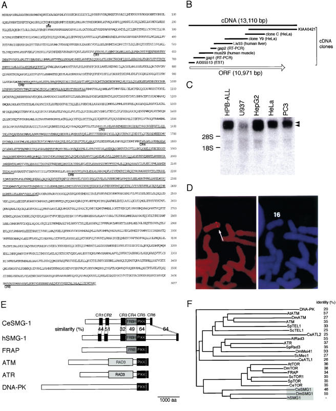
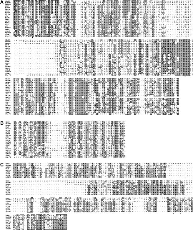
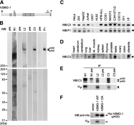
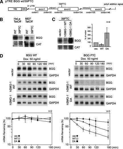
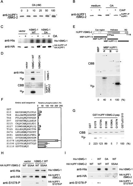
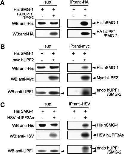
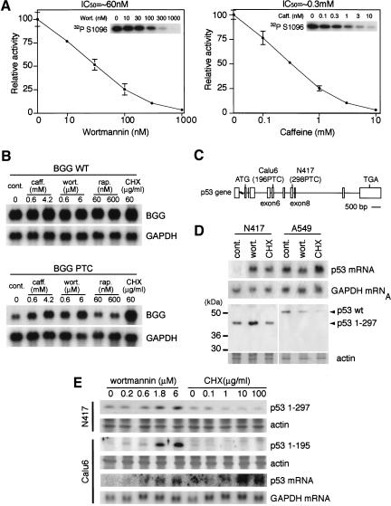
Nonsense-mediated mRNA decay (NMD) is a conserved surveillance mechanism that eliminates imperfect mRNAs that contain premature translation termination codons (PTCs) and code for nonfunctional or potentially harmful polypeptides. We show that a novel phosphatidylinositol 3-kinase-related protein kinase, hSMG-1, is a human ortholog of a product of Caenorhabditis elegans smg-1, one of seven smg genes involved in NMD. hSMG-1 phosphorylates hUPF1/SMG-2 in vivo and in vitro at specific serine residues in SQ motifs. hSMG-1 can associate with hUPF1/SMG-2 and other components of the surveillance complex. In particular, overexpression of a kinase-deficient point mutant of hSMG-1, hSMG-1-DA, results in a marked suppression of the PTC-dependent beta-globin mRNA degradation; whereas that of wild-type hSMG-1 enhances it. We also show that inhibitors of hSMG-1 induce the accumulation of truncated p53 proteins in human cancer cell lines with p53 PTC mutation. Taken together, we conclude that hSMG-1 plays a critical role in NMD through the direct phosphorylation of hUPF1/SMG-2 in the evolutionally conserved mRNA surveillance complex.


Figures

Figure 1
Figure 1
Structure of hSMG-1. (A) The predicted translation product of the human SMG-1 (hSMG-1) cDNA clones is shown. CR1–CR6 show regions conserved between CeSMG-1 and hSMG-1. (B) Schematic representation of the hSMG-1 ORF (open arrow) and cDNA clones used to analyze the complete structure. (C) Northern blotting analysis of hSMG-1 mRNA using total RNA from the indicated human cell lines. Arrowheads show the positions of two major signals. Positions for the 18S and 28S ribosomal RNAs are also shown. (D) FISH mapping of hSMG-1. Human mitotic chromosomes with the FISH signal (left panel). The arrow shows the position of the specific signal. In the right panel, the PI (propidium iodide) staining pattern of the same viewing field is shown to observe chromosome morphology. (E) Schematic representation of the members of mammalian PIK-related kinase family in comparison with CeSMG-1. The putative PIKK domain is shown as a dark gray box. The FRBH region is shown as medium gray and the RAD3 homology region is shown as light gray. GenBank accession nos. are as follows: hSMG-1,AB061371; FRAP, L34075; ATM, U33841; ATR, U76308; DNA-PK, U34994. (F) Molecular phylogenetic tree for putative catalytic domains of human PIKK family members.
Figure 2
Figure 2
Sequence comparison of human SMG-1 with other PIK-related protein kinase family members. (A) Alignment of catalytic core domains of PIKKs. Completely and partially conserved amino acids are boxed in dark and light gray, respectively. The amino acid sequence, DFG (asterisks), seen in several members of the PIKK family, is highly conserved between kinases. The solid triangle shows the aspartic acid residue completely conserved in essentially all protein kinases. (B) Alignment of the C-terminal homology region of PIKK. (C) Alignment of the FRBH domain. The tryptophan residue (solid triangle) shows a conserved residue essential for both kinase activity and binding to the FKBP12–rapamycin complex. The asterisks show residues essential for binding to the FKBP12–rapamycin complex.
Figure 3
Figure 3
Identification of human SMG-1 and its intrinsic protein kinase activity. (A) The schematic structure of hSMG-1 is shown with the regions (N, L, C, and P) used for antibody generation. CR1–CR6 are shown in boxes. (B) The HeLa cell lysate was analyzed by Western blotting using antisera shown on top and the corresponding pre-immune sera. Two bands, corresponding to 400- and 430-kD proteins (p400 and p430), are indicated by arrowheads. (C) hSMG-1 expression in various cell lines from human, simian, mouse, and rat was examined using C3 or P1 antiserum. The uppermost bands in two cell lines from mouse, NIH3T3 and C3H10T1/2, p460 (asterisk) were not stained with P1 and appeared to be a nonspecific band. (D) hSMG-1 expression in various tissues, mainly from rat, was examined with C3 antiserum. Only the placenta was from mouse, and the asterisk indicates a nonspecific band. (E) hSMG-1 expressed in HeLa cells has intrinsic autophosphorylation activity. The HeLa cell lysate was subjected to immunoprecipitation using N1, L2, or C3 antiserum or their corresponding pre-immune serum. Immunoprecipitated proteins were analyzed by Western blotting using C3 antiserum (top). In vitro kinase reaction with the immuno complex was performed without using any exogenous substrate. This was followed by 5.5% SDS-PAGE and autoradiography. (F) His-tagged hSMG-1 overexpressed in 293T cells has intrinsic autophosphorylation activity. 293T cells were transfected with SR6H (vector), SR6H hSMG-1, or SR6H hSMG-1-DA. Transfected cells were harvested and lysed, followed by immunoprecipitation using an anti-His-tag antibody and in vitro kinase assay. The autoradiogram is shown (bottom). Western blotting results of the immunoprecipitated His-tagged hSMG-1s are also shown (top).
Figure 4
Figure 4
SMG-1 is involved in the PTC-dependent degradation of β-globin mRNA. (A) Schematic representation of human β-globin (BGG) gene reporter constructs, BGG-WT and BGG-PTC. The ORF is represented by boxes, and introns and UTRs are represented by lines. (B) The BGG PTC transcript is less abundant than transcripts from the WT reporter. The WT or mutant reporter plasmid was transfected with the CAT plasmid into the HeLa Tet-Off or MEF Tet-Off cell lines. The same amount of total RNA from each cell was analyzed by Northern blotting using a BGG or a CAT probe. (C) Overexpression of hSMG-1 suppresses the accumulation of the BGG 39PTC transcript; whereas that of hSMG-1-DA enhances it. HeLa Tet-Off was transfected with the BGG WT reporter or BGG 39PTC (1.5 μg) with the hSMG-1-WT or hSMG-1-DA (3 μg) and a CAT construct (1.5 μg). Then the accumulated BGG transcripts were evaluated by Northern blotting using CAT as an internal control. The relative amount of transcripts is shown in the bar graph. (D) Overexpression of hSMG-1-WT enhances the decay of the BGG 39PTC transcript; whereas that of hSMG-1-DA suppresses it. The HeLa Tet-Off cell was transfected with the WT (left) or mutant (right) reporter gene, and cells were harvested at an indicated time point after the addition of 50 ng/mL doxycycline to the medium. Total RNA was isolated and analyzed by Northern blotting using the corresponding probe. The relative amount of transcripts is shown in the graph. The BGG RNA levels are normalized to those of GAPDH.
Figure 5
Figure 5
hSMG-1 phosphorylates hUPF1/SMG-2 in vivo and in vitro. (A) Upward-shift of hUPF1/SMG-2 induced by OA treatment. HeLa cells were treated with the indicated concentrations of OA for 4.5 h. The lysate was analyzed by Western blotting using an anti-hUPF1/SMG-2 antibody. Positions of the shifted bands are marked by asterisks. (B) Upward-shift of hUPF1/SMG-2 (marked by an asterisk) is due to phosphorylation. Immunoprecipitate prepared as in A was treated with CIAP (+) or without CIAP (−), followed by Western blotting using an anti-hUPF1/SMG-2 antibody. (C) Overexpression of hSMG-1-WT induces OA-dependent phosphorylation of hUPF1/SMG-2 in 293T cells. Cells were transfected with a WT or a DA mutant hSMG-1 expression plasmid (10 μg) or a vehicle (vector) with an HA-tagged hUPF1/SMG-2 construct (0.5 μg). The upward shift was evaluated as shown in B. The position of the shifted HA- hUPF1/SMG-2 is marked by an asterisk. (D) hSMG-1 directly phosphorylates hUPF1/SMG-2. 293T cells were transfected with the expression plasmid for WT or DA mutant His-tagged hSMG-1 or a vehicle (vector). Immunocomplex using an anti-His antibody was subjected to in vitro kinase assay using purified HA-tagged hUPF1/SMG-2 as an exogenous substrate. An autoradiogram showing hUPF1/SMG-2 phosphorylation is shown (bottom). (E) hSMG-1 directly phosphorylates hUPF1/SMG-2 at its N- and C-terminal regions. In vitro kinase assay was performed using recombinant MBP-fused hUPF1/SMG-2 proteins (MBP hUPF1/SMG-2-N, M and C). The pattern of CBB staining of the gel and its autoradiogram are shown. The extent of phosphorylation relative to MBP-hUPF1/SMG-2-C is shown as percentages at the bottom. Positions of the major bands of each recombinant protein are indicated by asterisks. (F) hSMG-1 phosphorylates GST-fused 14mer SQ- or TQcontaining peptides derived from hUPF1/SMG-2. In vitro kinase assay was performed as in E using GST-fused peptides. Numbers are of the serine or threonine residue adjacent to glutamine. Data in the graph are means ± SD from three to seven independent experiments. (G) Phosphorylation of the GST-hUPF1/SMG-2 peptides with mutations at S1096 was carried out according to a protocol similar to that described in C. (H) Phosphorylation of hUPF1/SMG-2 at S1078 in vitro. 293T cells were transfected with SR6H (vector) or SR6H-hSMG-1-WT. His-SMG-1 was immunoprecipitated from SR6H (vector) or SR6H-hSMG-1-WT-transfected 293T cells using a His-tag antibody. In vitro kinase assay was performed using purified HA-hUPF1/SMG-2-WT or SSAA mutant in the presence of 200 μM ATP. Phosphorylated HA-UPF1/SMG-2 was visualized by Western blotting using anti-S1078-P (bottom) and anti-HA (middle) antibodies. Immunoprecipitated His-hSMG-1 was also probed using an anti-His antibody (top). (I) Overexpression of hSMG-1-WT enhances the phosphorylation of hUPF1/SMG-2 S1078 and S1096; whereas that of hSMG-1-DA suppresses it. 293T cells were transfected with SR6H-SMG-1-WT or SR6H-SMG-1-DA (10 μg) in combination with either SRHA-hUPF1/SMG-2-WT or SRHA-hUPF1/SMG-2-SSAA (0.5 μg). HA-hUPF1/SMG-2 was immunoprecipitated using an anti-HA antibody and analyzed by Western blotting with anti-HA (top) and anti-S1078-P (bottom) antibodies.
Figure 6
Figure 6
Association of hSMG-1 with other NMD components. His-hSMG-1 (10 μg) was coexpressed with either HA-hUPF1/SMG-2 (5 μg), myc-hUPF2 (5 μg), or HSV-hUPF3As (5 μg) in 293T cells. HA-hUPF1/SMG-2 (A), myc-hUPF2 (B), or HSV-hUPF3As (C) was immunoprecipitated using the corresponding anti-tag antibody and the resulting immunoprecipitate was probed using an anti-His antibody to detect hSMG-1. Coimmunoprecipitated endogenous hUPF1/SMG-2 was also probed in B and C. In C, upward-shifted hUPF1/SMG-2 is indicated by an asterisk.
Figure 7
Figure 7
Inhibitors of hSMG-1 suppress NMD and cause accumulation of truncated p53 proteins in human cancer cell lines with PTC mutations. (A) Wortmannin or caffeine inhibits protein kinase activity of hSMG-1 in vitro. In vitro kinase assays were performed using immunoprecipitated His-hSMG-1 and a GST- hUPF1/SMG-2 fusion protein, hUPF1/SMG-2 S1096, as a substrate in the presence of an indicated concentration of wortmannin or caffeine. Autoradiograms of hUPF1/SMG-2 S1096 phosphorylation are indicated in the corresponding insets. (B) The BGG reporter mRNA with a PTC is specifically accumulated by inhibitors of hSMG-1. The MEF Tet-Off cells were transfected with a wild-type (upper) or mutant (lower) BGG reporter gene (Fig. 4C) and reseeded. Cells were treated for 2 h with caffeine (caff.), wortmannin (wort.), rapamycin (rap.), or cycloheximide (CHX) in the absence of doxycycline. Total RNAs were analyzed by Northern blotting using a BGG probe (top) or a GAPDH probe (bottom). (C) Schematic representation of p53 gene structure with PTC mutations in human lung cancer cell lines, Calu6 (196PTC) and N417 (298PTC). Boxes show exons. (D) Wortmannin induces the accumulation of transcripts and their corresponding proteins from the p53 gene with PTC. N417 and A549 cells were treated with or without (cont.) 10 μM wortmannin (wort.) or 100 μg/mL cycloheximide (CHX) for 4 h. Total RNA and cell lysates were analyzed by Northern blotting using a p53 probe and by Western blotting using an anti-p53 antibody, DO-1, respectively. The CBB image showing actin staining is also shown (bottom). (E) Wortmannin induces expression of p53 transcripts and p53 proteins of lower molecular weights in a dose-dependent manner. Calu6 or N417 cells were treated with an indicated concentration of wortmannin or CHX for 2 h. The total RNA and cell lysate were prepared and analyzed, as in E, by Northern blotting using a p53 probe and by Western blotting using DO-1, respectively.

References

    1. Beelman CA, Stevens A, Caponigro G, LaGrandeur TE, Hatfield L, Fortner DM, Parker R. An essential component of the decapping enzyme required for normal rates of mRNA turnover. Nature. 1996;382:642–646. - PubMed
    1. Bhattacharya A, Czaplinski K, Trifillis P, He F, Jacobson A, Peltz SW. Characterization of the biochemical properties of the human Upf1 gene product that is involved in nonsense-mediated mRNA decay. RNA. 2000;6:1226–1235. - PMC - PubMed
    1. Bodner SM, Minna JD, Jensen SM, D'Amico D, Carbone D, Mitsudomi T, Fedorko J, Buchhagen DL, Nau MM, Gazdar AF, et al. Expression of mutant p53 proteins in lung cancer correlates with the class of p53 gene mutation. Oncogene. 1992;7:743–749. - PubMed
    1. Brown EJ, Beal PA, Keith CT, Chen J, Shin TB, Schreiber SL. Control of p70 S6 kinase by kinase activity of FRAP in vivo. Nature. 1995;377:441–446. - PubMed
    1. Cali BM, Anderson P. mRNA surveillance mitigates genetic dominance in Caenorhabditis elegans. Mol Gen Genet. 1998;260:176–184. - PubMed

Publication types

MeSH terms