. 2001 Sep 1;15(17):2282-94.
doi: 10.1101/gad.205501.

Binding of transcriptional activators to sigma 54 in the presence of the transition state analog ADP-aluminum fluoride: insights into activator mechanochemical action

Affiliations

Binding of transcriptional activators to sigma 54 in the presence of the transition state analog ADP-aluminum fluoride: insights into activator mechanochemical action

M Chaney et al. Genes Dev. .

Abstract

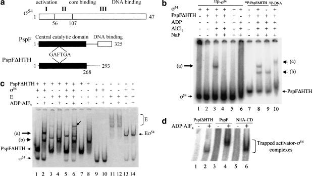
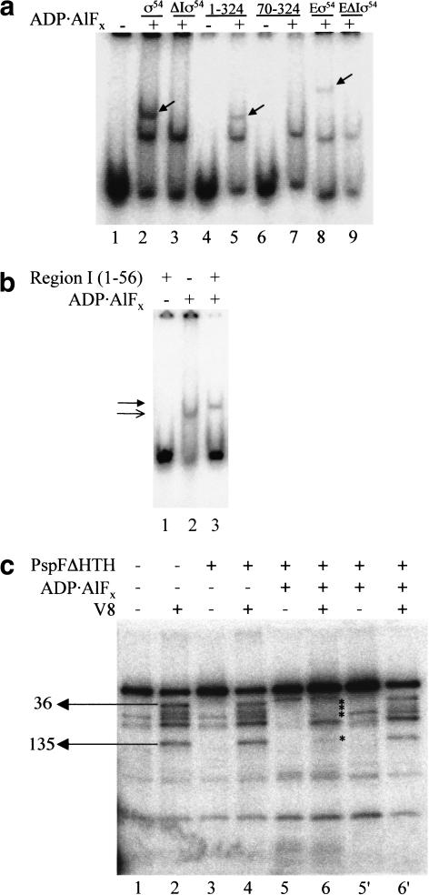
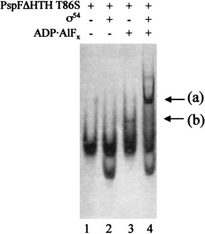
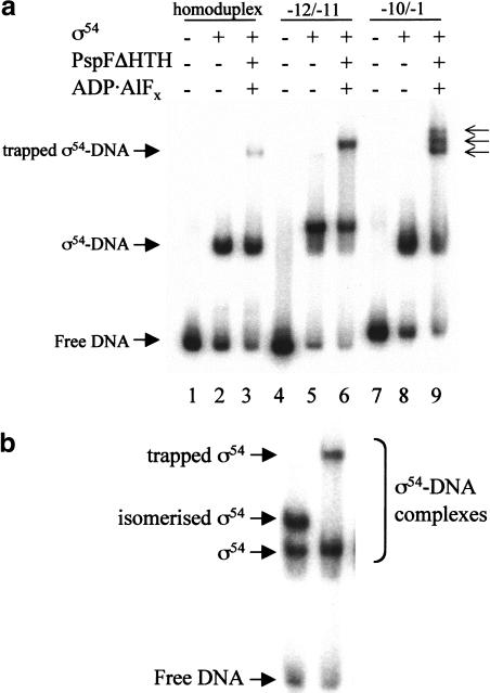
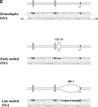
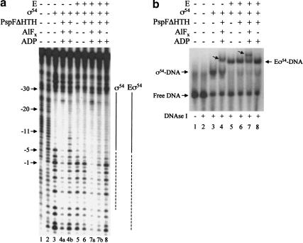
Conformational changes in sigma 54 (sigma(54)) and sigma(54)-holoenzyme depend on nucleotide hydrolysis by an activator. We now show that sigma(54) and its holoenzyme bind to the central ATP-hydrolyzing domains of the transcriptional activators PspF and NifA in the presence of ADP-aluminum fluoride, an analog of ATP in the transition state for hydrolysis. Direct binding of sigma(54) Region I to activator in the presence of ADP-aluminum fluoride was shown and inferred from in vivo suppression genetics. Energy transduction appears to occur through activator contacts to sigma(54) Region I. ADP-aluminum fluoride-dependent interactions and consideration of other AAA+ proteins provide insight into activator mechanochemical action.


Figures

Figure 1
Figure 1
(a) Schematics of ς54 and the activator PspF. The three regions of Klebsiella pneumoniae ς54 and their associated functions are indicated (Buck et al. 2000). The functional domains of the Escherichia coli ς54 activator PspF and its derivative PspFΔHTH deleted for its DNA-binding domain are shown (Jovanovic et al. 1999). The approximate position of the highly conserved GAFTGA motif implicated as part of the Switch 1 region in ς54 activators (Rombel et al. 1998; Yan and Kustu 1999) is indicated. (b) Gel mobility-shift assay for ADP–aluminum fluoride-dependent complex formation between PspFΔHTH and ς54 using 32P-HMK-tagged protein. Reactions were with 32P-HMK-tagged ς54 or 32P-HMK-tagged PspFΔHTH (100 nM), unlabeled PspFΔHTH (10 μM), and unlabeled ς54 (1 μM). Lane 10 contains ς54 (1 μM), PspFΔHTH (10 μM) with 32P-end-labeled Sinorhizobium meliloti nifH promoter 88-nt DNA (16 nM). Arrow (a) indicates position of complexes formed between ς54 and PspFΔHTH in the presence of ADP · AlFx; arrow (b) indicates the position of PspFΔHTH complex formed in the presence of ADP · AlFx, and arrow (c) indicates the position of DNA–ς54–PspFΔHTH complex in the presence of ADP · AlFx. (c) Gel mobility-shift assay for ADP · AlFx-dependent complex formation between PspFΔHTH and ς54 and ς54-holoenzyme detected by Coomassie staining. Reaction conditions were as in a except for PspFΔHTH (20 μM), ς54 (4 μM or 600 nM when present with core RNAP [E]), and core RNAP (300nM). The arrow on the gel indicates the holoenzyme trapped activator complex in lane 6. Arrows (a) and (b) point to complexes as indicated in a. (d) Wild-type PspF and the NifA central domain form an ADP · AlFx-dependent complex with ς54. Reaction conditions were as in b with 32P-HMK ς54 (50 nM), PspFΔHTH (10 μM), wild-type PspF (3 μM), and NifA central domain (3 μM). PspFΔHTH (∼36 kD), wild-type PspF (∼37.5 kD), and NifA-CD (∼32 kD).
Figure 2
Figure 2
(a) Gel mobility-shift assay for ADP · AlFx-dependent complex formation between PspFΔHTH and ς54, ς54 peptides, and ς54-holoenzyme with and without Region I. Reactions contained 32P-HMK-tagged PspFΔHTH (100 nM), ς54, and ΔIς54 (1 μM). Peptides 1–324 and 70–324 (50 μM). Eς54 and EΔIς54 were formed with ς54 (600 nM) and E (300 nM). Trapped activator–ς54 fragment complexes are marked with an arrow. (b) Gel mobility shift assay for ADP · AlFx-dependent complex formation between PspFΔHTH and ς54 Region I. Reactions contained 32P-HMK-tagged PspFΔHTH (100 nM) and ς54 Region I (50 μM). The lower, unfilled arrowhead indicates the PspFΔHTH–ADP · AlFx complex and the upper, filled arrowhead the trapped PspFΔHTH–Region I complex. (c) V8 footprinting of the trapped PspFΔHTH–ς54 complex. Reactions contained 32P-HMK-tagged ς54 (200nM) and PspFΔHTH (20 μM). V8-treated reactions are marked with + (lanes 2,4,6,6‘) and untreated reactions are marked with − (lanes 1,3,5,5′). Lanes 5′ and 6‘ contain the free ς54 isolated from reactions in lanes 5 and 6, respectively. V8 cleavage sites are as marked.
Figure 3
Figure 3
Gel mobility-shift assay for ADP · AlFx-dependent complex formation between the PspFΔHTH T86S GAFTGA mutant and ς54 detected by Coomassie staining. Reactions contained PspFΔHTH T86S (20 μM) and ς54 (2 μM). Arrow (a), trapped ς54–PspFΔHTH T86S complex; (b), PspFΔHTH T86S–ADP · AlFx complex.
Figure 4
Figure 4
Gel mobility shift assay for ς54 bound to DNA in the presence of PspFΔHTH ADP · AlFx. (a) Reactions contained 32P-end-labeled Sinorhizobium meliloti nifH promoter 88-nt DNA (16 nM), ς54 (1 μM), and PspFΔHTH (10 μM). The three arrows indicate the multiple bands obtained with the −10/−1 (late melted) heteroduplex DNA. (b) Mobility of the isomerized ς54–DNA supershifted complex (lane 1; Cannon et al. 2000) compared with the trapped PspFΔHTH–ς54–DNA complex (lane 2). ATP was used for isomerization. (c) DNA molecules used in this experiment. The consensus GG and GC of the ς54 binding sites are indicated by the vertical bars. Mismatched regions that create the early and late melted DNA templates are indicated.
Figure 4
Figure 4
Gel mobility shift assay for ς54 bound to DNA in the presence of PspFΔHTH ADP · AlFx. (a) Reactions contained 32P-end-labeled Sinorhizobium meliloti nifH promoter 88-nt DNA (16 nM), ς54 (1 μM), and PspFΔHTH (10 μM). The three arrows indicate the multiple bands obtained with the −10/−1 (late melted) heteroduplex DNA. (b) Mobility of the isomerized ς54–DNA supershifted complex (lane 1; Cannon et al. 2000) compared with the trapped PspFΔHTH–ς54–DNA complex (lane 2). ATP was used for isomerization. (c) DNA molecules used in this experiment. The consensus GG and GC of the ς54 binding sites are indicated by the vertical bars. Mismatched regions that create the early and late melted DNA templates are indicated.
Figure 5
Figure 5
DNase I footprints of trapped complexes bound to promoter DNA. (a) Complexes were formed with Escherichia coli glnHp2 homoduplex 88-nt DNA (100 nM, bottom strand end-labeled), ς54 (1 μM) or its holoenzyme (Eς54, 100 nM), and PspFΔHTH (20 μM) in the absence or presence of ADP · AlFx. Samples were treated with DNase I (1.75 × 10−3 units; Amersham Life Sciences) for 1 min and the reaction was stopped by the addition of EDTA (10 mM). Bound and unbound complexes were separated and excised from a native gel (b) and the DNA was eluted into H2O overnight at 37°C. Equal amounts of DNA were denatured and then electrophoresed through a 10% denaturing gel. Additions to each binding reaction are indicated above each lane. (Lane 1) Untreated DNA; (lane 2) DNA alone treated with DNase I. Lanes 4a and 7a show extended DNase I footprints (dashed lines) from the trapped ς54–DNA and ς54-holoenzyme–DNA complexes shown in b (marked with an arrow in lanes 4 and 7, respectively). Because of the fragment sizes migrating close to the gel front it was not possible to precisely define the downstream end of the extended footprint in lane 7a. (Lanes 4b,7b) Footprints of untrapped ς54–DNA and ς54-holoenzyme–DNA complexes shown in b (lanes 4 and 7, respectively). (b) Native gel showing DNase I-treated complexes described in a. Additions to each binding reaction are indicated above each lane.

References

    1. Buck M, Gallegos MT, Studholme DJ, Guo Y, Gralla JD. The bacterial enhancer-dependent ς54 (ςN) transcription factor. J Bacteriol. 2000;182:4129–4136. - PMC - PubMed
    1. Busby S, Ebright RH. Transcription activation by catabolite activator protein (CAP) J Mol Biol. 1999;293:199–213. - PubMed
    1. Cannon W, Missailidis S, Smith C, Cottier A, Austin S, Moore M, Buck M. Core RNA polymerase and promoter DNA interactions of purified domains of ςN: Bipartite functions. J Mol Biol. 1995;248:781–803. - PubMed
    1. Cannon W, Gallegos MT, Casaz P, Buck M. Amino-terminal sequences of ςN (ς54) inhibit RNA polymerase isomerization. Genes & Dev. 1999;13:357–370. - PMC - PubMed
    1. Cannon WV, Gallegos MT, Buck M. Isomerization of a binary ς–promoter DNA complex by transcription activators. Nat Struct Biol. 2000;7:594–601. - PubMed

Publication types

MeSH terms

LinkOut - more resources