. 2001 Sep;108(5):689-701.
doi: 10.1172/JCI12450.

Sphingosine 1-phosphate promotes endothelial cell barrier integrity by Edg-dependent cytoskeletal rearrangement

Affiliations

Sphingosine 1-phosphate promotes endothelial cell barrier integrity by Edg-dependent cytoskeletal rearrangement

J G Garcia et al. J Clin Invest. 2001 Sep.

Abstract

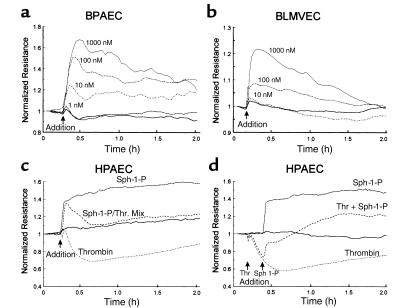
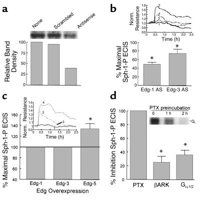
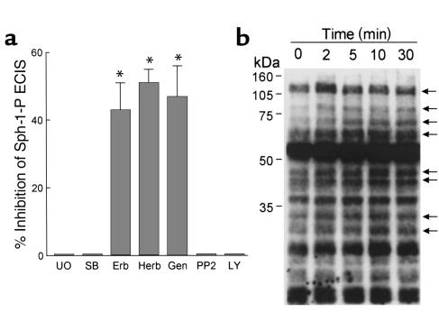
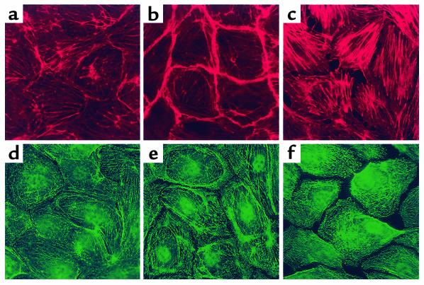
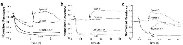
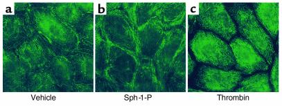
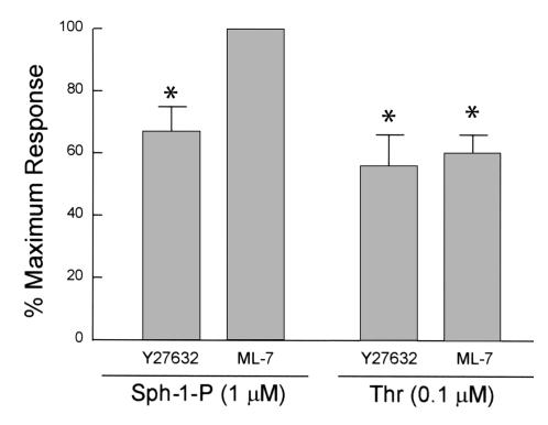
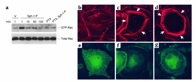
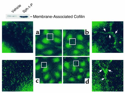
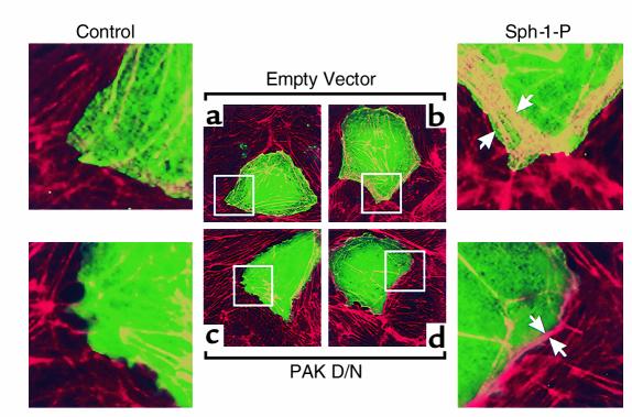
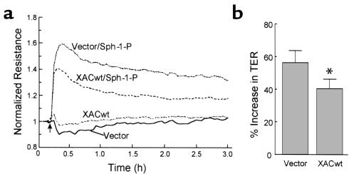
Substances released by platelets during blood clotting are essential participants in events that link hemostasis and angiogenesis and ensure adequate wound healing and tissue injury repair. We assessed the participation of sphingosine 1-phosphate (Sph-1-P), a biologically active phosphorylated lipid growth factor released from activated platelets, in the regulation of endothelial monolayer barrier integrity, which is key to both angiogenesis and vascular homeostasis. Sph-1-P produced rapid, sustained, and dose-dependent increases in transmonolayer electrical resistance (TER) across both human and bovine pulmonary artery and lung microvascular endothelial cells. This substance also reversed barrier dysfunction elicited by the edemagenic agent thrombin. Sph-1-P-mediated barrier enhancement was dependent upon G(ialpha)-receptor coupling to specific members of the endothelial differentiation gene (Edg) family of receptors (Edg-1 and Edg-3), Rho kinase and tyrosine kinase-dependent activation, and actin filament rearrangement. Sph-1-P-enhanced TER occurred in conjunction with Rac GTPase- and p21-associated kinase-dependent endothelial cortical actin assembly with recruitment of the actin filament regulatory protein, cofilin. Platelet-released Sph-1-P, linked to Rac- and Rho-dependent cytoskeletal rearrangement, may act late in angiogenesis to stabilize newly formed vessels, which often display abnormally increased vascular permeability.


Figures

Figure 1
Figure 1
Sph-1-P enhances endothelial cell barrier properties. Endothelial cells were plated on gold microelectrodes to measure TER and were cultured to confluence. Growth medium was replaced with serum-free M199, and after equilibration and stabilization, cells were challenged with agonist (Sph-1-P or thrombin). Data are representative of multiple independent experiments (minimum of three). Shown are the responses to increasing concentrations of Sph-1-P in bovine pulmonary artery endothelial cells (BPAEC) (a) and in bovine lung microvascular endothelial cells (BLMVEC) (b). (c) The effect of Sph-1-P on thrombin-induced barrier dysfunction in human pulmonary artery endothelial cells (HPAEC). In these experiments, endothelial cells were simultaneously treated with Sph-1-P (1 μM, 30 minutes) and with the potent edemagenic agent thrombin (100 nM) (38). Sph-1-P consistently elicits a significant increase in TER, which returns to baseline control values in the presence of thrombin but does not exhibit the 40% decline below control values produced by thrombin alone. Furthermore, Sph-1-P rapidly and consistently restores human endothelial cell barrier integrity in cells stimulated previously with thrombin (d).
Figure 2
Figure 2
Role of Edg family receptors and heterotrimeric G proteins in Sph-1-P–mediated endothelial cell barrier regulation. (a) Subconfluent bovine endothelial cells were incubated with Edg-1 antisense or their respective control scrambled oligonucleotides (10 μg/ml, 48 hours). This treatment reduced Edg expression by approximately 50%, whereas control oligonucleotide was without effect. (b) In similar experiments, Edg-1 and Edg-3 antisense (AS) oligonucleotides significantly attenuated the Sph-1-P–induced TER response. Sph-1-P–induced TER increased from baseline normalized resistance level of 1.0 to a level of 1.5 to 1.75 in control oligo-treated cells. Data are presented as percentages of Sph-1-P–induced TER in AS-treated cells to Sph-1-P–induced TER in control oligonucleotide–treated cells (mean ± SE, n = 3, P < 0.05). Inset: ECIS tracing reflecting the reduction of the Sph-1-P response in Edg-1–AS treated cells. Tracing number 1, cells with control oligo; number 2, cells with control oligo and stimulated with Sph-1-P; number 3, cells with Edg-1 antisense; and number 4, cells with Edg-1 antisense and stimulated with Sph-1-P. (c) Bovine endothelial cells were cotransfected with pEGFP and a plasmid encoding either Edg-1, Edg-3, Edg-5, or the empty vector. GFP-positive cells were isolated by flow cytometry (13) and subjected to TER measurements. The percentage of changes of resistance in Edg-transfected cells relative to the vector-transfected cells were calculated. Overexpression of Edg-5, but not Edg-1 or Edg-3, augmented Sph-1-P–induced increases in TER. Data are presented as percentage of Sph-1-P–induced TER in cells overexpressing Edg receptors to Sph-1-P–induced TER in cells transfected with vector control (mean ± SE, n = 3, P < 0.05). Inset: ECIS tracing to show the augmentation of Sph-1-P response in cells expressing Edg-5. In tracing number 1 cells are transfected with the empty vector; in tracing number 2 cells are transfected with the empty vector and stimulated with Sph-1-P; in tracing number 3 cells are transfected with Edg-5; and in tracing number 4 cells are transfected with Edg-5 and stimulated with Sph-1-P. (d) Bovine endothelium were either incubated with PTX (200 ng/ml, 2 hours) or transfected with plasmids encoding G protein inhibitory peptides βARK or Giα2 using pEGFP cotransfection and cell-sorting strategies described for c. Each G protein manipulation, that is, PTX, βARK, and Giα1/2 expression, significantly attenuated the Sph-1-P response (mean ± SE, n = 3, P < 0.05). Inset: Time-dependent ADP ribosylation of G protein in bovine endothelial monolayers incubated with PTX followed by in vitro ADP-ribosylation. PTX treatment before cell lysis completely inactivated Gi protein via ADP-ribosylation as we have described previously (27, 28).
Figure 3
Figure 3
Signaling events involved in Sph-1-P–mediated endothelial cell barrier enhancement. (a) Bovine endothelial cells were incubated with tyrosine kinase inhibitors genistein (Gen; 100 μM, 30 minutes), herbimycin A (Herb; 5 μM, 8 hours), or erbstatin analog (Erb; 20 μM, 1 hour); the PI-3′ kinase inhibitor LY294002 (LY; 25 μM, 30 minutes), p42/p44 MAP kinase inhibitor UO126 (UO; 10 μM, 30 minutes), the p38 MAP kinase inhibitor SB203580 (SB; 20 μM, 30 minutes), or p60 src inhibitor PP2 (10 μM, 30 minutes), followed by stimulation with Sph-1-P (1 μM). TER was continuously measured for 3 hours and the maximal Sph-1-P response (10–15 minutes after stimulation) were employed for determining the effect of the inhibitors on Sph-1-P–induced barrier enhancement. Data are presented as mean ± SE, n = 3, P < 0.05. Genistein, herbimycin A, and erbstatin analogues did not significantly alter the basal TER, but inhibited Sph-1-P–induced increases in TER, indicating the involvement of tyrosine kinases in Sph-1-P–mediated modulation of endothelial cell barrier integrity. (b) Endothelial cells were incubated with Sph-1-P (1 μM) for indicated times, followed by Western analysis using an phosphotyrosine Ab. Sph-1-P stimulated tyrosine phosphorylation of multiple proteins as indicated by arrows.
Figure 4
Figure 4
Effect of receptor agonists on bovine endothelial cell cytoskeletal structure. Cells were treated with either vehicle (a and d), Sph-1-P (1 μM for 5 minutes) (b and e), or thrombin (100 nM for 15 minutes) (c and f). F-actin staining was assessed with Texas red phalloidin (ac) and myosin staining evaluated with anti-myosin IIa polyclonal Ab (df). Thrombin induces dissolution of the cortical cytoskeleton, prominent stress fiber formation, and formation of intercellular gaps. In contrast, Sph-1-P significantly enhances cortical actomyosin staining, which correlates with enhancement of endothelial cell barrier function.
Figure 5
Figure 5
Effect of cytoskeletal alterations on Sph-1-P–induced endothelial cell barrier protection. (a) Bovine endothelium, grown on gold microelectrodes, was pretreated with either vehicle or cytochalasin B (CytB; 25 μg/ml, 60 minutes), followed by stimulation with Sph-1-P (1 μM). Actin depolymerization and disruption of microfilament structure decreases TER and completely prevented the effect of Sph-1-P on TER. (b) Human endothelial cells were pretreated with either vehicle or latrunculin A (Lat; 0.1 μM, 40 minutes), followed by stimulation with Sph- 1-P (1 μM). Inhibition of actin polymerization decreases basal level TER and prevented Sph-1-P–mediated TER. (c) Bovine endothelial cells were incubated with either vehicle or the microtubule-disrupting agent, nocodazole (ND; 2.5 μM, 30 minutes), followed by stimulation with Sph-1-P (1 μM). Disruption of microtubule decreases TER, but failed to alter Sph-1-P–induced increases in TER.
Figure 6
Figure 6
Effect of receptor agonists on bovine endothelial cell MLC phosphorylation. Cells were treated with either vehicle (a), Sph-1-P (1 μM for 5 minutes) (b), or thrombin (100 nM for 15 minutes) (c). Phospho-MLC staining was assessed with specific anti-monophospho MLC Ab. Sph-1-P challenge locally increases MLC phosphorylation in the cortical actin ring. In contrast, thrombin significantly enhances phospho MLC staining, which appears to colocalize with cytoplasmic actin stress fibers.
Figure 7
Figure 7
Effect of Rho kinase and MLCK inhibition on Sph-1-P–mediated changes in TER. (a) Confluent bovine pulmonary artery endothelium grown on gold microelectrodes were pretreated with either vehicle control, Rho kinase inhibitor, Y27632 (10 μM), or the MLCK inhibitor, ML-7 (20 μM) for 60 minutes, followed by stimulation with Sph-1-P (1 μM) or thrombin (Thr, 100 nM). The maximal Sph-1-P–induced endothelial barrier enhancement observed at 10–15 minutes was attenuated by the reduction of Rho kinase activity, but not affected by the inhibition of MLCK activity, whereas maximal thrombin-induced endothelial barrier disruption (30 minutes) was significantly blocked by both Rho kinase and MLCK inhibition. Data are mean ± SE, n = 3, P < 0.05.
Figure 8
Figure 8
Role of Rac GTPase in Sph-1-P–mediated endothelial actin rearrangement and barrier regulation. (a) Bovine pulmonary artery endothelium were incubated with vehicle (v) Sph-1-P (1 μM), for indicated periods of time, or pretreated with PTX (1 μg/ml, 2 hours) and subsequently incubated with Sph-1-P for 1 minute. Cells were lysed, centrifuged, and supernatants were collected. The activated GTP-bound Rac was precipitated by agarose-conjugated human PAK-1 p21-binding domain and immunoblotted by anti-Rac mAb as described in Methods. Total Rac content was detected using cell lysates. These results indicate rapid Sph-1-P–induced Rac activation, which was completely abolished by PTX. (bg) Bovine endothelium was transfected with either an empty vector (b and e) or a constitutively active HA-tagged Rac construct (Rac V12) (c, d, f, and g) as described in Methods. Shown are subsequent immunofluorescence images (×100) of endothelial cells stained with either Texas red phalloidin for F-actin (bd) or anti-HA tag Ab for identification of transfected cells (eg). b and e, c and f, d and g represent matched images. Overexpression of constitutively active Rac V12, but not control vector, significantly enhances polymerized actin staining (arrows) within the cortical ring with the degree of enhancement dependent upon the level of Rac V-12 overexpression.
Figure 9
Figure 9
Effect of Sph-1-P on cofilin and PAK subcellular localization. Human endothelial cells were treated by either media (two left panels), or Sph-1-P (two right panels) for 5 minutes. PAK-1 staining (a and b) was assessed with specific anti-PAK 1 Ab as described in Methods. Cofilin staining (c and d) was assessed with specific anti-cofilin Ab. Sph-1-P caused rapid translocation of both cofilin and PAK-1 to the cell periphery (translocated proteins shown by arrows). Inset: Western blot demonstrating increased cofilin content in the membrane fraction. Human endothelial cells were challenged with vehicle or Sph-1-P for 5 minutes. Cell lysates then underwent detergent fractionation and the membrane and cytosol fractions (10 μg/lane) separated by SDS PAGE and stained with anti-cofilin Ab.
Figure 10
Figure 10
Effect of PAK inhibition on Sph-1-P–induced endothelial cell cytoskeletal rearrangement. Bovine endothelial cells were transfected with either empty vector (a and b) or dominant negative c-myc-tagged PAK-1 construct (c and d) as described in Methods, followed by challenge with either media (a and c) or 1 μM Sph-1-P (b and d) for 5 minutes. Shown are subsequent merged immunofluorescence images (×100) of endothelial cells stained with Texas red phalloidin for F-actin (red) and anti–c-myc tag Ab for identification of PAK-1 overexpressing cells (green). Overexpression of dominant negative PAK-1 but not empty vector significantly inhibited Sph-1-P–induced actin cortical ring enhancement (F-actin staining in transfected cells after merging green and red images appears as yellow). Arrows point to the increased cortical actin band in the Sph-1-P–treated cells.
Figure 11
Figure 11
Cofilin overexpression significantly attenuates Sph-1-P–mediated increases in TER. Approximately 80% confluent human endothelial cells grown on gold microelectrodes were infected with recombinant adenovirus expressing a GFP-tagged wild-type xenopus ADF/cofilin (XACwt) or vector control that expresses GFP only. Cells were cultured in complete medium for 72 hours, followed by stimulation with Sph-1-P (1 μM). Shown is a representative tracing. Although basal TER was unaffected by cofilin overexpression, the increases in TER (percentage over basal) stimulated by Sph-1-P were significantly attenuated in the XACwt virus-infected cells. (b) The results pooled from four independent experiments. Data represent TER values obtained 10-15 minutes after Sph-1-P addition (mean ± SD, P < 0.05).

References

    1. Gimbrone MA, Jr, et al. Preservation of vascular integrity in organs perfused in vitro with a platelet-rich medium. Nature. 1969;221:33–36. - PubMed
    1. Roy AJ, Djerassi I. Effects of platelet transfusions: plug formation and maintenance of vascular integrity. Proc Soc Exp Biol Med. 1972;139:137–142. - PubMed
    1. Shepro D, Welles SL, Hechtman HB. Vasoactive agonists prevent erythrocyte extravasation in thrombocytopenic hamsters. Thromb Res. 1984;35:421–430. - PubMed
    1. Lo SK, Burhop KE, Kaplan JE, Malik AB. Role of platelets in maintenance of pulmonary vascular permeability to protein. Am J Physiol. 1988;254:H763–H771. - PubMed
    1. McDonagh PF. Platelets reduce coronary microvascular permeability to macromolecules. Am J Physiol. 1986;251:H581–H587. - PubMed

Publication types

MeSH terms