. 2001 Sep;159(3):861-73.
doi: 10.1016/S0002-9440(10)61762-2.

Thyroid autoimmune disease: demonstration of thyroid antigen-specific B cells and recombination-activating gene expression in chemokine-containing active intrathyroidal germinal centers

Affiliations

Thyroid autoimmune disease: demonstration of thyroid antigen-specific B cells and recombination-activating gene expression in chemokine-containing active intrathyroidal germinal centers

M P Armengol et al. Am J Pathol. 2001 Sep.

Abstract

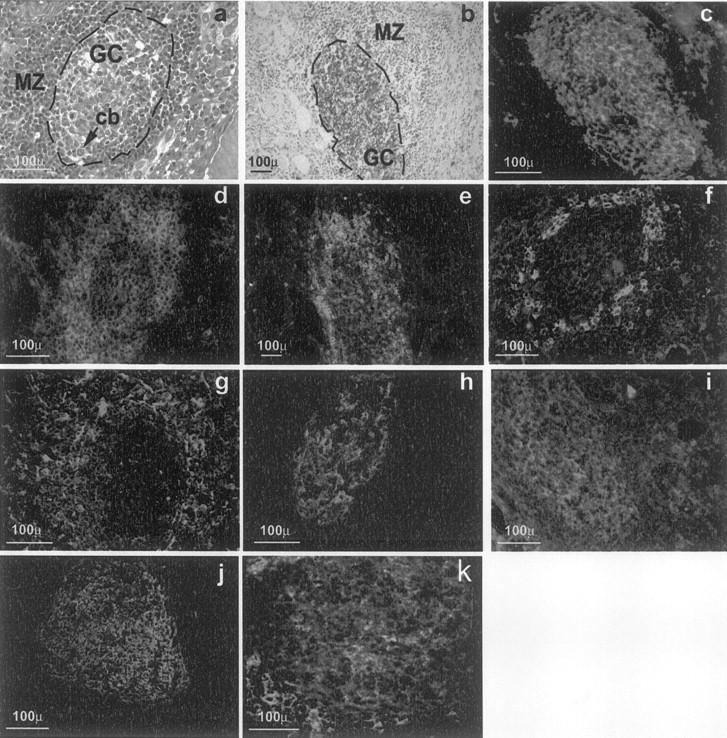
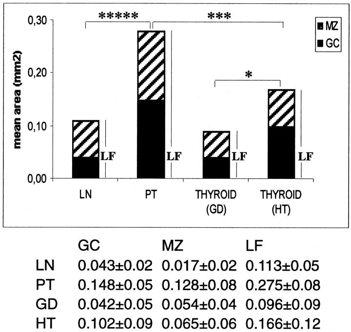
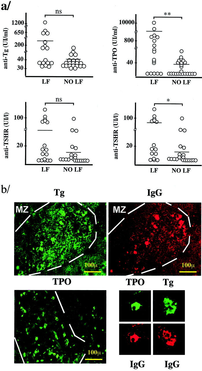
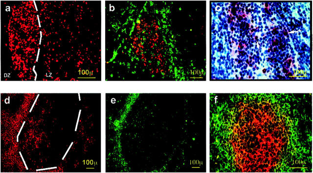
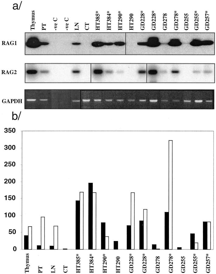
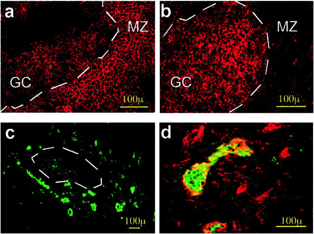
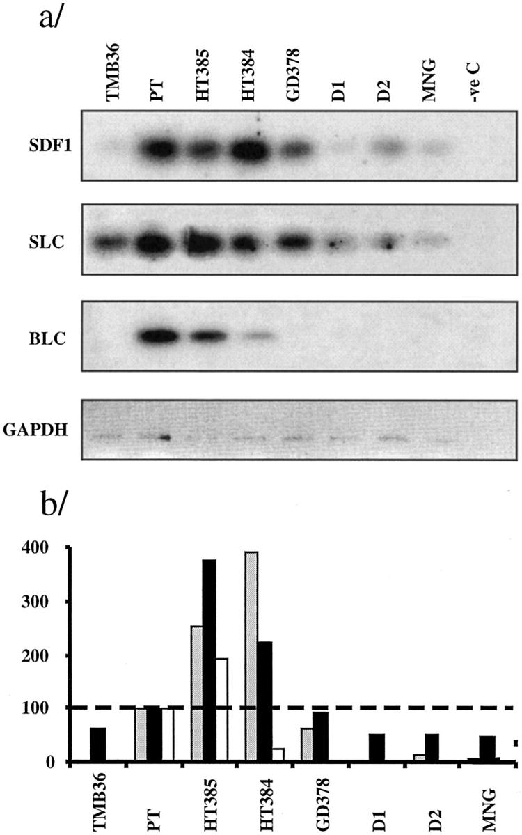
Autoimmune thyroid disease--Hashimoto thyroiditis and Graves' disease--patients produce high levels of thyroid autoantibodies and contain lymphoid tissue that resembles secondary lymphoid follicles (LFs). We compared the specificity, structure, and function of tonsil and lymph node LFs with those of the intrathyroidal LFs to assess the latter's capability to contribute to autoimmune response. Thyroglobulin and thyroperoxidase binding to LFs indicated that most intrathyroidal LFs were committed to response to thyroid self-antigens and were associated to higher levels of antibodies to thyroglobulin, thyroperoxidase, and thyroid-stimulating hormone receptor. Intrathyroidal LFs were microanatomically very similar to canonical LFs, ie, they had well-developed germinal centers with mantle, light, and dark zones and each of these zones contained B and T lymphocytes, follicular dendritic and interdigitating dendritic cells with typical phenotypes. Careful assessment of proliferation (Ki67) and apoptosis (terminal dUTP nick-end labeling) indicators and of the occurrence of secondary immunoglobulin gene rearrangements (RAG1 and RAG2) confirmed the parallelism. Unexpected high levels of RAG expression suggested that receptor revision occurs in intrathyroidal LFs and may contribute to generate high-affinity thyroid autoantibodies. Well-formed high endothelial venules and a congruent pattern of adhesion molecules and chemokine expression in intrathyroidal LFs were also detected. These data suggest that ectopic intrathyroidal LFs contain all of the elements needed to drive the autoimmune response and also that their microenvironment may favor the expansion and perpetuation of autoimmune response.


Figures

Figure 1.
Figure 1.
Morphology of intrathyroidal secondary LFs in AITD. a: A section from a formalin-fixed paraffin-embedded block from a HT gland. b to h: Sections from frozen blocks from GD glands. g was deconvolved to improve image definition (see text for details), the other images are standard micrographs. a: H&E staining showing a typical secondary follicle with a GC and a well-formed MZ (cb, centroblasts undergoing mitosis). b: CD20 on B cells of the GC (brown) using the immunoperoxidase technique and counterstained with hematoxylin (GD, case TB228). c: Direct immunofluorescence with peanut agglutinin-fluorescein isothiocyanate, showing positive staining of centroblasts and follicular dendritic cells (GD, case TB378). d: Staining for CD3+ reveals abundant T cells in the MZ with scattered cells inside the GC (GD, case TB228). e: Demonstration of abundant CD4+ T among the T cells in the MZ (GD, case TB228). f: Staining for CD8 shows moderately abundant CD8+ T lymphocytes in the MZ (GD, case TB378). g: Staining for CD 83 highlights the network of mature dendritic cells in the MZ (GD, case TB278). h: Staining for the long form of CD21 reveals the network of follicular dendritic cells in the CG and their polarization toward the light zone (GD, case TB373). i: CD38, as marker of centrocytes. j: CD23 staining revealing the area occupied by the GC. k: CD77 staining as an additional GC marker .
Figure 2.
Figure 2.
Morphometric analysis of LFs from HT and GD compared with LFs from LNs and PTs. Solid bar, area occupied by the GC; hatched bar, area occupied by the MZ. Number of LFs studied: LNs, 39; PTs, 26; GDs, 20; and HTs, 37. The asterisks indicate significant differences between total LF areas: ****, P < 0.001; ***, P < 0.00.5; *, P < 0.05. The table below gives numerical values ± SD.
Figure 3.
Figure 3.
a: Relationship between thyroid antibody titer and the presence of intrathyroidal LFs. TSHR, thyrotropin receptor; LF, group of thyroid glands containing LFs; NO LF, group of thyroid glands without LFs. The plotted data, except for those at bottom right, correspond to all of the patients in Table 2 ▶ . At the bottom right, only the data from GD patients were plotted (*, P < 0.05; **, P < 0.01; ns, not significant P value, Mann-Whitney test). b: Demonstration of the specificity of B and plasma cells in the intrathyroidal LFs. Double immunofluorescence using biotinylated Tg (green, top left) and anti-IgG (red, top right), demonstrating binding of Tg to the same cells that are stained for IgG. Positive lymphocytes only appear in the LFs, whereas plasma cells are present both in the LFs and in the diffuse infiltrate. Bottom left: Binding of biotinylated TPO to abundant cells in the LFs and also to some cells in the diffuse infiltrate. Note the presence of membrane and cytoplasmic staining that correspond to lymphocytes and plasma cells, respectively. Bottom right: Examples of plasma cells stained for TPO and Tg in double immunofluorescence with IgG (GD, case 378).
Figure 4.
Figure 4.
Proliferation and apoptosis in thyroid LFs. a: Immunofluorescence staining for Ki67 in an intrathyroidal LF, showing abundant proliferating cells polarized toward the dark zone (GD, case TB228). b: Double-immunofluorescence staining for IgD (green) and Ki67 (red), demonstrating that most proliferating cells are in the light zone of the GC and are either negative or slightly positive for IgD, as in the case of centroblasts. c: Demonstration of the existence of apoptotic cells by the terminal dUTP nick-end labeling technique and hematoxylin counterstain. Most positive cells were found in the GC area; no positive cells were seen in the epithelium of the thyroid follicles (data not shown). d and e: Double immunofluorescence for bcl-2 and IgD. The level of expression of apoptosis inhibitor bcl-2 is high in the MZ but is also expressed in the GC cells. The staining for IgD shows the distribution of mature B cells in the MZ. f: Double exposure of double-immunofluorescence staining for IgD (green) and CD95/Fas (red). Notice that Fas is mainly expressed in the CG, whereas IgD is expressed in the MZ, as is the case in normal LNs.
Figure 5.
Figure 5.
RAG1 and RAG2 mRNA expression measured by RT-PCR Southern blot in HT and GD glands. a: Southern blot of RT-PCR products amplified with RAG1 and RAG2 primers and hybridized with the corresponding oligoprobes; bottom, RT-PCR for control GAPDH; only partial normalization was achieved because of the small amount of available sample. Asterisks indicate samples that contained visible GCs. TMB, thymus; CT, esophagus. b: Graph representing the ratio of densitometry values: RAG1, GAPDH (solid bars); RAG2, GAPHDH (open bars), using an inverted image of the GAPDH gel image.
Figure 6.
Figure 6.
Distribution of adhesion molecules in the thyroidal LFs. a: ICAM-1 membrane staining is mainly present in the MZ cells. b: ICAM3 expression is higher in cells inside the GC and has a reticular pattern similar to that observed for the follicular dendritic cells. c: CD62L-positive HEVs are distributed around the LF. d: Picture resulting from superimposing digitized images for CLA (green) and Factor VIII (red) staining in the same section. Note that the large vessels are positive for both markers, thus indicating the location of the HEV. Cryostat sections from GD, case TB278.
Figure 7.
Figure 7.
Chemokine expression as assessed by RT-PCR-Southern blot and phosphorimager counting. a: RT-PCR-Southern blots. b: Ratio for normalized chemokine value in each gland: normalized tonsil value, ×100; the broken line at 100% represents the tonsil reference value. Gray bars, SDF1 (CXCL12); solid bars, SLC (CCL21); open bars, BLC (CXCL13). TMB, thymus; D1 and D2, normal donors.

Comment in

References

    1. Weetman AP, McGregor AM: Autoimmune thyroid disease: further developments in our understanding. Endocr Rev 1994, 15:788-830 - PubMed
    1. Ajjan RA, Kemp EH, Waterman EA, Watson PF, Endo T, Onaya T, Weetman AP: Detection of binding and blocking autoantibodies to the human sodium-iodide symporter in patients with autoimmune thyroid disease. J Clin Endocrinol Metab 2000, 85:2020-2027 - PubMed
    1. Rapoport B, Chazenbalk GD, Jaume JC, McLachlan SM: The thyrotropin (TSH) receptor: interaction with TSH and autoantibodies Endocr Rev 1998, 19:673-716 - PubMed
    1. Fukuma N, Petersen VB, McLachlan SM, Pegg CA, Rees Smith B: Human monoclonal thyroglobulin autoantibodies of high affinity. I. Production, characterisation and interaction with murine monoclonal thyroglobulin antibodies. Autoimmunity 1991, 10:291-295 - PubMed
    1. Prentice L, Kiso Y, Fukuma N, Horimoto M, Petersen V, Grennan F, Pegg C, Furmaniak J, Rees Smith B: Monoclonal thyroglobulin autoantibodies: variable region analysis and epitope recognition. J Clin Endocrinol Metab 1995, 80:977-986 - PubMed

Publication types

MeSH terms

LinkOut - more resources