

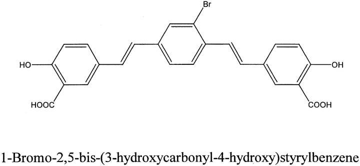
The fluorescent Congo red derivative, (trans, trans)-1-bromo-2,5-bis-(3-hydroxycarbonyl-4-hydroxy)styrylbenzene (BSB), labels diverse beta-pleated sheet structures in postmortem human neurodegenerative disease brains
- PMID: 11549586
- PMCID: PMC1850468
- DOI: 10.1016/s0002-9440(10)61769-5
The fluorescent Congo red derivative, (trans, trans)-1-bromo-2,5-bis-(3-hydroxycarbonyl-4-hydroxy)styrylbenzene (BSB), labels diverse beta-pleated sheet structures in postmortem human neurodegenerative disease brains
Abstract
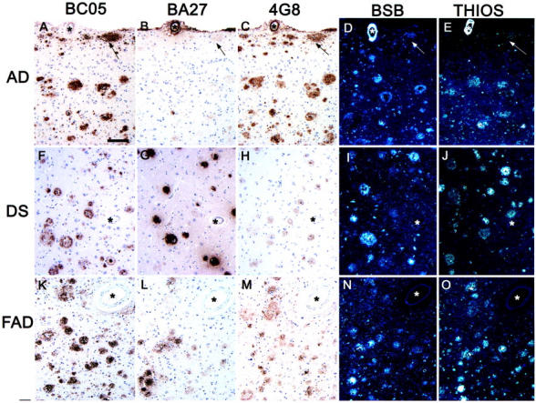
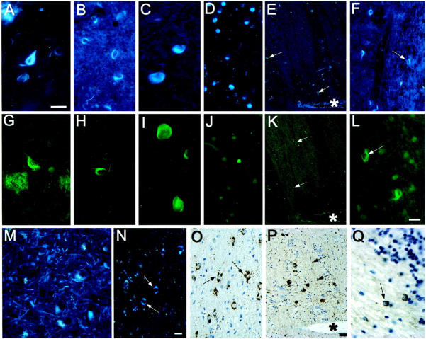
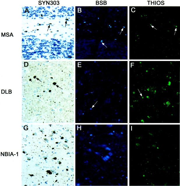
A novel Congo red-derived fluorescent probe (trans, trans),-1-bromo-2,5-bis-(3-hydroxycarbonyl-4-hydroxy)styrylbenzene (BSB) that binds to amyloid plaques of postmortem Alzheimer's disease brains and in transgenic mouse brains in vivo was designed as a prototype imaging agent for Alzheimer's disease. In the current study, we used BSB to probe postmortem tissues from patients with various neurodegenerative diseases with diagnostic lesions characterized by fibrillar intra- or extracellular lesions and compared these results with standard histochemical dyes such as thioflavin S and immunohistochemical stains specific for the same lesions. These data show that BSB binds not only to extracellular amyloid beta protein, but also many intracellular lesions composed of abnormal tau and synuclein proteins and suggests that radioiodinated BSB derivatives or related ligands may be useful imaging agents to monitor diverse amyloids in vivo.
Figures
References
-
- Klunk WE, Debnath ML, Pettegrew JW: Development of small molecule probes for the β-amyloid protein of Alzheimer’s disease. Neurobiol Aging 1994, 15:691-698 - PubMed
-
- Klunk WE, Debnath ML, Pettegrew JW: Chrysamine-G binding to Alzheimer and control brain: autopsy study of a new amyloid probe. Neurobiol Aging 1995, 16:541-548 - PubMed
-
- Styren SD, Hamilton RL, Styren GC, Klunk WE: X-34, a fluorescent derivative of Congo red: a novel histochemical stain for Alzheimer’s disease pathology. J Histochem Cytochem 2000, 48:1223-1232 - PubMed
-
- Iwatsubo T, Odaka A, Suzuki N, Mizusawa H, Nukina N, Ihara Y: Visualization of Aβ42(43)-positive and Aβ40-positive senile plaques with end-specific Aβ-monoclonal antibodies: evidence that an initially deposited Aβ species is Aβ1–42(43). Neuron 1994, 13:45-53 - PubMed
Publication types
MeSH terms
Substances
Grants and funding
LinkOut - more resources
Full Text Sources
Medical
Miscellaneous
