. 2001 Sep;159(3):1045-54.
doi: 10.1016/S0002-9440(10)61779-8.

Endothelial oxidative stress activates the lectin complement pathway: role of cytokeratin 1

Affiliations

Endothelial oxidative stress activates the lectin complement pathway: role of cytokeratin 1

C D Collard et al. Am J Pathol. 2001 Sep.

Abstract

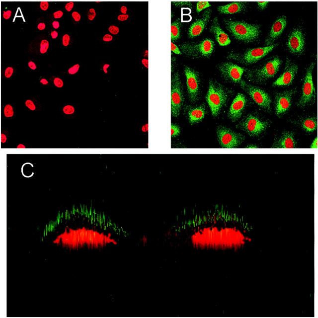
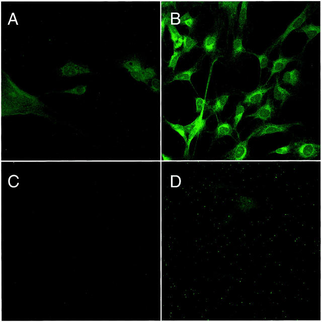
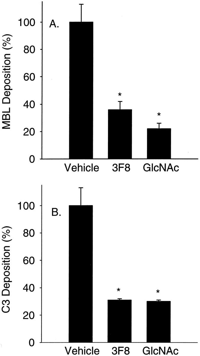
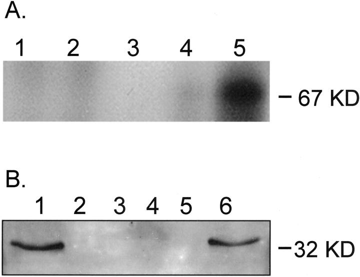
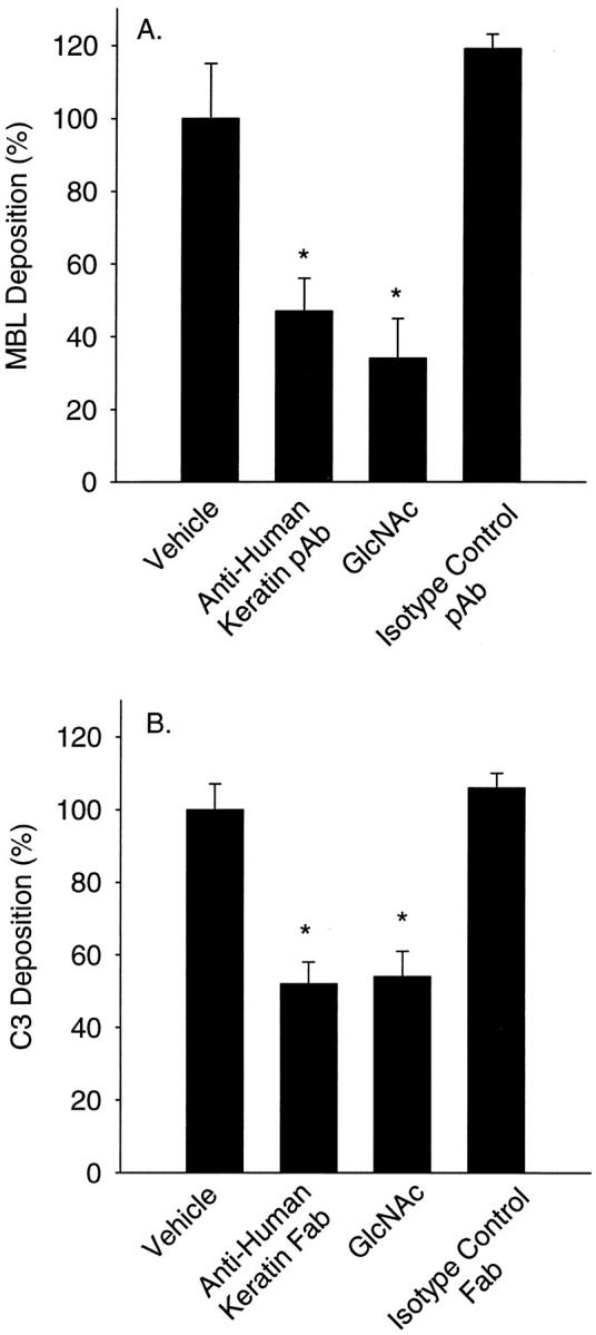
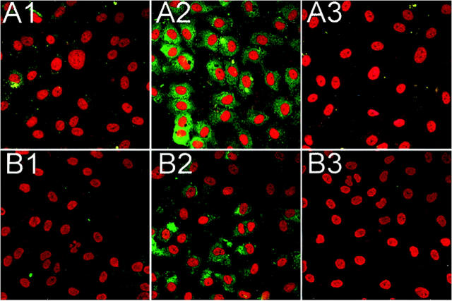
Oxidative stress increases endothelial mannose-binding lectin (MBL) binding and activates the lectin complement pathway (LCP). However, the molecular mechanism of MBL binding to the endothelium after oxidative stress is unknown. Intermediate filaments have been previously reported to activate the classical complement pathway in an antibody-independent manner. We investigated whether oxidative stress increases human umbilical vein endothelial cell (HUVEC) cytokeratin 1 (CK1) expression and activates the LCP via MBL binding to CK1. Reoxygenation (3 hours, 21% O(2)) of hypoxic HUVECs (24 hours, 1% O(2)) significantly increased CK1 mRNA (in situ hybridization) and membrane protein expression [enzyme-linked immunosorbent assay (ELISA)/confocal microscopy]. Incubating human serum (HS) with N-acetyl-D-glucosamine or anti-human MBL monoclonal antibody attenuated MBL and C3 deposition on purified CK1 (ELISA). CK1 and MBL were co-immunoprecipitated from hypoxic HUVECs reoxygenated in HS. Treatment with anti-human cytokeratin Fab fragments attenuated endothelial MBL and C3 deposition after oxidative stress (ELISA/confocal microscopy). We conclude that: 1) endothelial oxidative stress increases CK1 expression, MBL binding, and C3 deposition; 2) inhibition of MBL attenuates purified CK1-induced complement activation; and 3) anti-human cytokeratin Fab fragments attenuate endothelial MBL and C3 deposition after oxidative stress. These results suggest that MBL binding to endothelial cytokeratins may mediate LCP activation after oxidative stress.


Figures

Figure 1.
Figure 1.
Endothelial CK1 protein expression after oxidative stress. HUVEC CK1 expression after oxidative stress was determined by ELISA and Western blot (inset). CK1 expression was significantly increased after oxidative stress compared to normoxic HUVECs or hypoxic/reoxygenated HUVECs incubated with an isotype control pAb (n = 3; error bars = SEM; *, P < 0.05 compared to normoxia).
Figure 2.
Figure 2.
Endothelial CK1 protein expression after oxidative stress. HUVEC CK1 expression after oxidative stress was determined by immunofluorescent confocal microscopy. CK1 expression (green) was significantly increased after oxidative stress (B) compared to normoxic HUVECs (A). Z-section scanning further confirmed cell membrane CK1 expression after endothelial oxidative stress (C). These panels are representative of three different experiments (red = propidium iodide-stained endothelial nuclei).
Figure 3.
Figure 3.
Endothelial CK1 mRNA expression after oxidative stress. HUVEC CK1 mRNA expression after oxidative stress was determined by in situ hybridization. HUVECs were grown under normoxic (A) or hypoxic (B) conditions as described and hybridized with a CK1 cDNA probe. Analysis by confocal microscopy revealed a diffuse cytoplasmic staining pattern, leaving the nucleus devoid of staining. Oxidative stress (B) increased the level of fluorescent staining indicating an increase in CK1 mRNA. C demonstrates that normoxic HUVECs do not express porcine MBL mRNA (normoxia control). D shows hypoxic HUVECs do not express porcine MBL mRNA (hypoxia control). No fluorescence was observed when normoxic or hypoxic HUVECs were incubated with RNase A (data not shown). This figure is representative of three experiments.
Figure 4.
Figure 4.
MBL and C3 deposition on purified human CK1. Human MBL (A) and C3 (B) deposition on purified human dermal CK1 was determined by ELISA. Treatment of 2% HS with GlcNAc (100 mmol/L) or the functionally inhibitory anti-human MBL mAb, 3F8 (10 μg/ml), significantly attenuated C3 and MBL deposition compared to untreated HS (vehicle) (n = 3; data normalized to vehicle; error bars = SEM; *, P < 0.05 compared to vehicle).
Figure 5.
Figure 5.
Immunoprecipitation of human CK1 or MBL after endothelial oxidative stress. A: Western blot analysis of human CK1. Purified human MBL was used to immunoprecipitate HUVEC CK1. Western blot of reduced immunoprecipitates revealed a 67-kd band (lane 5) consistent with human CK1. Note that the 67-kd band was observed after endothelial oxidative stress (lane 5), but not in normoxic HUVECs (lane 4) or the controls (lane 1, normoxic HUVEC lysate preclear; lane 2, hypoxic HUVEC lysate preclear; lane 3, anti-human MBL mAb [1C10]-conjugated protein G and human MBL only). B: Western blot analysis of human MBL. To further confirm that MBL binds endothelial CK1 after oxidative stress, MBL was co-immunoprecipitated with CK1 from hypoxic HUVECs reoxygenated in HS. Western blot of HUVEC lysates immunoprecipitated with a monospecific anti-human CK1 pAb revealed a 32-kd band consistent with reduced human MBL (lane 6). Note that the 32-kd band was observed after endothelial oxidative stress (lane 6), but not in normoxic HUVECs (lane 5) or the controls (lane 1, purified human MBL standard; lane 2, normoxic HUVEC lysate preclear; lane 3, hypoxic HUVEC lysate preclear; lane 4, anti-human CK1 pAb and protein G only).
Figure 6.
Figure 6.
Anti-human keratin antibodies attenuate endothelial MBL and C3 deposition after oxidative stress. HUVEC MBL (A) and C3 (B) deposition after oxidative stress were measured by ELISA. MBL and C3 deposition after oxidative stress was significantly increased compared to normoxic HUVECs. Incubation of HS (vehicle) with the MBL inhibitory sugar, GlcNAc (100 mmol/L) or anti-human keratin pAb (50 μg/ml) significantly attenuated MBL deposition (A). Incubation of HS (vehicle) with GlcNAc (100 mmol/L) or anti-human keratin Fab fragments (20 μg/ml) significantly attenuated C3 deposition (B). Incubation of HS with species-matched, isotype-irrelevant control pAb (50 μg/ml) or Fab fragments (20 μg/ml) did not significantly attenuate MBL (A) or C3 deposition (B), respectively (n = 3; data normalized to vehicle; error bars = SEM; *, P < 0.05 compared to vehicle).
Figure 7.
Figure 7.
Anti-human keratin Fab fragments attenuate endothelial C3 and MBL deposition after oxidative stress. Immunofluorescent confocal microscopical demonstration of MBL (row A; green) and C3 (row B; green) deposition on normoxic (column 1) and hypoxic (24 hours) HUVECs reoxygenated (3 hours) in 30% HS (column 2) with and without anti-human keratin Fab fragments (20 μg/ml; column 3). MBL (A2) and C3 (B2) staining after oxidative stress was significantly increased compared to normoxic HUVECs (A1 and B1, respectively). Treatment of HS with anti-human keratin Fab fragments significantly decreased MBL (A3) and C3 (B3) staining. These panels are representative of three different experiments.

Similar articles

Cited by

References

    1. Thiel S, Vorup-Jensen T, Stover CM, Schwaeble W, Laursen SB, Poulsen K, Willis AC, Eggleton P, Hansen S, Holmskov U, Reid KB, Jensenius JC: A second serine protease associated with mannan-binding lectin that activates complement. Nature 1997, 386:506-510 - PubMed
    1. Turner MW: The lectin pathway of complement activation. Res Immunol 1996, 147:110-115 - PubMed
    1. Turner MW: Mannose-binding lectin: the pluripotent molecule of the innate immune system. Immunol Today 1996, 17:532-540 - PubMed
    1. Turner MW: Mannose-binding lectin (MBL) in health and disease. Immunobiology 1998, 199:327-339 - PubMed
    1. Collard CD, Väkevä A, Morrissey MA, Agah A, Rollins SA, Reenstra WR, Buras JA, Meri S, Stahl GL: Complement activation after oxidative stress: role of the lectin complement pathway. Am J Pathol 2000, 156:1549-1556 - PMC - PubMed

Publication types

MeSH terms