. 2001 Sep 15;21(18):7089-98.
doi: 10.1523/JNEUROSCI.21-18-07089.2001.

Caspase 3 deficiency rescues peripheral nervous system defect in retinoblastoma nullizygous mice

Affiliations

Caspase 3 deficiency rescues peripheral nervous system defect in retinoblastoma nullizygous mice

M T Simpson et al. J Neurosci. .

Abstract

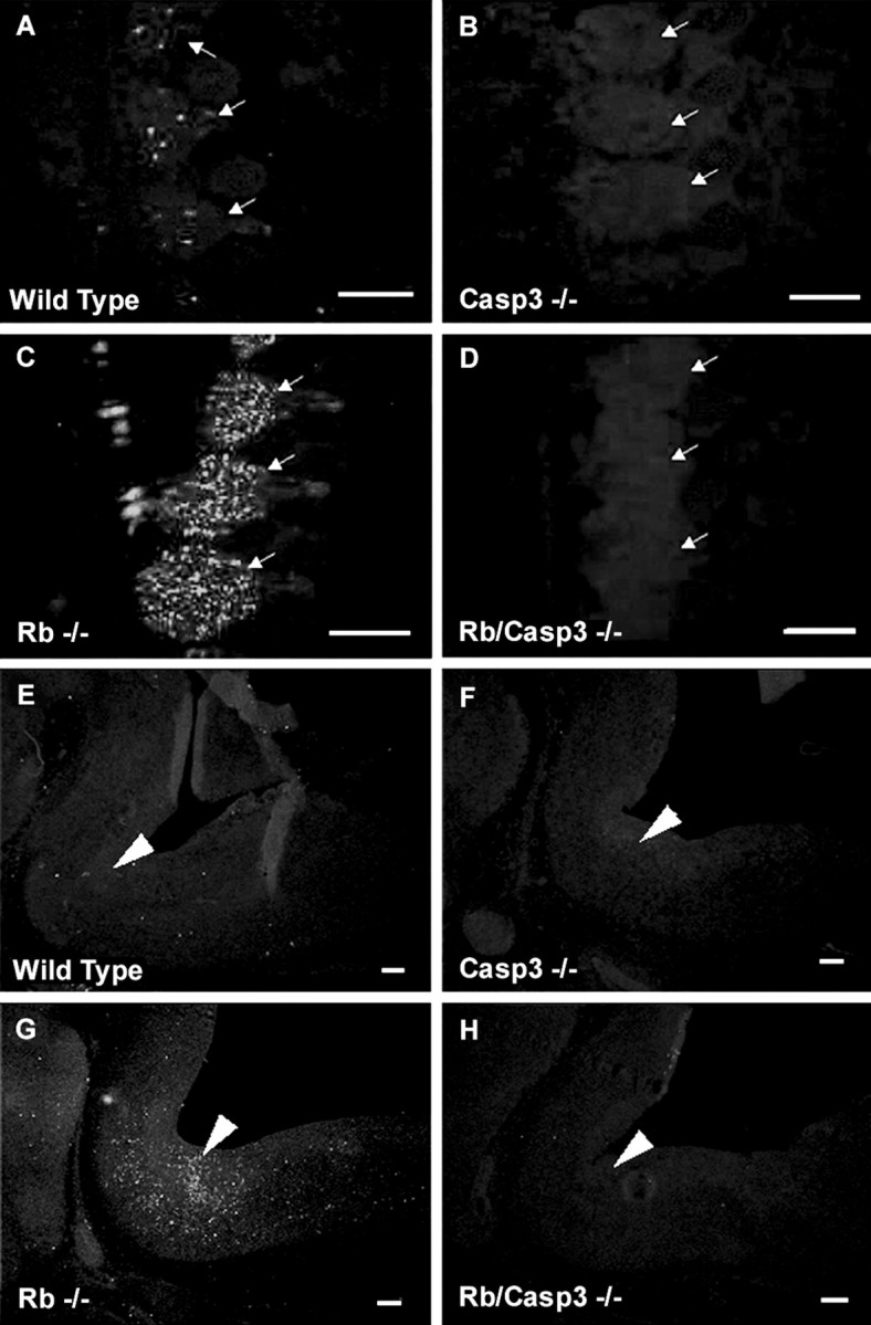
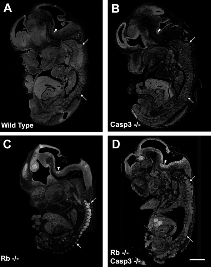
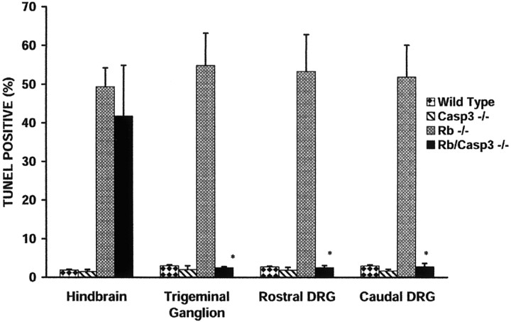
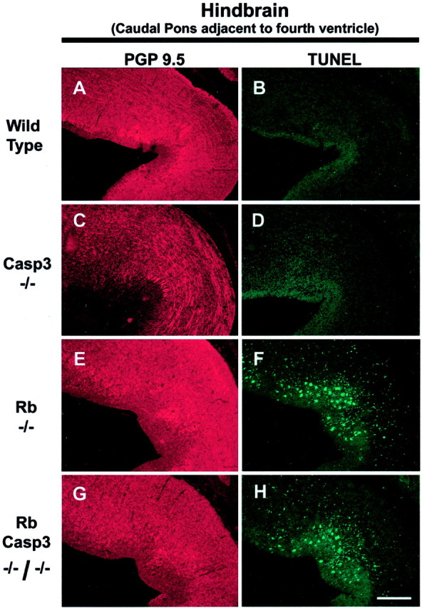
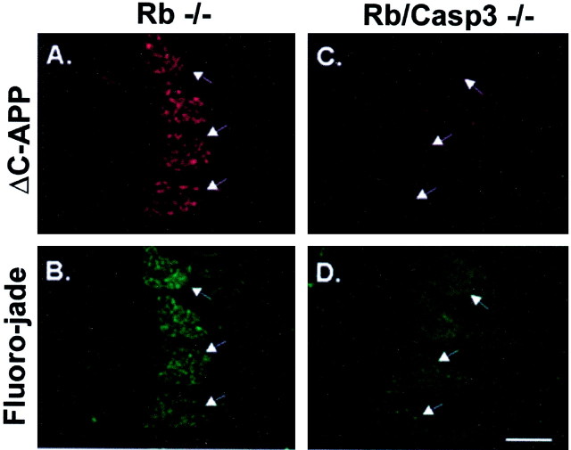
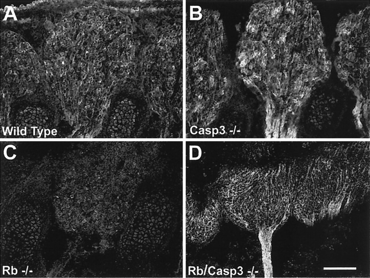
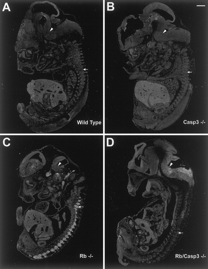
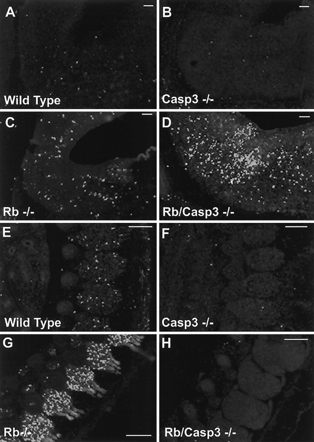
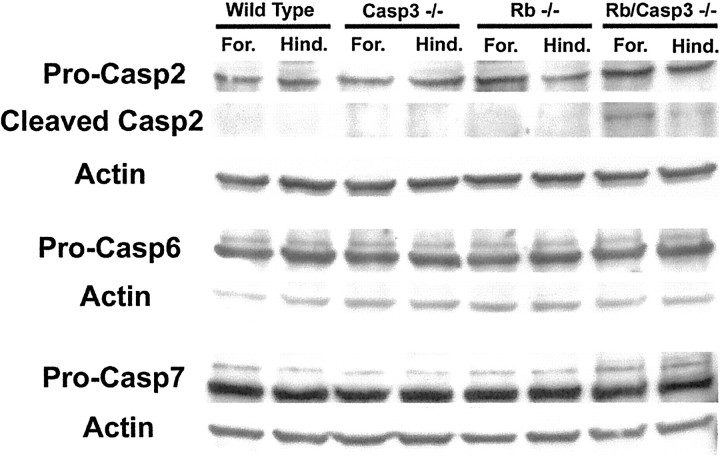
The retinoblastoma tumor suppressor protein, pRb, is a key regulator of cell cycle and has been implicated in the terminal differentiation of neuronal cells. Mice nullizygous for pRb die by embryonic day 14.5 from hematopoietic and neurological defects attributed to failed differentiation (Clarke et al., 1992; Jacks et al., 1992; Lee et al., 1992). Previous studies by MacLeod et al. (1996) have demonstrated that the loss of p53 protects Rb-deficient CNS neurons but not peripheral nervous system (PNS) neurons from cell death. Thus, the mechanisms by which PNS neurons undergo apoptosis in response to Rb deficiency remain unknown. In view of the pivotal role of caspase 3 in the regulation of neuronal apoptosis during development, we examined its function in the execution of the wide-spread neuronal cell death induced by Rb deficiency. Our results support a number of conclusions. First, we show that caspase 3 becomes activated in all neuronal populations undergoing apoptosis. Second, caspase 3 deficiency does not extend the life span of Rb null embryos, because double null mutants exhibit high rates of liver apoptosis resulting in erythropoietic failure. Third, Rb/caspase 3 double-mutant neurons of the CNS exhibit widespread apoptosis similar to that seen in Rb mutants alone; thus caspase 3 deficiency does not protect this population from apoptosis. Finally, in contrast to the CNS, neurons of the PNS including those comprising the trigeminal ganglia and the dorsal root ganglia are protected from apoptosis in Rb/caspase 3 double-mutant embryos. Examination of the mechanistic differences between these two cell types suggest that CNS neurons may invoke other caspases to facilitate apoptosis in the absence of caspase 3. These findings suggest that PNS neurons are dependent on caspase 3 for the execution of apoptosis and that caspase 3 may serve as a key therapeutic target for neuroprotection after injury of this cell type.


Figures

Fig. 1.
Fig. 1.
Caspase 3 is activated in the Rb-deficient mouse nervous system. Active caspase 3 immunostaining of DRG (AD) and hindbrain (EH) from E13.5 embryos is shown. Extensive caspase 3 activation is seen throughout the developing nervous system of the Rb−/− embryo (C,G) and is present at low levels in wild-type tissue (A, E) and undetectable in caspase 3−/− (B, F) or Rb−/−/caspase 3−/− (D, H) samples. Arrows indicate DRGs; arrowheadspoint to the pons. Scale bars, 300 μm.
Fig. 2.
Fig. 2.
Caspase 3 deficiency protects neurons of the PNS but not the CNS or liver from apoptosis induced by Rb deficiency. Sections from wild type (A), caspase 3−/− (B), Rb−/− (C), and Rb−/−/caspase 3−/− (D) were stained for TUNEL, and whole embryo images were captured. Arrowsindicate rostral and caudal DRGs. Arrowheads indicate hindbrain. Scale bar, 1.5 mm.
Fig. 3.
Fig. 3.
Caspase 3 deficiency results in decreased TUNEL labeling in the PNS but not the CNS of the Rb−/− mouse embryo. TUNEL-positive cells in the hindbrain, trigeminal ganglion, and caudal and rostral DRGs were quantified by examination at 40× magnification. The mean is expressed as a percentage of TUNEL-positive cells/total cell number as determined by Hoechst staining within a 100 μm2 area for each group. Counts were obtained from three independent embryos (n = 3), and error bars indicate SE. *p < 0.001.
Fig. 4.
Fig. 4.
Caspase 3 deficiency does not rescue CNS neurons of Rb-deficient embryos from apoptosis. Frozen sections of wild-type (A, B), caspase 3−/− (C, D), Rb−/− (E, F), and Rb−/−/caspase 3−/− (G, H) E13.5 mouse brain were stained for TUNEL. Scale bar, 150 μm.
Fig. 5.
Fig. 5.
Caspase 3 is required for apoptosis of the Rb-deficient trigeminal and dorsal root ganglia neurons. Frozen sections from wild type (AD), caspase 3−/− (EH), Rb−/− (IL), and Rb−/−/caspase 3−/− (MP) were stained for TUNEL and counterstained with PGP 9.5 to view neuronal cell bodies and axons.Panels on left show Trigeminal Ganglion, and panels onright show Rostral DRG. Scale bars, 160 μm.
Fig. 6.
Fig. 6.
Fluoro-jade labeling demonstrates caspase 3 requirement for neuronal cell death in the Rb-deficient peripheral nervous system. Shown are ΔC-APP (A, C) and Fluoro-jade (B, D) double labeling of degenerating DRG neurons in Rb−/− (A, B) and Rb/caspase 3 double knock-out embryos (C, D). Arrows indicate DRGs. Scale bar, 260 μm.
Fig. 7.
Fig. 7.
Rb−/−:caspase 3−/− double mutants exhibit normal levels of Trk A expression. Trk A is highly expressed in wild-type (A) and caspase 3−/− (B) DRGs by E13.5. This staining is dramatically reduced in Rb-deficient DRG neurons (C); however, Trk A staining is restored to wild-type levels when caspase 3 is also absent (Rb/Casp3−/−) (D). Scale bar, 150 μm.
Fig. 8.
Fig. 8.
Cleavage of the caspase substrate, APP, in E13.5 mouse embryos. Shown is immunodetection of ΔC-APP in wild-type (A), caspase 3−/− (B), Rb−/− (C), and Rb−/−/caspase 3−/− (D) E13.5 mouse embryos. The caspase 3 cleavage product, ΔC-APP, is detected in all neuronal populations undergoing apoptosis in the Rb null mouse (C). ΔC-APP is detected in the CNS but is significantly reduced in the PNS of Rb−/−/caspase 3−/− compound mutant embryos. Arrowsindicate DRGs. Arrowheads indicate hindbrain. Scale bar, 1.5 mm.
Fig. 9.
Fig. 9.
APP is cleaved in the CNS but not PNS neurons of Rb/caspase 3 double null mutants. Shown is immunodetection of cleaved caspase substrate, ΔAPP, in hindbrain (AD) and DRGs (EH) from E13.5 embryos.A, E, Wild type; B, F, caspase 3−/−; C, G, Rb−/−;D, H, Rb−/−/caspase 3−/−. Extensive ΔAPP is seen throughout the developing nervous system of the Rb−/− embryo, including hindbrain (C) and DRGs (G), and is present at low levels in corresponding wild-type tissue (A, E). ΔAPP is abundant in Rb−/−/caspase 3−/− hindbrain (D), showing compensatory caspase activation, but is undetectable above control levels in Rb−/−/caspase 3−/− DRGs (H), showing the lack of compensatory caspase-mediated ΔAPP cleavage. Scale bars, 300 μm.
Fig. 10.
Fig. 10.
Expression of other caspases in CNS tissue. CNS tissue obtained from embryos of the following genotypes, wild type, caspase 3−/−, Rb−/−, and Rb/caspase 3 double null, was extracted from forebrain (For.) and hindbrain (Hind.) and separated by SDS-PAGE. Protein was transferred to nitrocellulose and probed with antibodies directed against caspase 2 (Pro-Casp2), 6 (Pro-Casp6), and 7 (Pro-Casp7) as well as actin. A competing peptide was used to confirm specificity of caspase 2 bands and its cleavage product.

References

    1. Caccamo D, Katsetos CD, Herman MM, Frankfurter A, Collins VP, Rubinstein LJ. Immunohistochemistry of a spontaneous murine ovarian teratoma with neuroepithelial differentiation. Lab Invest. 1989;60:390–398. - PubMed
    1. Callaghan DA, Dong L, Callaghan SM, Hou YX, Dagnino L, Slack RS. Neural precursor cells differentiating in the absence of Rb exhibit delayed terminal mitosis and deregulated E2F1 and 3 activity. Dev Biol. 1999;207:257–270. - PubMed
    1. Clarke AR, Maandag ER, Vam Roon M, Van der Lugt NMT, Van der Valk M, Hooper MI, Berns A, Te Reile H. Requirement for a functional Rb-1 gene in murine development. Nature. 1992;359:328–330. - PubMed
    1. Clary DO, Weskamp G, Austin LR, Reichardt LF. TrkA cross-linking mimics neuronal responses to nerve growth factor. Mol Biol Cell. 1994;5:549–563. - PMC - PubMed
    1. Cohen GM. Caspases: the executioners of apoptosis. Biochem J. 1997;326:1–16. - PMC - PubMed

Publication types

MeSH terms