. 2001 Sep 15;21(18):7117-26.
doi: 10.1523/JNEUROSCI.21-18-07117.2001.

The Drosophila double-timeS mutation delays the nuclear accumulation of period protein and affects the feedback regulation of period mRNA

Affiliations

The Drosophila double-timeS mutation delays the nuclear accumulation of period protein and affects the feedback regulation of period mRNA

S Bao et al. J Neurosci. .

Abstract

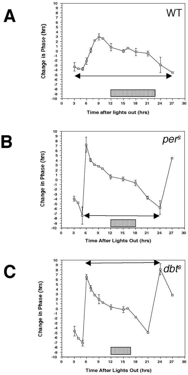
The Drosophila double-time (dbt) gene, which encodes a protein similar to vertebrate epsilon and delta isoforms of casein kinase I, is essential for circadian rhythmicity because it regulates the phosphorylation and stability of period (per) protein. Here, the circadian phenotype of a short-period dbt mutant allele (dbt(S)) was examined. The circadian period of the dbt(S) locomotor activity rhythm varied little when tested at constant temperatures ranging from 20 to 29 degrees C. However, per(L);dbt(S) flies exhibited a lack of temperature compensation like that of the long-period mutant (per(L)) flies. Light-pulse phase-response curves were obtained for wild-type, the short-period (per(S)), and dbt(S) genotypes. For the per(S) and dbt(S) genotypes, phase changes were larger than those for wild-type flies, the transition period from delays to advances was shorter, and the light-insensitive period was shorter. Immunohistochemical analysis of per protein levels demonstrated that per protein accumulates in photoreceptor nuclei later in dbt(S) than in wild-type and per(S) flies, and that it declines to lower levels in nuclei of dbt(S) flies than in nuclei of wild-type flies. Immunoblot analysis of per protein levels demonstrated that total per protein accumulation in dbt(S) heads is neither delayed nor reduced, whereas RNase protection analysis demonstrated that per mRNA accumulates later and declines sooner in dbt(S) heads than in wild-type heads. These results suggest that dbt can regulate the feedback of per protein on its mRNA by delaying the time at which it is translocated to nuclei and altering the level of nuclear PER during the declining phase of the cycle.


Figures

Fig. 1.
Fig. 1.
Both the perSand dbtS mutations increase the amplitude of the phase–response curve and shorten its period. After entrainment of flies with wild-type (A),perS (B), ordbtS (C) genotypes to at least three cycles of 12 hr LD, the LD cycle was terminated. A control group of flies with each genotype was left in constant darkness. Experimental groups were subjected to a 2 hr light pulse at the indicated time (Time after lights out) after termination of the last 12 hr photophase, but otherwise were treated the same as the control. The difference in the average activity offset time between experimental and control flies is plotted as a function of the time of the light pulse. A phase advance in the experimental group is plotted as a positive change in phase, whereas a phase delay is plotted as a negative change in phase. Error bars depict the SD for each point. The subjective day, or the interval during which light pulses do not reset the clock, is denoted by the hatched box under each PRC, whereas double-headed arrowslink time points with comparable phase shifts that define the period of the PRC. See Results for a more extensive discussion of the differences in these phase–response curves.
Fig. 2.
Fig. 2.
An increase in nuclear PER occurs later in the eyes of dbtS flies than in wild-type and perS flies. Heads from thepero mutant, wild-type (WT), perS, ordbtS mutant flies that had been entrained to a 12 hr LD cycle were removed at the indicated times (ZT; ZT0 = lights on, ZT12 = lights off), sectioned, and processed for detection of PER as described in Materials and Methods. Each panel is a section of an eye visualized with Nomarski optics at 200×; in the bottom right corner, a small region of the field (outlined with a black square on the larger image) is magnified an additional 3×. Thepero flies make no detectable levels of PER, so the level of diaminobenzidine chromogen in these eye sections is indicative of nonspecific detection. Some background staining is seen in the optic lobes, and sometimes lines of nonspecific staining are seen immediately under the lens tissue (blue arrow), but no punctate nuclear staining. By contrast,dbtS, perS, and wild-type eye sections can exhibit punctate staining at the surface of the eye in the nuclei of photoreceptor types 1–7 (red open arrows) and at the inside of the eye in the nuclei of photoreceptor type 8 (yellow triangles). Staining of eye sections was scored as none, weak, and strong; the panels here, for which the scores, genotypes, and collection times are given, are indicative of the level of staining observed for each class. A significant increase in the staining of eye sections was first observed in wild-type andperS eyes at ZT15. By contrast, mostdbtS eyes exhibited no staining at this time, with some showing weak staining. By ZT18, most wild-type eye sections were strongly stained (Table 2), whereasdbtS eye sections exhibited a mixture of weak and strong staining. A preponderance of strongly stained eye sections was not obtained until ZT21 indbtS (Fig. 3, Table 2). The overall staining scores of many sections are tabulated in Table 2.
Fig. 3.
Fig. 3.
Comparable levels of nuclear PER are found from late night to early morning in dbtSand wild-type eyes, with larger declines during the middle to the end of the day in dbtS eyes. Fly heads were collected, processed, and scored as described in the legend to Figure 2. Refer to the Figure 2 legend for an explanation of the labels in this figure. High levels of nuclear PER staining were observed in both wild-type and dbtS eyes from ZT21–3, with a mixture of weak and strong levels from ZT5–7 (see also Table 2). From ZT11–13 (ZT11 shown here; see also Table 2), weak or undetectable levels of PER immunoreactivity were obtained in both genotypes, with a higher proportion of unstaineddbtS eye sections than wild-type eye sections. The overall staining scores of many sections are tabulated in Table 2.
Fig. 4.
Fig. 4.
Immunoblot analysis of PER levels in head extracts demonstrates that the delay in accumulation of nuclear PER indbtS eyes is not the result of a delayed or reduced accumulation of total PER protein levels. Wild-type (W),dbtS (S), orpero(po) flies were frozen in liquid nitrogen at the indicated times (ZT; ZT0 = lights on, ZT12 = lights off). Extracts were prepared from the heads of these flies, electrophoresed on SDS-polyacrylamide gels, and subjected to immunoblot analysis with an antibody to PER (see Materials and Methods). Two representative experiments are shown. Sixty micrograms of extract were electrophoresed in each lane, except as indicated (3X = 180 μg, 0.3X= 20 μg). During the time at which PER accumulates in wild-type photoreceptor nuclei more than indbtS photoreceptor nuclei (ZT15–18), the total amount of PER in the dbtSheads is higher than or comparable to the amount in wild-type heads. Note in particular the higher level of PER at ZT15 indbtS, when immunohistochemical detection of nuclear PER is at its trough indbtS and significantly weaker than in wild-type eyes (Fig. 2, Table 2). By contrast, total PER levels are lower in dbtS at ZT21 than in wild type, although the amount of nuclear PER is indistinguishable (Fig. 3, Table 2). The gap in the bottom panelindicates that the ZT21 and perosamples were on a separate gel from the other samples (the relative amounts and mobilities of samples on different gels cannot be directly compared). The populations of flies that were processed for this immunoblot were also processed for immunohistochemical detection of PER at ZT15, and the immunohistochemical detection was weak indbtS and strong in wild type. (These sections are part of the data set tabulated in Table 2.)
Fig. 5.
Fig. 5.
RNase protection analysis of perRNA in heads demonstrates that per RNA accumulates later and declines sooner in dbtS heads than in wild-type heads. Wild-type (W) anddbtS (S) flies were frozen in liquid nitrogen at the indicated times (ZT; ZT0 = lights on, ZT12 = lights off). RNA was isolated from the heads of these flies, and 20 μg of RNA for each time point were analyzed for expression of per RNA and α-tubulin RNA (α-Tub, a constitutive control).A shows a representative analysis that was visualized by exposure to film, and B shows the quantitation of these signals by a phosphorimager analysis. For each time point, the normalized signal (per signal/α-tubulin signal) is plotted.

References

    1. Bae K, Lee C, Sidote D, Chuang KY, Edery I. Circadian regulation of a Drosophila homolog of the mammalian Clock gene: PER and TIM function as positive regulators. Mol Cell Biol. 1998;10:6142–6151. - PMC - PubMed
    1. Baylies MK, Bargiello TA, Jackson FR, Young MW. Changes in abundance or structure of the per gene product can alter periodicity of the Drosophila clock. Nature. 1987;326:390–392. - PubMed
    1. Cermakian N, Sassone-Corsi P. Multilevel regulation and the circadian clock. Nat Rev Mol Cell Biol. 2000;1:59–67. - PubMed
    1. Curtin KD, Huang ZJ, Rosbash M. Temporally regulated nuclear entry of the Drosophila period protein contributes to the circadian clock. Neuron. 1995;14:365–372. - PubMed
    1. Dunlap JC. Molecular bases for circadian clocks. Cell. 1999;96:271–290. - PubMed

Publication types

MeSH terms

LinkOut - more resources