. 2001 Sep 15;21(18):7143-52.
doi: 10.1523/JNEUROSCI.21-18-07143.2001.

Neuronal P2X7 receptors are targeted to presynaptic terminals in the central and peripheral nervous systems

Affiliations

Neuronal P2X7 receptors are targeted to presynaptic terminals in the central and peripheral nervous systems

S A Deuchars et al. J Neurosci. .

Abstract

The ionotropic ATP receptor subunits P2X(1-6) receptors play important roles in synaptic transmission, yet the P2X(7) receptor has been reported as absent from neurons in the normal adult brain. Here we use RT-PCR to demonstrate that transcripts for the P2X(7) receptor are present in extracts from the medulla oblongata, spinal cord, and nodose ganglion. Using in situ hybridization mRNA encoding, the P2X(7) receptor was detected in numerous neurons throughout the medulla oblongata and spinal cord. Localizing the P2X(7) receptor protein with immunohistochemistry and electron microscopy revealed that it is targeted to presynaptic terminals in the CNS. Anterograde labeling of vagal afferent terminals before immunohistochemistry confirmed the presence of the receptor in excitatory terminals. Pharmacological activation of the receptor in spinal cord slices by addition of 2'- and 3'-O-(4-benzoylbenzoyl)adenosine 5'-triphosphate (BzATP; 30 microm) resulted in glutamate mediated excitation of recorded neurons, blocked by P2X(7) receptor antagonists oxidized ATP (100 microm) and Brilliant Blue G (2 microm). At the neuromuscular junction (NMJ) immunohistochemistry revealed that the P2X(7) receptor was present in motor nerve terminals. Furthermore, motor nerve terminals loaded with the vital dye FM1-43 in isolated NMJ preparations destained after application of BzATP (30 microm). This BzATP evoked destaining is blocked by oxidized ATP (100 microm) and Brilliant Blue G (1 microm). This indicates that activation of the P2X(7) receptor promotes release of vesicular contents from presynaptic terminals. Such a widespread distribution and functional role suggests that the receptor may be involved in the fundamental regulation of synaptic transmission at the presynaptic site.


Figures

Fig. 1.
Fig. 1.
RT-PCR indicates expression of the P2X7 receptor in spinal cord and medulla oblongata. Agarose gel electrophoresis of PCR products obtained after reverse transcription of total RNA. RT-PCR analysis revealed the presence of P2X7 transcripts in RNA extracted from spinal cord (lane 1) and medulla (lane 2). No amplified products were detected when using water or RNA as template (lanes 3, 4, 5,8). Veracity of the amplified products was confirmed by cloning the PCR product into pGEM T-Easy followed by DNA sequencing. As a positive control, the PCR products for the housekeeping gene hprt were also detected in both tissues (lanes 6,7). Products resulting from a 1 kb molecular weight marker are indicated in lane M, and the bands correspond to the calculated size for all PCR products.
Fig. 2.
Fig. 2.
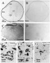
In situ hybridization reveals that messenger RNA coding for the P2X7 receptor is present in neurons throughout the spinal cord and medulla oblongata.A, Spinal cord section indicating positive signal after hybridization with a DIG-labeled antisense probe specific to the P2X7 receptor, visualized with alkaline phosphatase. The signal is present throughout the gray matter and is easily seen in the large motoneurons of the ventral horn (VH) as well as in dorsal horn (DH) neurons. The white matter (WM) is sparsely labeled. B, Spinal cord section indicating lack of signal when tissue was incubated with a sense probe to the P2X7 receptor. C, Medulla oblongata section indicating the widespread positive signal obtained after hybridization with the DIG-labeled antisense probe specific to the P2X7 receptor. Positive signal is visible in labeled neurons throughout the medulla. D, Medulla oblongata section indicating lack of signal when tissue was incubated with a sense probe to the P2X7 receptor. E, Larger magnification of the dorsal vagal complex of the medulla oblongata, indicating that hybridization reaction product can be observed in neuronal structures (arrows). F, Larger magnification of the ventral horn of the spinal cord indicating that hybridization reaction product can be observed in the cytoplasm of large ventral horn motoneurons (arrows).G, Neurons (arrow) in the dorsal horn of the spinal cord also contain hybridization reaction product in their cytoplasm.
Fig. 3.
Fig. 3.
Western blotting indicates the tissue specificity of the antibody to P2X7 receptors. Western blotting of rat brainstem resulted in staining of a major band running at ∼69 kDa when detected with the anti-P2X7 receptor antibody. This is the predicted molecular weight of the P2X7 receptor. Preabsorption of the antiserum with the cognate peptide abolished staining of the membrane.
Fig. 4.
Fig. 4.
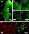
Widespread P2X7 receptor immunoreactivity in the spinal cord, medulla oblongata, and at the neuromuscular junction is localized to punctate structures.A1, Half section of the spinal cord stained for P2X7 immunoreactivity and detected with Alexa488 (green). Labeling is dense in the neuropil of the spinal cord, but less so in the white matter. CC, Central canal; VH, central horn; DH, dorsal horn; WM, white matter. Areas indicated in boxes are illustrated at higher magnification as appropriate. Dense fiber staining is evident in the dorsal horn (A2), intermediolateral cell column (A3), and the ventral horn, where labeled structures appear to surround the somatic membrane of large motoneurons (A4). In the white matter (A5), occasional labeled fibers can be observed. B,P2X7 immunoreactivity is also ubiquitous throughout the medulla oblongata. In this high-magnification view, P2X7immunoreactivity (detected with Cy3, red) is observed in punctate structures in the neuropil as well as surrounding the somatic membrane of neurons in the hypoglossal nucleus. C,Mammalian motor nerve terminals are also immunoreactive for P2X7 receptors. Transversus abdominis muscle from adult mice contained P2X7 receptor immunoreactivity localized to the motor nerve terminal (green, detected with FITC), which appear to be distinct from the postsynaptic receptors (red, labeled withTRITC-α-bungarotoxin).
Fig. 5.
Fig. 5.
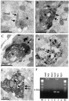
P2X7 receptors are localized presynaptically to terminal boutons in the CNS displaying characteristics of excitatory synapses and to the motor nerve terminal at the neuromuscular junction. A, A presynaptic terminal (asterisk) in the nucleus tractus solitarius of the medulla oblongata contains electron-dense DAB reaction product (dark gray). The terminal forms a synapse (arrows) of the asymmetric type with a dendritic structure (Dend). B, A presynaptic terminal (asterisk) in the intermediolateral cell column of the spinal cord forms an asymmetric type synapse (arrows) with a dendritic structure (Dend). C, At the neuromuscular junction, immunoreactivity was also located presynaptically in the motor nerve terminal (asterisk). Arrows indicate a region of synaptic specialization at the endplate. D, To determine whether P2X7 receptor containing terminals are excitatory, a population containing purely excitatory fibers (vagal nerve afferent fibers) was anterogradely labeled before immunohistochemistry. The electron-dense DAB labeling in this case is the result of the reaction to localize the anterograde tracer, whereas the gold deposits (open arrow) indicate the presence of P2X7 receptor immunoreactivity in these excitatory terminals. E, Another example of a DAB-labeled vagal afferent terminal that contains P2X7 receptor immunoreactivity. F, P2X7 receptor mRNA was detected in the nodose ganglion, the location of the neurons giving rise to central vagal afferent projections. Agarose gel electrophoresis of PCR products obtained after reverse transcription of total RNA. RT-PCR analysis revealed the presence of P2X7 transcripts in RNA extracted from medulla (lane 1) and nodose ganglion (lane 2). No amplified products were detected when using water as template (lanes 3,6). As a positive control, the PCR products for the housekeeping gene hprt were also detected in both tissues (lanes 2, 5). Products resulting from a 1 kb molecular weight marker are indicated in lane M, and the bands correspond to the calculated size for all PCR products.
Fig. 6.
Fig. 6.
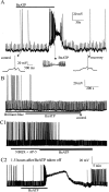
Activation of the P2X7 receptor in CNS slices elicits depolarizations in neurons attributable to release of glutamate. A, The P2X7 receptor agonist BzATP depolarizes neurons. In this example the cell was at a potential of −60 mV, and a depolarizing current of +20 pA, 1 sec duration was applied every 7 sec, which on occasion caused the neuron to reach the threshold for firing (shown on a faster time base below). Application of BzATP (30 μm) depolarized the neuron, resulting in discharge of action potentials and eventual depolarizing block. Recovery after washout is shown in the inset.B, The effects of BzATP were antagonized by appropriate antagonists. When preincubated in the P2X7 receptor antagonist Brilliant Blue G (2 μm) for 30 min, BzATP failed to depolarize neurons. This neuron was also held at −60 mV, and depolarizing current pulses of +30 pA were applied. C,BzATP-evoked depolarizations were blocked by excitatory amino acid receptor antagonists. Superfusion of the non-NMDA receptor antagonist NBQX (20 μm) and the NMDA receptor antagonist AP-5 (50 μm) prevented BzATP (30 μm)-evoked depolarization even after prolonged application (C1). After 90 min washout, application of BzATP caused a large depolarization and increase in firing rate that again resulted in depolarizing block. This neuron was held at −60 mV with current pulses of +15 pA applied every 7 sec.
Fig. 7.
Fig. 7.
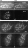
Activation of P2X7 receptors promotes nerve terminal destaining associated with transmitter release at the neuromuscular junction. Nerve terminals were loaded with FM1–43 or RH414 and transferred to the microscope stage. Terminals were visualized, using a ND25 filter at all times, at t= 0 (a, c, e) and every 5 min untilt = 30 min (b, d, f). Control preparations were repeatedly imaged and showed modest photobleaching (a, b). In an identical experiment but with 30 μm BzATP added at t = 0 (c, d), terminals destained by ∼50%, over 30 min. In a repeat experiment in which the preparation was preincubated with 100 μm oxidized ATP (e, f) or 1 μm Brilliant Blue G (g, h) before and during BzATP addition, BzATP-mediated destaining was prevented and terminals again demonstrated some photobleaching comparable with control over 30 min.
Fig. 8.
Fig. 8.
Quantification of P2X7receptor-mediated motor nerve terminal destaining. Plots of mean pixel intensity from multiple regions of interest on multiple nerve terminals. Values are background-subtracted and expressed as mean fractions of the value at t = 0 ± SEM. Control values (open circles) showed a moderate degree of photobleaching (10%). In the presence of BzATP (30 μm;filled squares), terminals destained by 50% over 30 min. Pretreatment with oxidized ATP (100 μm;filled circles) or Brilliant Blue G (1 μm;filled triangles) completely blocked the BzATP-evoked destain. The BzATP evoked destain is significantly different from control, oxidized ATP, or Brilliant Blue G-blocked preparations.

References

    1. Afework M, Burnstock G. Distribution of P2X receptors in the rat adrenal gland. Cell Tissue Res. 1999;298:449–456. - PubMed
    1. Armstrong JN, MacVicar BA. P2X7-receptor activation depresses synaptic transmission at hippocampal mossy fiber-CA3 synapses. Soc Neurosci Abstr. 2000;26:884.
    1. Atkinson L, Batten TF, Deuchars J. P2X(2) receptor immunoreactivity in the dorsal vagal complex and area postrema of the rat. Neuroscience. 2000;99:683–696. - PubMed
    1. Ballerini P, Rathbone MP, Di Iorio P, Renzetti A, Giuliani P, D'Alimonte I, Trubiani O, Caciagli F, Ciccarelli R. Rat astroglial P2Z (P2X7) receptors regulate intracellular calcium and purine release. NeuroReport. 1996;7:2533–2537. - PubMed
    1. Bardini M, Lee HY, Burnstock G. Distribution of P2X receptor subtypes in the rat female reproductive tract at late pro-oestrus/early oestrus. Cell Tissue Res. 2000;299:105–113. - PubMed

Publication types

MeSH terms

LinkOut - more resources