. 2001 Sep 15;21(18):7153-60.
doi: 10.1523/JNEUROSCI.21-18-07153.2001.

Astrocytes give rise to new neurons in the adult mammalian hippocampus

Affiliations

Astrocytes give rise to new neurons in the adult mammalian hippocampus

B Seri et al. J Neurosci. .

Abstract

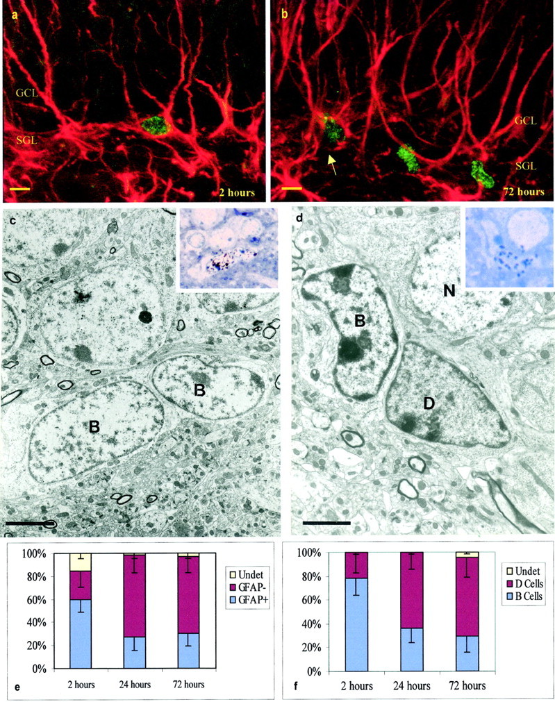
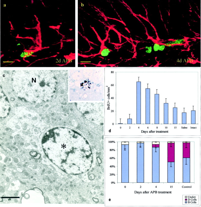
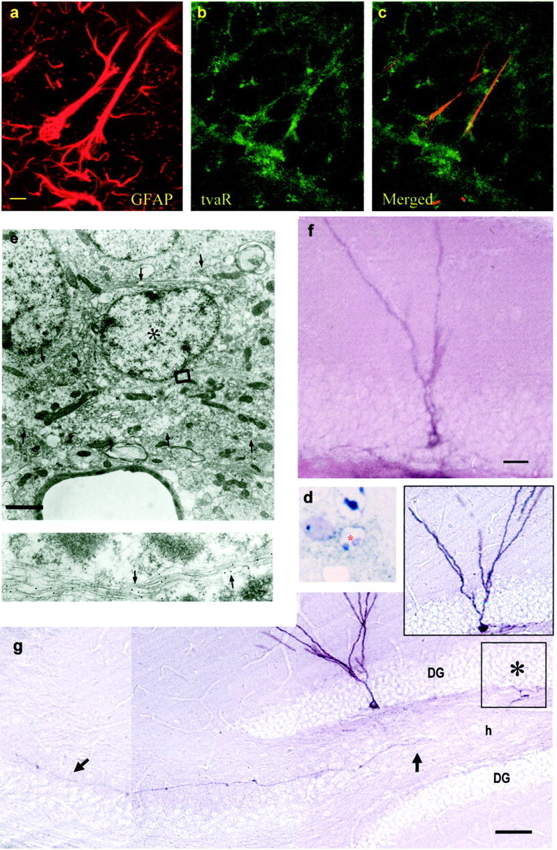
Neurogenesis in the dentate gyrus of the hippocampus persists throughout life in many vertebrates, including humans. The progenitors of these new neurons reside in the subgranular layer (SGL) of the dentate gyrus. Although stem cells that can self-renew and generate new neurons and glia have been cultured from the adult mammalian hippocampus, the in vivo primary precursors for the formation of new neurons have not been identified. Here we show that SGL cells, which express glial fibrillary acidic protein and have the characteristics of astrocytes, divide and generate new neurons under normal conditions or after the chemical removal of actively dividing cells. We also describe a population of small electron-dense SGL cells, which we call type D cells and are derived from the astrocytes and probably function as a transient precursor in the formation of new neurons. These results reveal the origins of new neurons in the adult hippocampus.


Figures

Fig. 1.
Fig. 1.
Dividing cells in the SGL. a, Confocal micrograph of BrdU-labeled GFAP-positive SGL astrocyte 2 hr after BrdU injection. Note the BrdU-labeled nuclei (green) and multiple GFAP-positive processes (red) penetrating the GCL. b, Confocal micrograph of BrdU-labeled GFAP-positive (arrow) and GFAP-negative cells in the SGL 72 hr after BrdU injection. c, Electron micrograph of two B cells labeled with [3H] 2 hr after [3H]thymidine injection. The insetshows the nuclei of these two B cells overlaid by autoradiographic silver grains in a 1.5 μm section used to prepare the ultrathin section shown below. d, [3H]-labeled D cell 72 hr after [3H]thymidine injection. Note the dark cytoplasm of D cells in contrast to the light cytoplasm of B cells. Theinset shows the nuclei of the D cell overlaid by autoradiographic silver grains in a 1.5 μm section used to prepare the ultrathin section shown below. e, Percentage of BrdU-labeled cells in the SGL 2, 24, and 72 hr after BrdU injection. This analysis was done at the confocal microscope; 108 cells in three animals were analyzed for each survival group. f, Percentage of [3H]-labeled cells in the SGL 2, 24, and 72 hr after [3H]thymidine injection; 25 cells in three animals were studied at the electron microscope for each survival group. In e and f, error bars indicate SD. Scale bars: a, b, 5 μm;c, 3.6 μm; d, 2 μm.
Fig. 2.
Fig. 2.
Cell reappearance after APB treatment.a, Cell labeled with BrdU (green) and GFAP (red) 2 d after termination of APB.b, Four days after APB, BrdU–GFAP-labeled cells remained, but some GFAP-negative BrdU-labeled cells were also present.c, Ultrastructural analysis of a [3H]-labeled cell (see autoradiogram in theinset) 2 hr after [3H]thymidine injection and 2 d after termination of APB treatment. This cell corresponds to a B cell. Note the light cytoplasm and irregular contours of the plasma membrane. d, Time course of reappearance of BrdU-labeled cells after termination of APB treatment;n = 5; mean ± SD. e, Cellular composition (EM) of the SGL at multiple survivals after APB termination; n = 3; 250 cells per animal; mean ± SD. Scale bars: a, b, 8 μm;c, 2 μm.
Fig. 3.
Fig. 3.
Retrovirally labeled SGL astrocytes give rise to new neurons in the granule cell layer. Double staining for GFAP and tvaR in the SGL of normal Gtva mice (a–c).a, Staining for GFAP. b, Staining for the tvaR. c, Merged fields showing combined immunostaining for GFAP and the tvaR. AP-positive cells identified after injection of RCAS-AP virus into the dentate gyrus of Gtva mice in eand d 2 d after viral injection cells were analyzed at the electron microscope. d, Plastic section (1.5 μm) of an AP-positive cell (asterisk) stained with NBT-BCIP (purple deposit; bright field). The cell was reembedded, resectioned, and stained with gold-conjugated antibodies to GFAP for ultrastructural analysis (e). Labeled cell corresponded to a B cell that contained intermediate filaments decorated by gold particles (arrows in the inset below). Thearrowheads in e show the irregular contours of the plasma membrane of this cell. f, Immature granule neurons start appearing 8 d after viral injection. g, Thirty days after RCAS-AP injection into SGL of Gtva mice, AP-labeled granule neurons are present in the dentate gyrus. These cells have the characteristic dendritic arbors in the molecular layer and an axon (arrows) that projects to CA3. The cell in the inset gives rise to the mossy fiber shown in g. This cell body is in the adjoining section in the approximate location shown by the asterisk. Scale bars: a, 5 μm (for a–c);e, 2 μm; f, 10 μm; g, 100 μm.

References

    1. Altman J. Autoradiographic and histological studies of postnatal neurogenesis. IV. Cell proliferation and migration in the anterior forebrain, with special reference to persisting neurogenesis in the olfactory bulb. J Comp Neurol. 1969;137:433–458. - PubMed
    1. Altman J, Das GD. Autoradiographic and histological evidence of postnatal hippocampal neurogenesis in rats. J Comp Neurol. 1965;124:319–336. - PubMed
    1. Altman J, Das GD. Autoradiographic and histological studies of postnatal neurogenesis. I. A longitudinal investigation of the kinetics, migration and transformation of cells incorporating tritiated thymidine in neonate rats, with special reference to postnatal neurogenesis in some brain regions. J Comp Neurol. 1966;126:337–390. - PubMed
    1. Alvarez-Buylla A, Nottebohm F. Migration of young neurons in adult avian brain. Nature. 1988;335:353–354. - PubMed
    1. Alvarez-Buylla A, Theelen M, Nottebohm F. Birth of projection neurons in the higher vocal center of the canary forebrain before, during, and after song learning. Proc Natl Acad Sci USA. 1988;85:8722–8726. - PMC - PubMed

Publication types

MeSH terms

LinkOut - more resources