. 2001 Sep 15;21(18):7236-46.
doi: 10.1523/JNEUROSCI.21-18-07236.2001.

The Meissner corpuscle revised: a multiafferented mechanoreceptor with nociceptor immunochemical properties

Affiliations

The Meissner corpuscle revised: a multiafferented mechanoreceptor with nociceptor immunochemical properties

M Paré et al. J Neurosci. .

Abstract

Meissner corpuscles (MCs) in the glabrous skin of monkey digits have at least three types of innervation as revealed by immunofluorescence. The previously well known Aalphabeta-fiber terminals are closely intertwined with endings from peptidergic C-fibers. These intertwined endings are segregated into zones that alternate with zones containing a third type of ending supplied by nonpeptidergic C-fibers. Although MCs are widely regarded as low-threshold mechanoreceptors, all three types of innervation express immunochemical properties associated with nociception. The peptidergic C-fiber endings have readily detectable levels of immunoreactivity (IR) for calcitonin gene-related peptide (CGRP) and substance P (SP). The Aalphabeta endings have relatively lower levels of IR for CGRP and SP as well as the SP neurokinin 1 receptor and vanilloid-like receptor 1. Both the Aalphabeta and peptidergic C-fiber endings were also labeled with antibodies for different combinations of adrenergic, opioid, and purinergic receptors. The nonpeptidergic C-fiber endings express IR for vanilloid receptor 1, which has also been implicated in nociception. Thus, MCs are multiafferented receptor organs that may have nociceptive capabilities in addition to being low-threshold mechanoreceptors.


Figures

Fig. 1.
Fig. 1.
Schematic drawing of an MC shown in a plane perpendicular to the skin surface and illustrating three different types of innervation: 1, NF-positive Aαβ-fiber innervation (dark gray); 2, varicose CGRP-positive C-fiber innervation (black withdots); and 3, nonpeptidergic VR1-positive C-fiber innervation (black). The MCs are located in dermal papillae that protrude into the epidermis. The unmyelinated CGRP-positive C-fiber innervation is closely affiliated with the Aαβ endings, although some distributes independently around the contour of the MC. The nonpeptidergic VR1 innervation is segregated to zones interdigitated between the intertwined Aαβ and unmyelinated CGRP-positive C-fiber innervation. Schwann cells (medium gray) are distributed between the innervation and at the base and apex of each MC. The additional immunochemical characteristics for each type of innervation are listed below the drawing.
Fig. 2.
Fig. 2.
Immunofluorescence labeling of MCs in 14-μm-thick sections cut perpendicular to the surface of the glabrous skin of a distal digit pad. The antigens for the primary antibodies are indicated. ep, Epidermis; d, dermis.A, MCs have a stratified appearance, as seen with rabbit anti-PGP9.5. The most intense PGP9.5-IR (arrowheads) occurs on processes of relatively thick-caliber Aαβ-fiber innervation that were labeled with anti-NF in other double-labeled preparations (Figs. 3A–D, 4). Intense PGP9.5-IR also occurs on thinner beaded profiles (straight arrows) that were labeled with anti-CGRP and anti-SP in other double-labeled preparations (Fig. 3). The large straight arrowindicates likely source axons; the small straight arrowsindicate presumptive terminals. Relatively medium-intensity PGP9.5-IR (curved arrows) occurs on the innervation that labels with anti-VR1 in other preparations (Fig. 4) and is restricted to zones between the Aαβ-fiber and CGRP-positive C-fiber innervation.B, Rabbit anti-S100 labels flattened presumptive Schwann cells (small broad arrows) between the axon terminals and teardrop-shaped, presumptive Schwann cells (large broad arrows) at the base and apex of the MCs. The Schwann cells also express low levels of PGP9.5-IR (A, broad open arrows).C, D, Double labeling with rabbit anti-nonphosphorylated 200 kDa neurofilament protein (NFn) and mouse anti-MBP reveals that myelin basic protein is expressed along the Aαβ-derived processes (arrowheads) within the MC. Labeling with mouse anti-phosphorylated 200 kDa neurofilament protein (NFp) is similar to labeling with rabbit anti-NFn (C, inset, arrowheads).Asterisks indicate NF-negative zones where VR1-positive C-fiber innervation terminates as revealed by double-label combinations (Fig. 4).
Fig. 3.
Fig. 3.
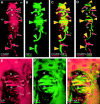
The close relationship is shown between the peptidergic C-fiber (arrows) and Aαβ-fiber (arrowheads) MC innervation as seen in immunofluorescence digital images of 14-μm-thick double-labeled sections cut perpendicular to the skin surface. ep, Epidermis; d, dermis. A–D, The same MC is shown labeled with sheep anti-CGRP and rabbit anti-NFn. InE–F, another MC is labeled with guinea pig anti-SP and rabbit anti-NK1. The antigens labeled by the primary antibodies are indicated at the bottom. The color barbeneath each antigen indicates that the primary antibody was revealed by secondary antibodies conjugated to Cy-3 (red) or Cy-2 (green). In images captured with just the Cy-3 filter (A, E), the labeled innervation isred and is indicated by red symbols. In images captured with just the Cy-2 filter (B, F), the labeled innervation is green and is indicated bygreen symbols. In digitally merged double-labeled images (C, D, G), yellow symbols indicate the innervation definitively labeled with both primary antibodies.Asterisks indicate zones where VR1-positive C-fiber innervation is interdigitated, as seen in other double-label preparations (Fig. 4). A–C, Deconvoluted digital images of conventional epifluorescence for anti-CGRP and anti-NFn. In the tissue section, the maximum intensity of anti-CGRP labeling was lower, and the background was higher than anti-NFn labeling. For illustration purposes, the intensity of the anti-CGRP image has been digitally increased approximately twofold so that the maximum intensity was comparable with the maximum intensity of the anti-NFn image. The higher background of the CGRP labeling was partially suppressed by increasing the contrast and digitally subtracting lower signals. Resulting images in A and B are digitally merged inC. The NFn labeling is in relatively thicker profiles that colabel with anti-MBP in other sections (Fig.2C,D), indicating that the detectable NF expression is in Aαβ innervation. The intensity of NFn labeling is very high and uniform in the Aαβ axons (large arrowheads) as well as in their presumptive endings (small arrowheads). In contrast, the intensity of anti-CGRP labeling varies and is highest only in relatively thin, varicose axons (large arrow) that fail to colabel with anti-MBP in other double-labeled preparations, indicating that they are C-fibers. Other thin, varicose axons are faintly labeled with anti-CGRP (chevrons). CGRP-IR is coexpressed at relatively low levels on the anti-NFn-labeled axons (large arrowheads) and at fairly high levels on anti-NFn-labeled presumptive endings (small arrowheads), especially along the lower border of the endings. D, Confocal image showing double labeling of the same MC as inA–C. Separate confocal images of the CGRP-IR and NF-IR were captured; the intensity of the anti-CGRP labeling was increased so that the maximum was comparable with the maximum for anti-NFn; and the two images were merged. Low- to medium-range signals were subtracted for both anti-CGRP and anti-NFn so that only sites with the most intense labeling remain. With the elimination of medium-intensity CGRP-IR from the NF-positive endings, the confocal images revealed that innervation having high levels of CGRP (small red arrows) is intertwined with the NF-positive innervation and is especially located along the inferior border of the NF-positive endings. E–G, Deconvoluted conventional epifluorescence images of an MC double-labeled for SP and NK1. Original immunofluorescence signals with anti-SP and anti-NK1 were much lower than those with anti-CGRP and anti-NF. For illustration purposes, the intensity of the images in E and F have been digitally increased threefold to fourfold so that the maximum signals are comparable with each other and with the maximum signals inA and B. Consequently, background labeling is also increased especially for anti-SP, which has relatively low signal to high background labeling particularly in the epidermis. Some amplified background labeling was reduced by increasing the contrast and digitally subtracting lower signals. Comparable with the pattern of labeling observed with anti-CGRP and anti-NF (A, B), SP- and NK1-IR are coexpressed on the presumptive endings (arrowheads) of Aαβ axons confirmed by double-label combinations with anti-NF (results not shown). The Aαβ axons lack detectable SP-IR and have relatively low levels of NK1-IR. Merged images in G reveal separate SP-positive innervation (long arrows) along the inferior border of the Aαβ endings. A few profiles that only label with anti-SP are independently present in the MC (chevrons) and in the epidermis (broad arrows).
Fig. 4.
Fig. 4.
Segregated, interdigitated relationship between the VR1-positive C-fiber innervation (curved arrows) and NF-positive Aαβ-fiber innervation (arrowheads) as seen in double-labeled conventional immunofluorescence digital images of 14-μm-thick sections cut perpendicular to the skin surface.ep, Epidermis; d, dermis. The sections were double-labeled with guinea pig anti-VR1 revealed with a Cy-3-conjugated secondary antibody (red labeling indicated by red arrows) and rabbit anti-NFn revealed with a Cy-2-conjugated secondary antibody (greenlabeling indicated by green arrows andarrowheads). In original images captured separately for Cy-3 and Cy-2, anti-VR1 labeling was lower, and background was higher than anti-NFn labeling. For illustration purposes, the intensity of the anti-VR1 image was digitally increased approximately threefold so that the maximum intensity was comparable with the maximum intensity of the anti-NFn image. The higher background of the VR1 labeling was partially suppressed by increasing the contrast and digitally subtracting lower signals. The resulting separate images are digitally merged inA and B. No innervation was definitively double-labeled in the MCs or epidermis. VR1-positive innervation in the MCs is segregated from and interdigitated with NF-positive innervation. Other double-label combinations revealed that the anti-NFn labeling is on myelinated Aαβ-fiber innervation (Fig. 2C,D) and that the VR1 innervation is on unmyelinated C-fiber innervation that lacks CGRP-IR (Fig. 5A). A, inset, Drawing by Dogiel (1892) illustrating a similar segregation of thin and thick fiber innervation in human MCs based on a reduced silver stain. In A, anti-VR1 and anti-NFn label separate thin-caliber innervation terminating in the epidermis (red andgreen broad arrows). Asterisks indicate individual melanocytes in the lamina basalis of the epidermis. Thewhite bracket indicates a gap in the row of melanocytes. The gap is occupied by melanin-lacking Merkel cells, which are labeled by anti-CGRP in other preparations (results not shown). The Merkel cells are innervated by anti-NFn labeled endings (green bent arrows) supplied by Aαβ-fibers. In B, the right MC only has the VR1-positive innervation.
Fig. 5.
Fig. 5.
Additional immunochemical characteristics of MC innervation as seen in conventional immunofluorescence digital images of MCs in 14-μm-thick sections cut perpendicular to the skin surface.ep, Epidermis; d, dermis. Each panel is an image double-labeled with two primary antibodies made in different species against the antigens indicated at the bottom. The fluorophore conjugated to the secondary antibodies is indicated by a red bar (Cy-3) or green bar (Cy-2) located beneath the antigen of the correspondingly labeled primary antibody. Based on morphology, location, and the total results of all double-label combinations used in the study (Figs. 3, 4), the likely NF-positive Aαβ-fiber innervation is indicated byarrowheads; likely CGRP-positive C-fiber innervation is indicated by long straight arrows; and likely VR1-positive C-fiber innervation is indicated by curved arrows. Epidermal innervation is indicated by broad arrows in some panels. Red and green arrowheads and arrows indicate processes definitively labeled only by the antibody for the antigen listed over the red and green bars, respectively.Yellow arrowheads and arrows indicate definitively double-labeled innervation. Large arrowsand arrowheads indicate likely source axons;small arrows and arrowheads indicate presumptive terminals. For illustration purposes, the image intensities were digitally adjusted so that the maximum labeling intensity for each primary–secondary antibody combination was approximately the same. In many cases, contrast was increased, and low-end signals were subtracted to partially reduce the relatively high background labeling that occurs with some antibodies, the increased background caused artificially by digitally enhancing the overall image intensity, or both.A, Guinea pig anti-VR1 labeled C-fiber innervation (red curved arrows) is segregated from innervation labeled with rabbit anti-CGRP-IR. On the basis of other preparations (Fig. 3A–D), the segregated CGRP-positive innervation includes intermingled CGRP-positive endings of Aαβ-fibers (green arrowheads) and C-fibers (green long straight arrows). Some epidermal innervation expresses only VR1-IR (broad red arrow) or CGRP-IR (results not shown). B, Rabbit anti-P2X2 labeling (green arrows andarrowheads) is segregated from innervation labeled with guinea pig anti-VR1 (red curved arrows). As determined from other preparations (e.g., G), P2X2-IR is expressed on both the CGRP-positive C-fiber innervation (green arrows) and Aαβ-fiber innervation (green arrowheads). C, guinea pig anti-VR1 (red curved arrows) and rabbit anti-μOR (green straight arrows) label separate sets of innervation in the MC. On the basis of other label combinations (results not shown), the μOR-IR was definitive only on the CGRP-positive C-fiber innervation. A few spots of μOR-IR coincide with the VR1-positive innervation, but this was not consistent. Anti-μOR and anti-VR1 label mostly separate epidermal innervation (red and green broad arrows). D, Rabbit anti-δOR labels innervation (green arrows and arrowheads) in zones between the guinea pig anti-VR1-positive innervation (red curved arrows). Other label combinations (results not shown) revealed that the δOR-IR is expressed on the Aαβ-fiber innervation and on the CGRP-positive C-fiber innervation. A few spots of δOR-IR coincide with the VR1-positive innervation, but this was not consistent. E, Rabbit anti-NOCI labeling is coexpessed (yellow curved arrows) on guinea pig anti-VR1-positive innervation in the MCs. As determined from other preparations (e.g., K), other anti-NOCI labeling (red straight arrows) is likely on the CGRP-positive C-fiber innervation. Asterisks indicate VR1-negative zones where the Aαβ-fiber and CGRP-positive C-fibers terminate (Figs. 3, 4). Some epidermal innervation is VR1-positive and NOCI-negative (green broad arrow).F, Rabbit anti-P2X1 labeling is highly punctate. Some is detectable on guinea pig VR1-positive innervation (yellow curved arrows) in the MCs. As determined in other double-label combinations (results not shown), P2X1-IR is also expressed on CGRP-positive C-fiber innervation (red straight arrows).Asterisks indicate VR1-negative zones where the Aαβ-fiber and CGRP-positive C-fibers terminate. VR1-IR and P2X1-IR can be expressed on separate epidermal innervation (redand green broad arrows). G, H, Rabbit anti-P2X2 and anti-P2X3 colabels with sheep CGRP-IR both on the CGRP-positive C-fiber innervation (yellow straight arrows) and on the Aαβ-fiber innervation (yellow arrowheads), as determined from other preparations. Both P2X2-IR and P2X3-IR are relatively fainter on the C-fiber innervation. Asterisks indicate CGRP-, P2X2-, and P2X3-negative zones where the VR1-positive innervation terminates (Fig. 4). Anti-P2X3 labeling is more readily detectable than anti-P2X2 on epidermal innervation, where it can be independent of CGRP expression (red and green broad arrows).I, Rabbit α2A-IR is coexpressed on sheep anti-CGRP-labeled C-fiber (yellow long arrows) as well as coexpressed more intensely on the Aαβ-fiber innervation (yellow arrowheads), as determined from other double-label combinations (results not shown). The α 2A-IR and CGRP-IR can be expressed on separate axons terminating in the epidermis (red and green broad arrows).J, Rabbit anti-α 2C labeling is coexpressed only on the CGRP-positive profiles that are the thicker endings of the Aαβ innervation (yellow arrowheads). CGRP-positive C-fiber innervation is present without α2C-IR in and around the MCs (green long arrows) and supplying the epidermis (green broad arrow). Asterisksindicate the CGRP and α2C-negative zones where the VR1-positive innervation would be located. K, Aαβ innervation labeled with mouse anti-NFp (green arrowheads) has little if any definitive rabbit anti-NOCI-IR. Anti-NOCI labels innervation (red curved arrows) between the Aαβ endings, which is where the VR1 innervation terminates (seeE), as well as innervation (red straight arrows) closely juxtaposed to the Aαβ endings, which is where the CGRP-positive C fibers terminate. L, Rabbit anti-VRL1-IR is only definitively colocalized with mouse anti-NFp labeling, indicative of the Aαβ-fiber innervation.

References

    1. Arvidsson U, Dado RJ, Riedl M, Lee J-H, Law P-Y, Loh HH, Elde R, Wessendorf MW. Delta-opioid receptor immunoreactivity: distribution in brainstem and spinal cord, and relationship to biogenic amines and enkephalin. J Neurosci. 1995a;15:1215–1235. - PMC - PubMed
    1. Arvidsson U, Riedl M, Chakrabarti S, Lee J-H, Nakano AH, Dado RJ, Loh HH, Law P-Y, Wessendorf MW, Elde R. Distribution and targeting of a μ-opioid receptor (MOR1) in brain and spinal cord. J Neurosci. 1995b;15:3328–3341. - PMC - PubMed
    1. Arvidsson U, Riedl M, Chakrabarti S, Vulchanova L, Lee J-H, Nakano AH, Lin X, Loh HH, Law P-Y, Wessendorf MW, Elde R. The κ-opioid receptor (KOR1) is primarily postsynaptic: combined immunohistochemical localization of the receptor and endogenous opioids. Proc Natl Acad Sci USA. 1995c;92:5062–5066. - PMC - PubMed
    1. Ballet S, Mauborgne A, Benoliel J-J, Bourgoin S, Hamon M, Cesselin F, Collin E. Polyarthritis-associated changes in the opioid control of spinal CGRP release in the rat. Brain Res. 1998;796:198–208. - PubMed
    1. Björklund H, Dalsgaard CJ, Jonsson C-E, Hermasson A. Sensory autonomic innervation of non-hairy skin and hairy human skin. Cell Tissue Res. 1986;243:51–57. - PubMed

Publication types

MeSH terms