. 2001 Sep;13(9):1987-2004.
doi: 10.1105/tpc.010056.

PDE1 encodes a P-type ATPase involved in appressorium-mediated plant infection by the rice blast fungus Magnaporthe grisea

Affiliations

PDE1 encodes a P-type ATPase involved in appressorium-mediated plant infection by the rice blast fungus Magnaporthe grisea

P V Balhadère et al. Plant Cell. 2001 Sep.

Abstract

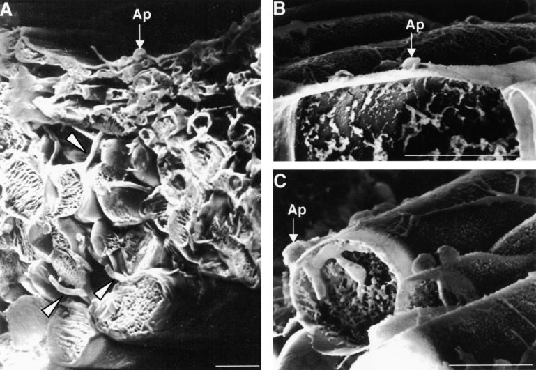
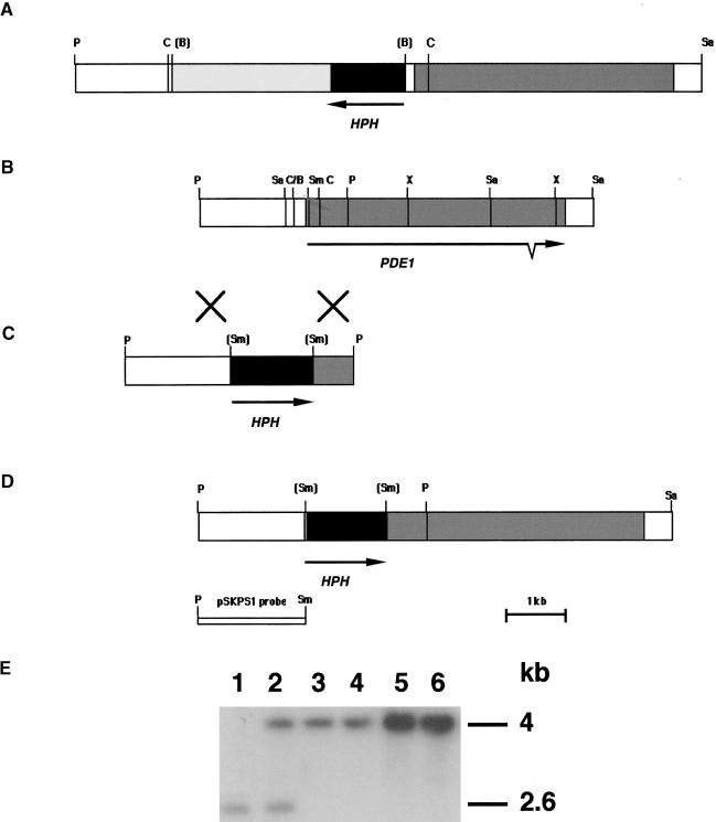
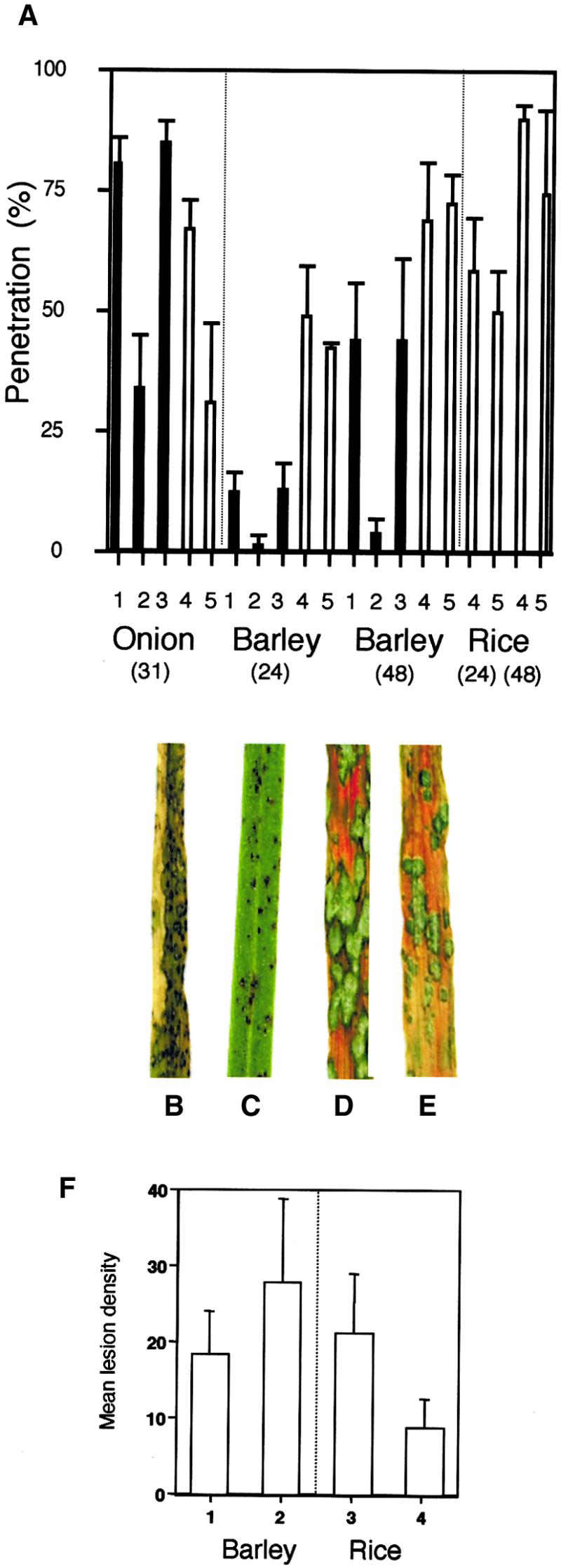
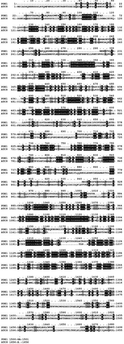
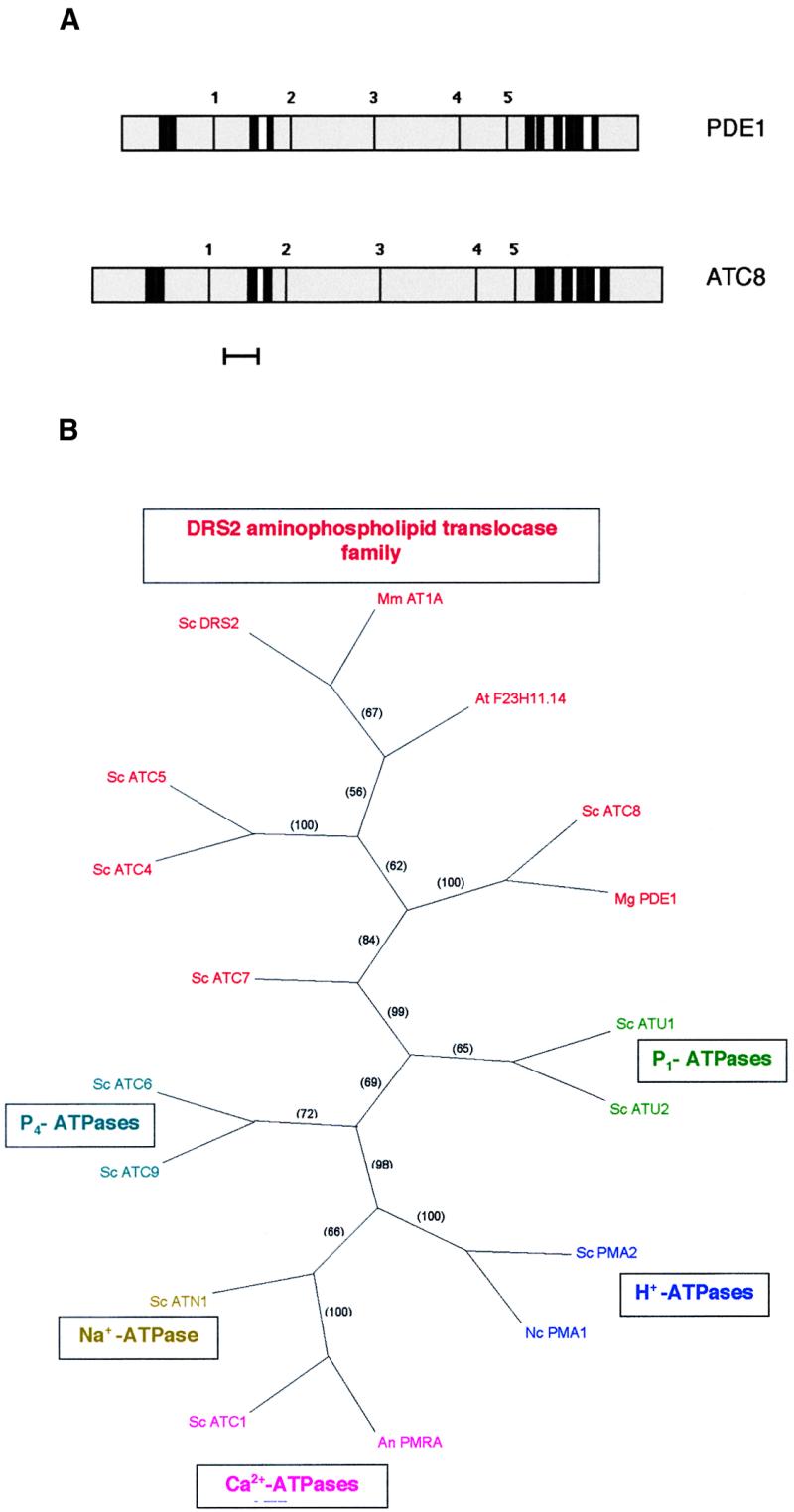
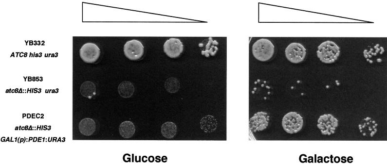
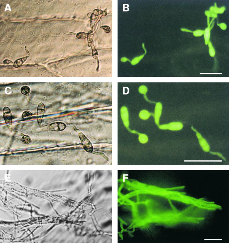
Plant infection by the rice blast fungus Magnaporthe grisea is brought about by the action of specialized infection cells called appressoria. These infection cells generate enormous turgor pressure, which is translated into an invasive force that allows a narrow penetration hypha to breach the plant cuticle. The Magnaporthe pde1 mutant was identified previously by restriction enzyme-mediated DNA integration mutagenesis and is impaired in its ability to elaborate penetration hyphae. Here we report that the pde1 mutation is the result of an insertion into the promoter of a P-type ATPase-encoding gene. Targeted gene disruption confirmed the role of PDE1 in penetration hypha development and pathogenicity but highlighted potential differences in PDE1 regulation in different Magnaporthe strains. The predicted PDE1 gene product was most similar to members of the aminophospholipid translocase group of P-type ATPases and was shown to be a functional homolog of the yeast ATPase gene ATC8. Spatial expression studies showed that PDE1 is expressed in germinating conidia and developing appressoria. These findings implicate the action of aminophospholipid translocases in the development of penetration hyphae and the proliferation of the fungus beyond colonization of the first epidermal cell.


Figures

Figure 1.
Figure 1.
Infection of Barley by Wild-Type and pde1 Mutant Strains of Magnaporthe. Conidia were inoculated on excised barley leaves (cv Golden Promise) and incubated at 24°C for 72 hr. Leaves were plunge frozen in nitrogen slush and then fractured at low temperature on the stage of a scanning electron microscope to reveal internal detail. Bars = 25 μm. (A) Barley leaf inoculated with the wild-type Magnaporthe strain 35-R-24. An appressorium (Ap) is visible on the leaf surface, and infection hyphae are present throughout the epidermal and mesophyll cell layers (arrowheads). (B) Barley leaf inoculated with pde1 mutant 2029. An appressorium is visible on the leaf surface (Ap) but has not developed a penetration hypha. Approximately 90% of the appressoria observed showed this appearance after 72 hr. (C) Barley leaf inoculated with pde1 mutant 2029. An appressorium (Ap) has penetrated the leaf and formed an infection hypha that has arrested growth within the first epidermal cell.
Figure 2.
Figure 2.
Organization of the PDE1 Locus and Targeted Gene Disruption. (A) Restriction map of the insertional mutant locus of pde1 mutant 2029. A copy of plasmid pCB1003 was introduced into Magnaporthe by REMI to create the hygromycin B–resistant mutant 2029 (Balhadère et al., 1999). The integration resulted in loss of the BamHI sites at each end of the integrated plasmid. The position and orientation of the hygromycin B resistance gene cassette (Carroll et al., 1994) are indicated (HPH). pCB1003 integration was found to have occurred 165 bp upstream of a large open reading frame (gray shading). ClaI sites found on either side of the integrated plasmid were used to excise a fragment of the insertion locus. (B) Restriction map of the PDE1 locus isolated as a 6.94-kb PstI-SalI fragment from a Magnaporthe strain Guy11 genomic library. The PDE1 locus contains a 4575-bp open reading frame interrupted by a single intron at positions 3927 to 3997. The arrow indicates the orientation of the open reading frame and the position of the intron. (C) Targeted gene disruption vector pPde1::Hph. The vector was made by excising a 2.6-kb PstI fragment at the 5′ end of PDE1 and inserting the hygromycin B resistance gene cassette (HPH) in a SmaI site. (D) Restriction map of the pde1::Hph disruption allele showing the position of the hygromycin resistance gene cassette (HPH) at the 5′ end of the PDE1 open reading frame. (E) DNA gel blot analysis of pPde1::Hph transformants. Genomic DNA was prepared from the wild-type strain Guy11 (lane 1), the ectopic integration transformant PV9 (lane 2), and four pde1::Hph mutant transformants, PV1, PV2, PV3, and PV4 (lanes 3 to 6, respectively). Genomic DNA was digested with PstI and separated on a 0.8% agarose gel. The blot was probed with pSKPS1. The Xs between (B) and (C) indicate a crossover event. B, BamHI; C, ClaI; P, PstI; Sa, SalI; Sm, SmaI; X, XhoI.
Figure 3.
Figure 3.
Appressorium-Mediated Penetration and Plant Infection Assays. Appressorium-mediated penetration assays were performed using a modification of the method of Chida and Sisler (1987). Magnaporthe conidial suspensions were incubated on sterilized onion epidermis or intact barley and rice epidermis, and the percentage of appressoria that had penetrated the cuticle by elaboration of a penetration hypha was recorded after 31 hr for onion epidermis and after 24 and 48 hr for barley and rice epidermis. For plant infection assays, barley seedlings (cv Golden Promise) and rice seedlings (cv CO-39) were spray inoculated with conidial suspensions of Magnaporthe, and representative leaves were collected 72 hr after inoculation. (A) Bar charts showing the percentage of appressorium penetration. 1, inoculation with wild-type strain 35-R-24; 2, pde1 mutant 2029-32; 3, pPDE1 transformant PV6.1; 4, Guy11; 5, pde1::Hph mutant PV1. (B) Rice leaf from a plant inoculated with wild-type strain Guy11. (C) Rice leaf inoculated with a pde1::Hph mutant of Guy11, PV1. (D) Barley leaf from a plant inoculated with wild-type strain Guy11. (E) Barley leaf inoculated with a pde1::Hph mutant of Guy11, PV1. (F) Bar chart showing the results of quantitative analysis of barley and rice infection assays. Mean lesion density values were recorded from 10 randomly chosen 5-cm leaf tips. 1, inoculation of barley with Guy11; 2, inoculation of barley with the pde1Δ mutant of Guy11, PV1; 3, inoculation of rice with Guy11; 4, inoculation of rice with the pde1Δ mutant, PV1. Bars indicate ±sd.
Figure 4.
Figure 4.
Predicted Amino Acid Sequence of the Magnaporthe PDE1 Gene. Amino acid sequence alignment of the predicted Magnaporthe PDE1 gene product with S. cerevisiae ATC8. Sequences were aligned with the ClustalW program (Thompson et al., 1994). Identical amino acids are shown on a black background, and similar amino acids are shown on a light gray background. Gaps in the sequences are indicated by dashes. PDE1 has GenBank accession number AY026257.
Figure 5.
Figure 5.
Predicted Topology of the Magnaporthe PDE1 and S. cerevisiae ATC8 Gene Products and Phylogenetic Tree of P-Type ATPases. (A) The PredictProtein program (Rost et al., 1995) was used to make predictions of the transmembrane helices and topology of the putative PDE1 and ATC8 gene products. Transmembrane spans are indicated by thick black lines. Putative cytoplasmic domains are shown in gray, and extracytoplasmic regions are shown in white. The P-type consensus motifs are indicated by thin black lines: 1, LTGET motif; 2, DKTGTLT phosphorylation site; 3, KGA nucleotide binding site; 4, mLTGD ATP binding motif; 5, GDGXND hinge motif (Catty et al., 1997). Bar = 100 amino acids. (B) Strict consensus phylogram of the most parsimonious tree showing relationships between P-type ATPases based on amino acid sequence. Relationships were determined by maximum parsimony using the heuristic search program with branch swapping and total branch recombination in PAUP version 4.0 (Swofford, 2000). Branch strengths were tested by 100 repetitions of the bootstrap algorithm with branch swapping. Numbers in parentheses are percentage bootstrap values. DRS2 aminophospholipid translocase family ATPases are shown in red: Mm AT1A, Mus musculus chromaffin granule ATPase II AT1A (SWISS-PROT accession number P70704); At F23H11.14, Arabidopsis thaliana putative ATPase F23H11.14 (GenBank accession number AC007258); Sc ATC8, Saccharomyces cerevisiae ATC8 (SWISS-PROT accession number Q12674); Mg PDE1, Magnaporthe grisea PDE1 (this study); Sc DRS2, S. cerevisiae DRS2 (SWISS-PROT accession number P39524); Sc ATC4, S. cerevisiae ATC4 (SWISS-PROT accession number Q12675); Sc ATC5, S. cerevisiae ATC5 (SWISS-PROT accession number P32660); Sc ATC7, S. cerevisiae ATC7 (SWISS-PROT accession number P40527). P1-ATPases are shown in green: Sc ATU1, S. cerevisiae copper-transporting ATPase ATU1 (SWISS-PROT accession number P38360); Sc ATU2, S. cerevisiae copper-transporting ATPase ATU2 (SWISS-PROT accession number P38995). H+-ATPases are shown in blue: Sc PMA2, S. cerevisiae plasma membrane proton ATPase PMA2 (SWISS-PROT accession number P19657); Nc PMA1, Neurospora crassa plasma membrane proton ATPase PMA1 (SWISS-PROT accession number P07038). Ca2+-ATPases are shown in pink: An PMRA, Aspergillus niger secretory pathway calcium ATPase PMRA (GenBank accession number AF232827); Sc ATC1, S. cerevisiae calcium-transporting ATPase ATC1 (SWISS-PROT accession number P13586). Na+-ATPases are shown in dark yellow: Sc ATN1, S. cerevisiae sodium-transporting ATPase ATN1 (SWISS-PROT accession number P13587). P4-ATPases are shown in turquoise: Sc ATC 6, S. cerevisiae cation-transporting ATPase ATC6 (SWISS-PROT accession number P39986); Sc ATC9, S. cerevisiae cation-transporting ATPase ATC9 (SWISS-PROT accession number Q12697).
Figure 6.
Figure 6.
The Magnaporthe PDE1 Gene Is a Functional Homolog of the S. cerevisiae P-Type ATPase Gene ATC8. Growth of S. cerevisiae on glucose- or galactose-amended growth medium plates. Plate cultures were inoculated with 10-μL droplets containing 2 × 10⁴, 104, 5 × 10³, and 103 cells and were left to grow for 17 hr before examination. Triangles show the decreasing concentration of inoculum. Strain YB322 is a wild-type S. cerevisiae strain. YB853 is an atc8Δ P-type ATPase mutant. Strain PDEC2 is a transformant of YB853 expressing Magnaporthe PDE1 under the control of the S. cerevisiae galactose-inducible GAL1 promoter.
Figure 7.
Figure 7.
Alignment of the Predicted Amino Acid Sequence of PDE1 Alleles from Magnaporthe Strains Guy11 and 35-R-24. Sequences were aligned with the ClustalW program (Thompson et al., 1994). Identical amino acids are shown on a black background, and similar amino acids are shown on a light gray background. A high degree of amino acid conservation is present. In total, 45 nucleotide differences were observed between the alleles within the 4.6-kb region of the PDE1 gene. This resulted in five nonsilent mutations, as indicated. All of the nonsilent mutations reside in the region corresponding to the large cytoplasmic loop between transmembrane domains 4 and 5. Each of the five conserved P-type ATPase consensus sequences is conserved in each allele. The 35-R-24 PDE1 allele has GenBank accession number AF408935.
Figure 8.
Figure 8.
Spatial Regulation of PDE1 Expression during Plant Infection and Vegetative Growth by Magnaporthe. Conidia of a PDE1(p)::sGFP::Hph transformant PVG3 were incubated on sterile onion epidermis and allowed to form appressoria. Micrographs in (A), (C), and (E) at left are bright-field images viewed with Hoffman modulation optics (Nikon). GFP fluorescence is shown in (B), (D), and (E) at right. (A) and (B) Germinated conidia with extended germ tubes 4 hr after inoculation. (C) and (D) Conidia with fully formed appressoria 24 hr after inoculation. (E) and (F) Hyphae grown in standard minimal medium for 48 hr. Bars = 50 μm for all panels.

References

    1. Adachi, K., and Hamer, J.E. (1998). Divergent cAMP signaling pathways regulate growth and pathogenesis in the rice blast fungus Magnaporthe grisea. Plant Cell 10 1361–1373. - PMC - PubMed
    1. Adams, T.H., Wieser, J.K., and Yu, J.H. (1998). Asexual sporulation in Aspergillus nidulans. Microbiol. Mol. Biol. Rev. 62 35–54. - PMC - PubMed
    1. Altschul, S.F., Gish, W., Miller, W., Myers, C.W., and Lipman, D.L. (1990). Basic local alignment search tool. J. Mol. Biol. 215 403–410. - PubMed
    1. Ashrafi, K., Farazi, T.A., and Gordon, J.I. (1998). A role for Saccharomyces cerevisiae fatty acid activation protein 4 in regulating protein N-myristoylation during entry into stationary phase. J. Biol. Chem. 273 25864–25874. - PubMed
    1. Balhadère, P.V., Foster, A.J., and Talbot, N.J. (1999). Identification of pathogenicity mutants of the rice blast fungus Magnaporthe grisea by insertional mutagenesis. Mol. Plant-Microbe Interact. 12 129–142.

Publication types

MeSH terms

Substances

LinkOut - more resources