. 2001 Sep;13(9):2085-98.
doi: 10.1105/tpc.010113.

Functional analysis of the conserved domains of a rice KNOX homeodomain protein, OSH15

Affiliations

Functional analysis of the conserved domains of a rice KNOX homeodomain protein, OSH15

H Nagasaki et al. Plant Cell. 2001 Sep.

Abstract

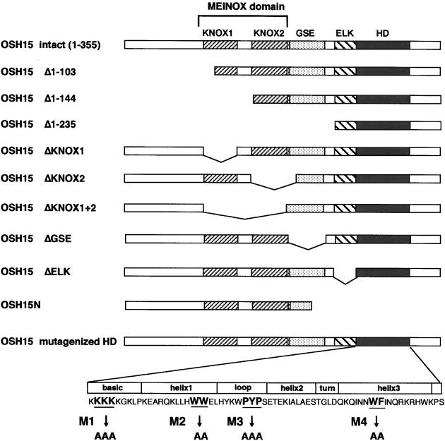
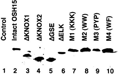
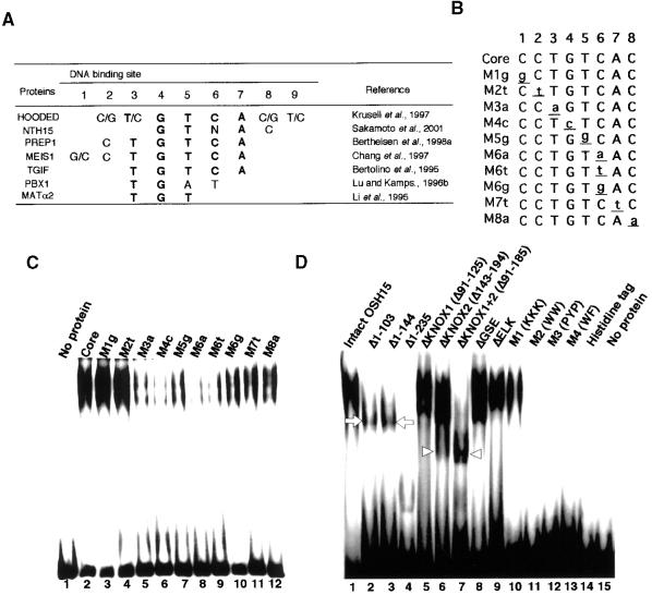
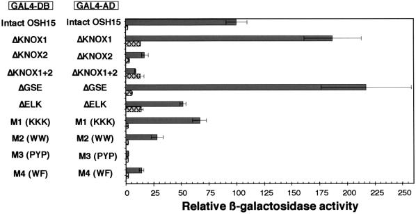
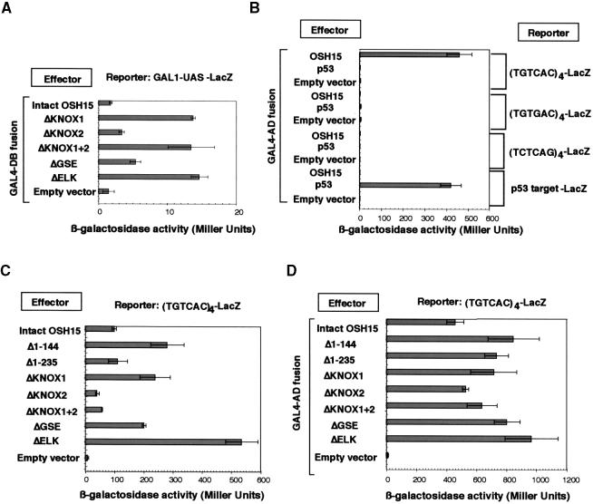
The rice KNOX protein OSH15 consists of four conserved domains: the MEINOX domain, which can be divided into two subdomains (KNOX1 and KNOX2); the GSE domain; the ELK domain; and the homeodomain (HD). To investigate the function of each domain, we generated 10 truncated proteins with deletions in the conserved domains and four proteins with mutations in the conserved amino acids in the HD. Transgenic analysis suggested that KNOX2 and HD are essential for inducing the abnormal phenotype and that the KNOX1 and ELK domains affect phenotype severity. We also found that both KNOX2 and HD are necessary for homodimerization and that only HD is needed for binding of OSH15 to its target sequence. Transactivation studies suggested that both the KNOX1 and ELK domains play a role in suppressing target gene expression. On the basis of these findings, we propose that overproduced OSH15 probably acts as a dimer and may ectopically suppress the expression of target genes that induce abnormal morphology in transgenic plants.


Figures

Figure 1.
Figure 1.
Scheme of the Mutagenized OSH15 Proteins. The full-length OSH15 (amino acids 1 to 355) contains four putative functional domains that are conserved in plant KNOX proteins: the MEINOX domain (which can divided into two subdomains, KNOX1 and KNOX2); the GSE domain; the ELK domain; and the HD. Deletions from the N- or C-terminal ends and internal deletions of each domain are indicated. In the HD, four sites of highly conserved amino acid residues in the basic, helix1, loop, and helix3 regions were replaced with alanine residues (M1, M2, M3, and M4, respectively).
Figure 2.
Figure 2.
Scheme of the Lamina Joint Region of Rice Leaf in the Wild- Type Plant. (A) Abaxial view. (B) Adaxial view.
Figure 3.
Figure 3.
Typical Phenotypes of Rice Leaves in Transgenic Plants That Overproduce the OSH15 Derivatives. (A) and (G) Wild-type phenotype (I): adaxial (A) and abaxial (G) views show the lamina joint region of a developed leaf. (B) and (H) Asymmetry phenotype (II): abaxial (B) and adaxial (H) views around the lamina joint region. The arrows in (B) indicate the split ligule, and the arrowheads in (H) indicate asymmetrical formation of auricles. (C) and (I) Knot phenotype (III): close-up (C) and abaxial (I) views of a leaf blade with a knot. (D) and (J) Ligule-less phenotype (IV): adaxial (D) and abaxial (J) views around the putative lamina joint region. There was no typical lamina joint, only the development of malformed auricles indicated by the arrow in (J). (E) and (K) Blade-less phenotype (V): whole plant (E) and abaxial (K) views of a blade-less leaf. (F) and (L) Multiple shoot phenotype (VI). Bars in (A) to (E) and (G) to (J) = 5 mm; bars in (F), (K), and (L) = 1 mm.
Figure 4.
Figure 4.
Morphology of Blade-Less Leaves. (A) Close-up view of the top of a blade-less leaf. The arrow indicates a tongue-like pale green organ. (B) Boundary region between the tongue-like organ and the leaf sheath of a blade-less leaf. The arrow indicates the auricle-like organ. (C) Close-up view of the auricle of a wild-type plant. (D) Transverse section of the leaf from a blade-less plant. (E) Transverse section of the leaf sheath from a wild-type plant. Bars = 1 mm.
Figure 5.
Figure 5.
Immunodetection of OSH15 Derivative Proteins. Total protein was extracted from 10 hygromycin-resistant calli transformed with the intact OSH15 transformants (lane 2), ΔKNOX1 (lane 3), ΔKNOX2 (lane 4), ΔGSE (lane 5), ΔELK (lane 6), M1 (lane 7), M2 (lane 8), M3 (lane 9), M4 (lane 10), or the pBI empty vector (lane 1) as a negative control. Twenty micrograms of total protein was subjected to SDS-PAGE, electroblotted onto nitrocellulose membrane, and probed with antiserum against OSH15.
Figure 6.
Figure 6.
Subcellular Localization of OSH15 Derivatives in Onion Cells in a Transient Assay. (A) Intact OSH15-GFP. (B) ΔKNOX1+2-GFP. (C) ΔELK-GFP. (D) OSH15N-GFP. (E) PEPC-GFP. Each panel shows a confocal image. Bars = 50 μm.
Figure 7.
Figure 7.
DNA Binding Property of OSH15. (A) Target sequences of TALE HD proteins that have been reported. Most TALE HD proteins share the same target sequence (i.e., the TGTCAC motif). (B) The TGTCAC motif and its derivative sequences. Oligonucleotides containing these sequences were used in this study. (C) Recombinant histidine-tagged OSH15 was subjected to EMSA using different DNA probes as indicated above each lane. Probe sequences are shown in (B). (D) EMSA was performed using histidine-tagged OSH15 derivatives, as indicated above the gel lanes. Both the histidine tag and no protein were used as negative controls. The arrowheads indicate the positions of the faster mobility bands (lanes 6 and 7), in contrast to the shifted bands with slower mobility, which are indicated by arrows (lanes 2 and 3).
Figure 8.
Figure 8.
Interaction between OSH15 and Rice KNOX Proteins. (A) Quantitative yeast two-hybrid assay with OSH15 and rice KNOX (OSH) proteins. For the bait construct, OSH15 was fused to the GAL4 DNA binding domain (GAL4-DB). For the prey constructs, OSH proteins were fused to the GAL4 activation domain (GAL4-AD). The pairwise combination of the bait and prey constructs for each row is shown at left. Relative β-galactosidase (LacZ) activity was calculated by standardizing each LacZ activity with that of intact OSH15/OSH15. We set the activity of OSH15/OSH15 at 100. Error bars represent the standard deviations calculated from three independent yeast clones. The combinations of OSH15–empty vector and OSH15–T antigen are negative controls of interaction, whereas the combination of p53–T antigen is a positive control. (B) In vitro pull-down assay with 35S-methionine–labeled OSH15. The fusion proteins consisted of OSH proteins and GST: GST-OSH1 (lane 3); GST-OSH15 (lane 4); GST-OSH43 (lane 5); GST-OSH6 (lane 6); and GST-OSH71 (lane 7). The input protein, 35S-methionine–labeled OSH15, was loaded onto the gel alone as a control (lane 1). GST alone was used as negative control for interaction (lane 2).
Figure 9.
Figure 9.
Homodimer Formation of OSH15 in a Quantitative Yeast Two-Hybrid Assay. Homodimer formation of OSH15 derivatives in yeast. Shown at left is the pairwise combination of OSH15 derivatives. The shaded bars show the relative LacZ activity of each pairwise combination of the OSH15 derivatives. The cross-hatched bars show the relative LacZ activity of GAL4-DB fused with each OSH15 derivative protein alone used as negative control. Error bars represent standard deviations. Relative LacZ activity was calculated by standardizing each LacZ activity with that of intact OSH15/OSH15. We set the activity of OSH15/OSH15 at 100.
Figure 10.
Figure 10.
Transcriptional Activity of OSH15 Derivatives. (A) Transcriptional activity only of GAL4-DB–fused OSH15 derivatives in yeast. GAL1-UAS stands for the enhancer-like sequence controlled by GAL4-DB. (B) DNA binding activity of GAL4-AD–fused OSH15 in yeast. Each reporter gene ([TGTCAC]4-LacZ; [TGTGAC]4-LacZ; [TCTCAG]4-LacZ; or p53 target–LacZ) was introduced into yeast strain YM4271. GAL4-AD–fused OSH15 was introduced into each yeast strain sequentially. The empty vector or GAL4-AD–fused p53 served as a negative control, whereas the combination of GAL4-AD–fused p53 with the p53 target–LacZ reporter was used as a positive control. (C) Transcriptional activity of OSH15 derivatives depending on their DNA binding context in yeast. OSH15 derivatives were introduced into the (TGTCAC)4-LacZ reporter strain. The empty vector served as a negative control. (D) DNA binding activity of GAL4-AD–fused OSH15 derivatives in yeast. GAL4-AD–fused OSH15 derivatives were introduced into the (TGTCAC)4-LacZ reporter strain. The empty vector served as a negative control. The bars show the LacZ activity, presented as the measured value in Miller units, of each of the effector proteins. Error bars represent standard deviations.

References

    1. Abu-Shaar, M., Ryoo, H.D., and Mann, R.S. (1999). Control of the nuclear localization of Extradenticle by competing nuclear import and export signals. Genes Dev. 13 935–945. - PMC - PubMed
    1. Affolter, M., Schier, A., and Gehring, W.J. (1990). Homeodomain proteins and regulation of gene expression. Curr. Opin. Cell Biol. 2 485–495. - PubMed
    1. Berthelsen, J., Zappavigna, V., Ferretti, E., Mavilio, F., and Blasi, F. (1998. a). The novel homeoprotein Prep1 modulates Pbx-Hox protein cooperativity. EMBO J. 17 1434–1445. - PMC - PubMed
    1. Berthelsen, J., Zappavigna, V., Mavilio, F., and Blasi, F. (1998. b). Prep1, a novel functional partner of Pbx proteins. EMBO J. 17 1423–1433. - PMC - PubMed
    1. Berthelsen, J., Kilstrup-Nielsen, C., Blasi, C., Mavilio, F., and Zappavigna, V. (1999). The subcellular localization of PBX1 and EXD proteins depends on nuclear import and export signals and is modulated by association with PREP1 and HTH. Genes Dev. 13 946–953. - PMC - PubMed

Publication types

MeSH terms