. 2001 Sep;13(9):2099-114.
doi: 10.1105/tpc.010098.

The Arabidopsis TT2 gene encodes an R2R3 MYB domain protein that acts as a key determinant for proanthocyanidin accumulation in developing seed

Affiliations

The Arabidopsis TT2 gene encodes an R2R3 MYB domain protein that acts as a key determinant for proanthocyanidin accumulation in developing seed

N Nesi et al. Plant Cell. 2001 Sep.

Abstract

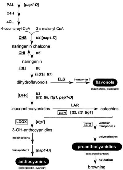
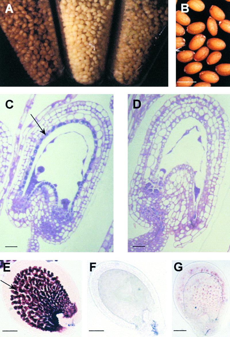
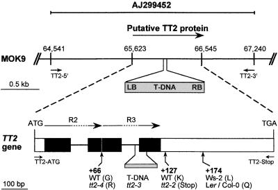
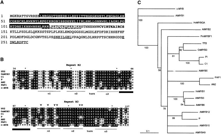
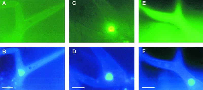
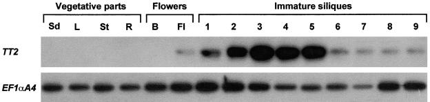
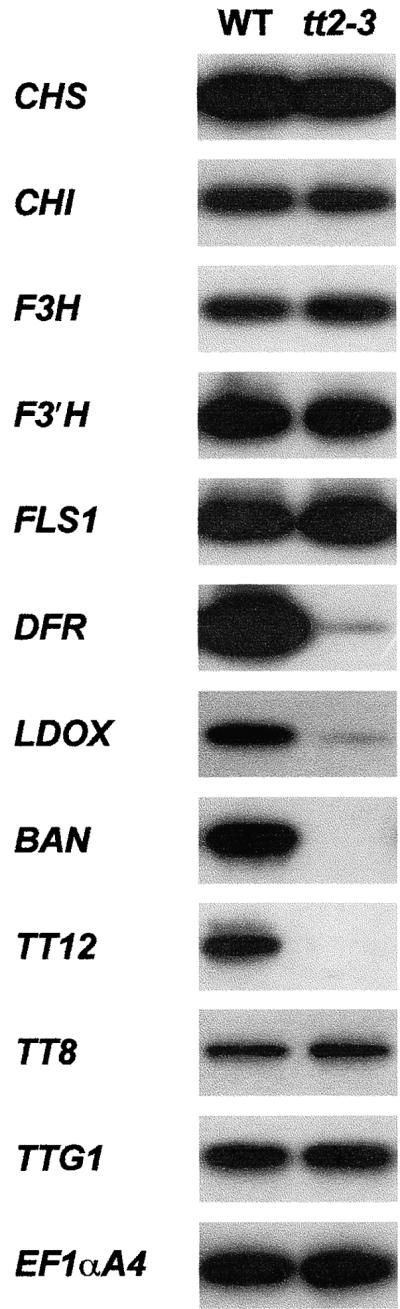
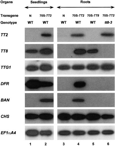
In Arabidopsis, proanthocyanidins specifically accumulate in the endothelium during early seed development. At least three TRANSPARENT TESTA (TT) genes, TT2, TT8, and TTG1, are necessary for the normal expression of several flavonoid structural genes in immature seed, such as DIHYDROFLAVONOL-4-REDUCTASE and BANYULS (BAN). TT8 and TTG1 were characterized recently and found to code for a basic helix-loop-helix domain transcription factor and a WD-repeat-containing protein, respectively. Here the molecular cloning of the TT2 gene was achieved by T-DNA tagging. TT2 encoded an R2R3 MYB domain protein with high similarity to the rice OsMYB3 protein and the maize COLORLESS1 factor. A TT2-green fluorescent protein fusion protein was located mostly in the nucleus, in agreement with the regulatory function of the native TT2 protein. TT2 expression was restricted to the seed during early embryogenesis, consistent with BAN expression and the proanthocyanidin deposition profile. Finally, in gain-of-function experiments, TT2 was able to induce ectopic expression of BAN in young seedlings and roots in the presence of a functional TT8 protein. Therefore, our results strongly suggest that stringent spatial and temporal BAN expression, and thus proanthocyanidin accumulation, are determined at least partially by TT2.


Figures

Figure 1.
Figure 1.
Scheme of the Flavonoid Biosynthetic Pathway of Arabidopsis Leading to the Synthesis of Anthocyanins, Flavonols, and Proanthocyanidins. Enzymes are indicated in uppercase letters, with the corresponding genetic loci given in lowercase italic letters. Mutants for regulatory genes are indicated in brackets. Enzymes encoded by flavonoid EBGs are underlined, and those encoded by flavonoid LBGs are boxed. Abbreviations are as follows: ban, banyuls; CHI, chalcone isomerase; CHS, chalcone synthase; C4H, cinnamate 4-hydroxylase; 4CL, 4-coumarate:CoA ligase; DFR, dihydroflavonol 4-reductase; F3H, flavanone 3-hydroxylase; F3′H, flavonoid 3′-hydroxylase; FLS, flavonol synthase; LAR, leucoanthocyanidin reductase; LDOX, leucoanthocyanidin dioxygenase; PAL, phenylalanine ammonia-lyase; pap1-D, production of anthocyanin pigment1-dominant; tt, transparent testa; ttg1, transparent testa glabrous1.
Figure 2.
Figure 2.
Seed Phenotypes. (A) Seed from wild-type plants and from tt2-3 and tt2-2 mutants (left to right). Seed of the tt2-3 mutant are golden yellow, whereas the tt2-2 mutation gives the seed a buff color. (B) T2 seed from a tt2-3 homozygous mutant transformed with the TT2 cDNA under the control of a double enhanced 35S promoter. The brown pigmentation of transgenic seed demonstrates the phenotypic complementation of the tt2-3 mutation by overexpression of the TT2 cDNA. Bar = 450 μm. (C) and (D) Localization of phenolic compounds in the testa of immature seed from a wild-type plant (C) and the tt2-3 mutant (D) after staining with toluidine blue. The flavonoids, stained blue, form granules that are localized in vacuoles of the endothelium cells in the wild-type seed coat (arrow in [C]). (D) shows the absence of blue granules in the tt2-3 endothelium. Bars = 20 μm. (E) to (G) Detection of proanthocyanidins and their precursors in immature seed treated with vanillin HCl. The vanillin test stains the proanthocyanidins and their precursors (leucoanthocyanidins and catechins) red in the endothelium of the wild type (arrow in [E]). The striking difference is the complete absence of these compounds in tt2-3 (F), whereas a slight red color is observed in the tt2-2 seed coat (G). Bars = 50 μm.
Figure 3.
Figure 3.
Isolation of the TT2 Gene. Top, physical map of the TT2 genomic region on the P1 clone MOK9 (chromosome 5). The arrow indicates the orientation of the putative TT2 protein. The T-DNA insertion is shown in the gray box. Numbers above the scheme indicate nucleotide positions on the MOK9 sequence (in base pairs). Bottom, exon-intron structure of the Arabidopsis TT2 gene. Boxes represent exons and lines represent introns. The region encoding the R2R3 MYB DNA binding domain is shown by black boxes. Numbering of amino acid residues is given from the predicted translation start codon and is shown in boldface below the scheme. The T-DNA copy (7 kb) in tt2-3 is inserted between intron 2 and exon 3 and causes a 45-bp deletion. Mutations in tt2-2 and tt2-4 are localized, with amino acid substitutions indicated in parentheses. Position 174 shows an amino acid polymorphism between Arabidopsis ecotypes. The different primers and probes used for molecular analyses are noted. Col-0, Columbia-0; LB, left border; Ler, Landsberg erecta; RB, right border; R2, repeat 2; R3, repeat 3; Ws-2, Wassilewskija-2; WT, wild type.
Figure 4.
Figure 4.
TT2 Shows Features of an R2R3 MYB DNA Binding Domain Protein. (A) Deduced amino acid sequence of TT2. The R2R3 MYB DNA binding domain is boxed in black. The amino acid residues conserved between the TT2 and OsMYB3 C-terminal halves are shown in boldface. The dotted line denotes the putative NLS found by the PSORT prediction program. The two putative α-helices found in the C-terminal sequence of TT2 are underlined. Numbers at left indicate amino acid positions (from the translation start codon). (B) Sequence comparison of the conserved MYB DNA binding domain of TT2 with other MYB-related proteins from rice, maize C1, maize P, petunia AN2, Arabidopsis PAP1, and human c-MYB (for GenBank accession numbers see Methods). Identical amino acids are boxed in black, and similar amino acids are boxed in gray. The dash in the c-MYB sequence indicates a gap introduced to perform the alignment. Asterisks denote the conserved W residues. Arrowheads indicate the amino acid residues in the C1 MYB domain that determine interaction with bHLH-related factors, according to Grotewold and co-workers (Grotewold et al., 2000). The three putative α-helices are noted below the diagram, and the closed box corresponds to the linker sequence between the R2 and R3 repeats. Amino acid residues in TT2 are numbered from the translation start codon. (C) Dendrogram of relationships among the R2R3 domains from several MYB-related proteins. For construction of the tree, we used only the R2R3 MYB domain sequence (104 amino acid residues; see [B]) of each selected MYB-related protein. The matrix of sequence similarities was calculated with the CLUSTAL program from the CLUSTAL X package (Thompson et al., 1997) and submitted to a neighbor-joining analysis to generate a branching pattern. The numbers below the branches indicate the percentage of bootstrap support after 1000 replicates. Nodes with bootstrap support of <50% were discarded. The human c-MYB sequence was included as an outgroup. The consensus tree was drawn using the TreeView program (version 1.5.3, Roderic D.M. Page, University of Glasgow, UK). Sequences used are human c-MYB, maize C1, maize Pl, maize P, rice OsMYB3, spruce PmMYBF1, barley HvMYBGA, barley HvMYB1, petunia AN2, Arabidopsis AtMYB1, Arabidopsis AtMYB2, Arabidopsis AtMYB3, Arabidopsis AtMYB5, Arabidopsis AtMYB6, Arabidopsis AtMYB12, Arabidopsis AtMYB15, Arabidopsis AtMYB49, Arabidopsis PAP1, and Arabidopsis TT2.
Figure 5.
Figure 5.
Targeting of the TT2-GFP Fusion Protein to Arabidopsis Cell Nuclei. Shown are trichome cells from 2-week-old Arabidopsis plants. The positions of the nuclei in (A), (C), and (E) are deduced from the comparison with UV light images after 4′,6-diamidino-2-phenylindole staining shown below in (B), (D), and (F), respectively. (A) and (B) Trichome cells of a wild-type nontransformed Arabidopsis plant used as a negative control. (C) and (D) Trichome cells of a TT2-GFP–expressing Arabidopsis transgenic plant. (E) and (F) Trichome cells of a GFP-expressing Arabidopsis transgenic plant. Bars = 50 μm for (A) to (F).
Figure 6.
Figure 6.
Expression Pattern of the TT2 Gene. The accumulation of TT2 transcript was measured by quantitative RT-PCR with RNA from 4-day-old seedlings (Sd), rosette leaves (L), stems (St), 10-day-old roots (R), flower buds (B), flowers (Fl), and immature siliques at different developmental stages (1 to 9) as indicated. Silique samples were numbered according to the prevailing embryo stage they contained: 1, one cell; 2, one to four cells; 3, early globular to globular; 4, heart; 5, late heart to torpedo; 6, late torpedo to curled cotyledons; 7, late curled cotyledons; 8, green cotyledons; 9, mature embryo. The expression profile of the Arabidopsis EF1αA4 gene was determined as an mRNA loading control.
Figure 7.
Figure 7.
TT2 Is Essential for the Activation of Flavonoid LBGs. Transcripts for five flavonoid EBGs (CHS, CHI, F3H, F3H, and FLS1), four flavonoid LBGs (DFR, LDOX, BAN, and TT12), two regulatory genes (TT8 and TTG1), and, as a control, EF1αA4 were detected by quantitative RT-PCR in immature siliques of wild-type (WT) and tt2-3 plants. In these experiments, siliques from approximately the unicellular to the torpedo stage of embryo development (stages 1 to 5 in Figure 6) were pooled and examined.
Figure 8.
Figure 8.
Gene Expression Analysis in Transgenic Plants That Overexpress TT2 or TT8. Transcripts for the TT2, TT8, TTG1, DFR, BAN, CHS, and EF1αA4 genes were detected by RT-PCR with RNA from 4-day-old seedlings (lanes 1 and 2) and 10-day-old roots (lanes 3 to 6) of wild-type (WT) nontransformed plants (lanes 1 and 3), wild-type plants harboring the 70S-TT2 transgene (lanes 2 and 4), wild-type plants harboring the 70S-TT8 transgene (lane 5), and tt8-3 plants harboring the 70S-TT2 transgene (lane 6). N, none.

References

    1. Aastrup, S., Outtrup, H., and Erdal, K. (1984). Location of the proanthocyanidins in the barley grain. Carlsberg Res. Commun. 49 105–109.
    1. Albert, S., Delseny, M., and Devic, M. (1997). BANYULS, a novel negative regulator of flavonoid biosynthesis in the Arabidopsis seed coat. Plant J. 11 289–299. - PubMed
    1. Bechtold, N., Ellis, J., and Pelletier, G. (1993). In planta Agrobacterium-mediated gene transfer by infiltration of adult Arabidopsis thaliana plants. C. R. Acad. Sci. Paris 316 1194–1199.
    1. Becker, D. (1990). Binary vectors which allow the exchange of plant selectable markers and reporter genes. Nucleic Acids Res. 18 203. - PMC - PubMed
    1. Borevitz, J.P., Xia, Y., Blount, J., Dixon, R.A., and Lamb, C. (2000). Activation tagging identifies a conserved MYB regulator of phenylpropanoid biosynthesis. Plant Cell 12 2383–2393. - PMC - PubMed

Publication types

MeSH terms