. 2001 Sep;13(9):2143-58.
doi: 10.1105/tpc.010114.

A 90-kD phospholipase D from tobacco binds to microtubules and the plasma membrane

Affiliations

A 90-kD phospholipase D from tobacco binds to microtubules and the plasma membrane

J C Gardiner et al. Plant Cell. 2001 Sep.

Abstract

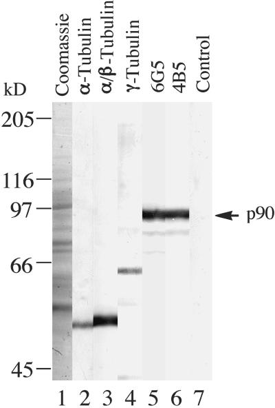
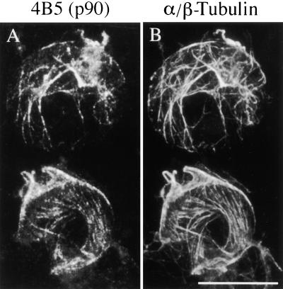
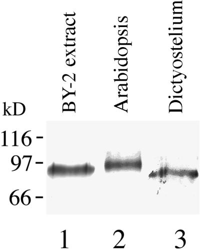
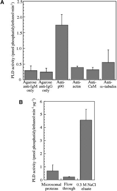
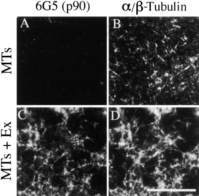
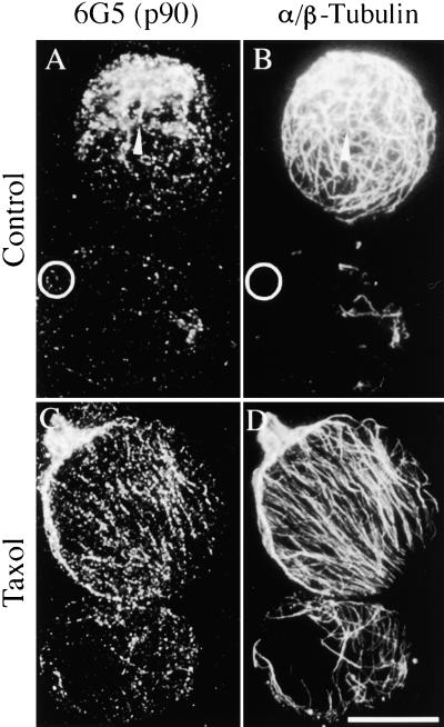
The organization of microtubule arrays in the plant cell cortex involves interactions with the plasma membrane, presumably through protein bridges. We have used immunochemistry and monoclonal antibody 6G5 against a candidate bridge protein, a 90-kD tubulin binding protein (p90) from tobacco BY-2 membranes, to characterize the protein and isolate the corresponding gene. Screening an Arabidopsis cDNA expression library with the antibody 6G5 produced a partial clone encoding phospholipase D (PLD), and a full-length gene was obtained by sequencing a corresponding expressed sequence tag clone. The predicted protein of 857 amino acids contains the active sites of a phospholipid-metabolizing enzyme and a Ca(2+)-dependent lipid binding domain and is identical to Arabidopsis PLD delta. Two amino acid sequences obtained by Edman degradation of the tobacco p90 are identical to corresponding segments of a PLD sequence from tobacco. Moreover, immunoprecipitation using the antibody 6G5 and tobacco BY-2 protein extracts gave significant PLD activity, and PLD activity of tobacco BY-2 membrane proteins was enriched 6.7-fold by tubulin-affinity chromatography. In a cosedimentation assay, p90 bound and decorated microtubules. In immunofluorescence microscopy of intact tobacco BY-2 cells or lysed protoplasts, p90 colocalized with cortical microtubules, and taxol-induced microtubule bundling was accompanied by corresponding reorganization of p90. Labeling of p90 remained along the plasma membrane when microtubules were depolymerized, although detergent extraction abolished the labeling. Therefore, p90 is a specialized PLD that associates with membranes and microtubules, possibly conveying hormonal and environmental signals to the microtubule cytoskeleton.


Figures

Figure 1.
Figure 1.
Immunoblot Detection of p90 in Tobacco BY-2 Membrane Proteins. Membrane proteins, prepared by extracting microsomal fractions of tobacco BY-2 cells with the detergent 3-([3-cholamidopropyl]dimethylamino)-1-propanesulfonate (Chaps), were separated by electrophoresis on 8% (w/v) SDS–polyacrylamide gels, transferred onto a nitrocellulose membrane, and probed with antibodies. Lane 1, Coomassie blue–stained gel; lane 2, monoclonal antibody against α-tubulin; lane 3, polyclonal serum against soybean α/β-tubulin; lane 4, peptide serum against a conserved region of γ-tubulin; lane 5, monoclonal antibody 6G5; lane 6, monoclonal antibody 4B5; lane 7, control using a preheated antibody 6G5. Immunoreactions were detected with alkaline phosphatase. The arrow at right indicates the position of the immunoreactive 90-kD polypeptide (p90). Bars at left indicate the positions of molecular mass standards.
Figure 2.
Figure 2.
p90 Colocalizes with Cortical MTs in Protoplasts. Tobacco BY-2 protoplasts were processed for immunofluorescence microscopy by using mouse monoclonal antibody 4B5 together with rabbit polyclonal serum against soybean α/β-tubulin, followed by a mixture of Texas Red–conjugated anti-mouse and fluorescein isothiocyanate (FITC)-conjugated anti-rabbit secondary antibodies. (A) p90 visualized with the antibody 4B5 and Texas Red–conjugated secondary antibody. (B) α/β-Tubulin visualized with serum against soybean tubulin and FITC-conjugated secondary antibody. Bar in (B) = 10 μm for (A) and (B).
Figure 3.
Figure 3.
p90 Is Present in Plants and Lower Eukaryotes. Protein extracts from plant tissue and Dictyostelium were separated on 10% (w/v) SDS–polyacrylamide gels (10 to 20 μg protein/lane), transferred onto a nitrocellulose membrane, and probed with the 6G5 antibody followed by alkaline phosphatase detection. Lane 1, tobacco BY-2 Chaps extract; lane 2, Arabidopsis seedlings; lane 3, Dictyostelium. Bars at left indicate the positions of molecular mass standards.
Figure 4.
Figure 4.
Alignment of Amino Acid Sequences Encoded by PLD cDNA Clones from Arabidopsis and Tobacco. Shown is an alignment of Arabidopsis PLD98 (AtPLD98) with Arabidopsis PLDδ (AtPLDδ) and a partial cDNA clone of tobacco PLD (NtPLD) prepared using the CLUSTAL W algorithm (Thompson et al., 1994). Shaded boxes show sequence identity, and dashes indicate gaps introduced to optimize the sequence alignment. Putative functional domains are indicated by lines above the sequences as follows: C2, a Ca2+-dependent lipid binding domain; pas, phospholipid-metabolizing enzyme active sites. An amino acid microsequence from tobacco p90, BY2seq1, is highly homologous with AtPLD98 residues 314 to 328 and identical to the corresponding region in the tobacco PLD.
Figure 5.
Figure 5.
PLD Activity in Tobacco BY-2 Proteins. Protein samples were incubated with a reaction mixture containing fluorescent phosphatidylcholine and phosphatidylethanol, the products were separated by thin-layer chromatography, and the fluorescence of phosphatidylethanol was measured with a spectrophotometer. (A) PLD activity in immunoprecipitated pellets. A significant (P < 0.01) PLD activity was measured in pellets immunoprecipitated with antibody 6G5 against p90 and agarose-conjugated secondary antibody (anti-IgM). Only background levels of PLD activity were detected in pellets obtained with antibodies against actin, calmodulin (CaM), and α-tubulin, followed by agarose-conjugated secondary antibody (anti-IgG). The data show the mean values ±sem from at least three independent experiments for each point. (B) PLD activity in tobacco BY-2 protein fractions from tubulin-affinity chromatography. The PLD activity in detergent (Chaps) extract of tobacco BY-2 microsomes was depleted by ∼67% in the column flow-through fraction and enriched 6.7-fold in tubulin binding proteins eluted with 0.3 M NaCl. The data show the mean values ±sem obtained from one set of the three protein fractions, each analyzed by four independent PLD assays.
Figure 6.
Figure 6.
p90 Decorates MTs in Vitro. Taxol-stabilized neuronal MTs were incubated with a Chaps extract (Ex) of tobacco BY-2 membrane proteins and sedimented by centrifugation. The pelleted protein complexes were spread on microscope slides, fixed, and double labeled using mouse monoclonal antibody 6G5 together with rabbit serum against soybean α/β-tubulin, followed by a mixture of Cy3-conjugated anti-mouse ([A] and [C]) and FITC-conjugated anti-rabbit ([B] and [D]) secondary antibodies. (A) and (B) Control MTs incubated in buffer alone, showing the absence of labeling with 6G5 antibody. (C) and (D) MTs decorated with p90 after incubation with membrane proteins. Bar in (D) = 10 μm for (A) to (D).
Figure 7.
Figure 7.
Colocalization of p90 with Cortical MTs in Protoplasts Accompanies Taxol-Induced Reorganization. Protoplasts were prepared from tobacco BY-2 cells with or without preincubation in 10 μM taxol and then processed for immunofluorescence microscopy. The preparations were double labeled using antibody 6G5 together with rabbit serum against soybean α/β-tubulin, followed by a mixture of Cy3-conjugated anti-mouse ([A] and [C]) and FITC-conjugated anti-rabbit ([B] and [D]) secondary antibodies. (A) and (B) Control protoplasts prepared without taxol. p90 associates with MTs in a punctate manner, although some spots appear in MT-free areas of the protoplast ghost (circles). (C) and (D) Protoplasts with taxol-induced parallel MT arrays and matching punctate arrays of p90. Bar in (D) = 20 μm for (A) to (D).
Figure 8.
Figure 8.
p90 Associates with Protoplast Plasma Membrane in the Absence of MTs but Can Be Extracted with a Detergent. Tobacco BY-2 protoplasts, prepared after preincubation with 10 μM taxol, were treated with calcium and detergent and then processed for immunofluorescence microscopy using double labeling with antibodies against p90 and α/β-tubulin as in Figure 7. (A) and (B) Cortical MTs were removed from the protoplast ghosts by cold treatment in the presence of 5 mM CaCl2 for 30 min. Nonetheless, numerous randomly dispersed dots labeled with antibody 6G5 remained attached to the plasma membrane. (C) and (D) Protoplast ghosts were additionally extracted with 0.05% (w/v) Triton X-100 (TX-100) for 10 min. This treatment removed most of the p90 labeling. Bar in (D) = 20 μm for (A) to (D).
Figure 9.
Figure 9.
p90 Colocalizes with MT Arrays throughout the Cell Cycle. Tobacco BY-2 cells were fixed and processed for immunofluorescence microscopy by using double labeling with antibodies against p90 and α/β-tubulin as in Figure 7. At all stages of the cell cycle, the MT arrays were matched with punctate arrays of p90. (A) and (B) Interphase cortical array. (C) and (D) Preprophase band. (E) and (F) Metaphase spindle. (G) and (H) Cytokinetic phragmoplast. Bar in (H) = 20 μm for (A) to (H).

References

    1. Akashi, T., and Shibaoka, H. (1991). Involvement of transmembrane proteins in the association of cortical microtubules with the plasma membrane in tobacco BY-2 cells. J. Cell Sci. 98 169–174.
    1. Akashi, T., Kawasaki, S., and Shibaoka, H. (1990). Stabilization of cortical microtubules by the cell wall in cultured tobacco cells: Effects of extensin on the cold-stability of cortical microtubules. Planta 182 363–369. - PubMed
    1. Altschul, S.F., Gish, W., Miller, W., Myers, E.W., and Lipman, D.J. (1990). Basic local alignment search tool. J. Mol. Biol. 215 403–410. - PubMed
    1. Asada, T., and Collings, D. (1997). Molecular motors in higher plants. Trends Plant Sci. 2 29–37.
    1. Assmann, S.M., and Baskin, T.I. (1998). The function of guard cells does not require an intact array of cortical microtubules. J. Exp. Bot. 49 163–170.

Publication types

MeSH terms