Review
. 2001 Aug;48(6):403-75.
doi: 10.1046/j.1439-0450.2001.00473.x.

Viruses of lower vertebrates

Affiliations
Review

Viruses of lower vertebrates

S Essbauer et al. J Vet Med B Infect Dis Vet Public Health. 2001 Aug.

Abstract

Viruses of lower vertebrates recently became a field of interest to the public due to increasing epizootics and economic losses of poikilothermic animals. These were reported worldwide from both wildlife and collections of aquatic poikilothermic animals. Several RNA and DNA viruses infecting fish, amphibians and reptiles have been studied intensively during the last 20 years. Many of these viruses induce diseases resulting in important economic losses of lower vertebrates, especially in fish aquaculture. In addition, some of the DNA viruses seem to be emerging pathogens involved in the worldwide decline in wildlife. Irido-, herpes- and polyomavirus infections may be involved in the reduction in the numbers of endangered amphibian and reptile species. In this context the knowledge of several important RNA viruses such as orthomyxo-, paramyxo-, rhabdo-, retro-, corona-, calici-, toga-, picorna-, noda-, reo- and birnaviruses, and DNA viruses such as parvo-, irido-, herpes-, adeno-, polyoma- and poxviruses, is described in this review.
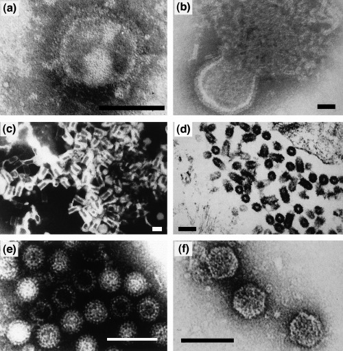
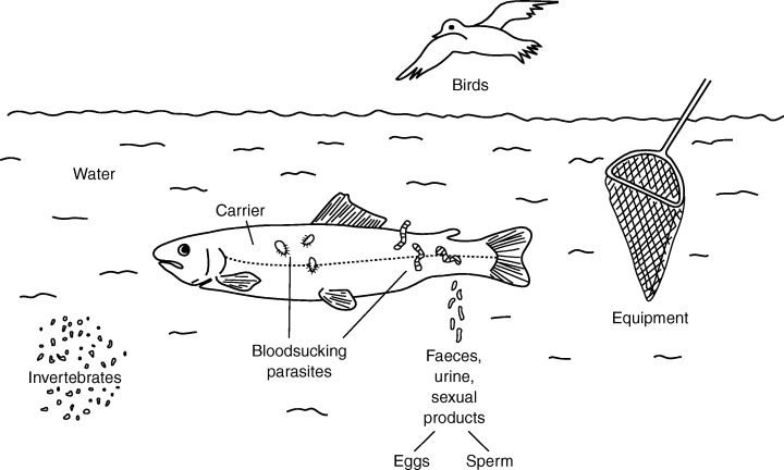
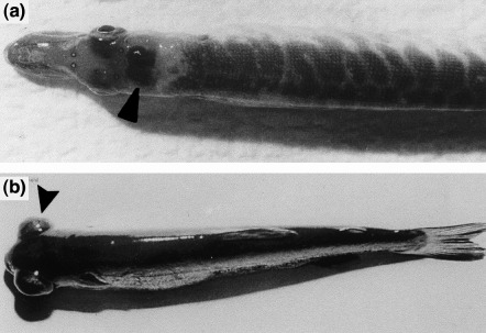
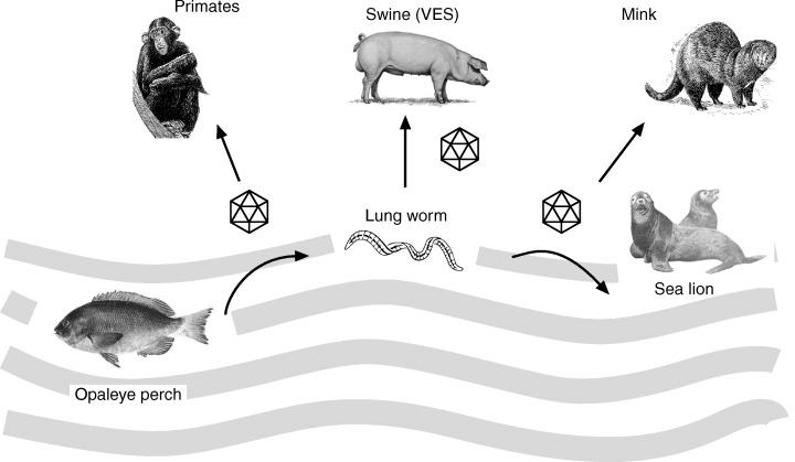

Figures

Figure 1
Figure 1
RNA viruses occurring in lower vertebrates. 1 Arthropod‐borne viruses termed ‘arboviruses’ in the review.
Figure 2
Figure 2
Electron microscopic images of some representative RNA viruses from poikilothermic animals: (a) orthomyxovirus‐like particles from eels (bar: 75 nm); (b) snake paramyxovirus (bar: 60 nm); (c) viral haemorrhagic septicaemia virus, VHSV (a rhabdovirus; bar: 60 nm); (d) spring viraemia of carp virus, SVCV (a rhabdovirus; bar: 150 nm); (f) grass carp reovirus (bar: 150 nm); (g) infectious pancreatic necrosis virus, IPNV (a birnavirus; bar: 80 nm). (a)–(c), (e) and (f) negative staining; (d) ultrathin section.
Figure 8
Figure 8
DNA viruses occurring in poikilothermic vertebrates.
Figure 9
Figure 9
Electron microscopy of DNA viruses detected in poikilothermic vertebrates: (a) corn snake parvovirus (bar: 100 nm); (b) tadpole oedema virus, TEV (a frog iridovirus; bar: 200 nm) (kindly provided by K. Wolf, USA); (c) European catfish iridovirus (ECV) budding from infected BF‐2 cells (bar: 250 nm); (d) toad herpesvirus (Pelobates fucus, bar: 75 nm) (F.T. Just, unpublished); (e) corn snake adenovirus (bar: 75 nm).
Figure 3
Figure 3
Phenogram showing the genetic relationships between 16 reptilian paramyxoviruses and Sendai virus, based on analysis of part of the L‐gene (nucleotides 9648–10 201). Bootstrap values >700 for the major branches are shown. Place of isolation is indicated by EUR (Europe)/USA, with the numbers following EUR/USA indicating years of isolation. Hosts: Bitis=Bitis sp.; Boa=Boa constrictor; Cro1–3=Crotalus spp. Trim=Trimeresurus spp.; Call=Callopistes maculatus; Both=Bothrops atrox; Ela 1–2=Elaphe guttata; Gono=Gonosoma oxycephala; Lamp=Lampropeltis sp.; Pyth=Python regius; More=Morelia argus (Ahne et al., 1999).
Figure 7
Figure 7
Transmission of fish viruses and their vectors.
Figure 5
Figure 5
Uptake and spreading of spring viraemia of carp virus (SVCV) in carp fry infected by bath exposure.
Figure 4
Figure 4
Fish infected with viral haemorrhagic septicaemia virus (VHSV): (a) pike fry showing bleeding in the brain (arrow); (b) rainbow trout fry showing exophthalmia (arrow).
Figure 6
Figure 6
Scheme showing multiple hosts of San Miguel sea lion virus (SMSV5–7), which crosses the water–land environment to affect different animal species.
Figure 10
Figure 10
Replication scheme for frog virus 3, FV‐3 (after Goorha, 1982). C=cytoplasm; N=nucleus.
Figure 11
Figure 11
Lymphocystis disease virus (LCDV) infection in flounder. (a) Skin of LCDV‐infected flounder exhibiting multiple lymphocystis nodules (see arrows). (b) Explantats of LCDV‐infected flounder skin: lymphocystis cells are round and hypertrophied (see arrow), and 1000 times the size of uninfected cells (× 22). (The fish was kindly provided by Dr Steinhagen, Faculty of Veterinary Medicine, Hannover, Germany.)
Figure 12
Figure 12
Phylogenetic tree of piscine and amphibian iridoviruses based on eIF‐2α homologous sequences. Three hundred and fifty nucleotides of frog virus 3 (FV‐3), European sheatfish virus (ESV), European catfish virus (ECV), epizootic haematopoietic necrosis virus (EHNV), and Rana esculenta iridovirus (REIR) eIF‐2α genes were compared with partial eIF‐2α genes of humans, Drosophila, and yeast, and complete K3L genes of Variola major virus, Vaccinia strain Copenhagen, and swine poxvirus. Figures on each branch represent percentage bootstrap support for maximum parsimony (S. Essbauer unpublished data).

References

    1. Adkison, A. M. , Cambre M., Hedrick R. P., 1998: Identification of an iridovirus in Russian sturgeon (Acipenser guldenstadi) from northern Europe. Bull. Eur. Assoc. Fish Pathol. 18 , 29–32.
    1. Aguirre, A. A. , Spraker T. R., Chaves A., DuToit L., Eure W., Balazs G. H., 1999: Pathology of fibropapillomatosis in olive ridley turtles Lepidochelys olivacea nesting in Costa Rica. J. Aquat. Anim. Health 11 , 283–289.
    1. Ahne, W. , 1978a: Uptake and multiplication of spring viremia of carp virus in carp (Cyprinus carpio). J. Fish Dis. 1 , 265–268.
    1. Ahne, W. , 1978b: Isolation and characterization of infectious pancreatic necrosis virus from pike (Esox lucius). Arch. Virol. 58 , 65–69. - PubMed
    1. Ahne, W. , 1985a: Argulus foliaceus L., and Piscicola geometra L. as mechanical vectors of spring viraemia of carp virus (SVCV). J. Fish Dis. 8 , 241–242.