. 2001 Sep 17;154(6):1117-23.
doi: 10.1083/jcb.200105020. Epub 2001 Sep 10.

The tandem C2 domains of synaptotagmin contain redundant Ca2+ binding sites that cooperate to engage t-SNAREs and trigger exocytosis

Affiliations

The tandem C2 domains of synaptotagmin contain redundant Ca2+ binding sites that cooperate to engage t-SNAREs and trigger exocytosis

C A Earles et al. J Cell Biol. .

Abstract

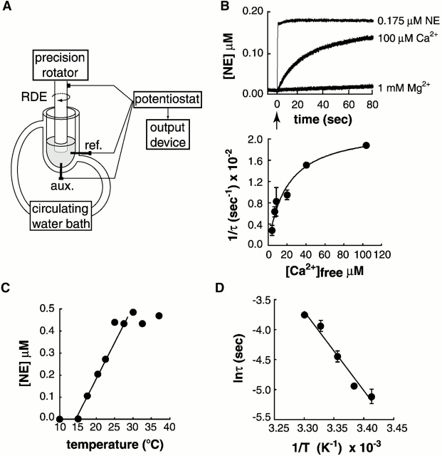
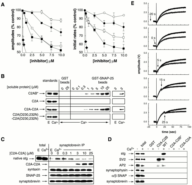
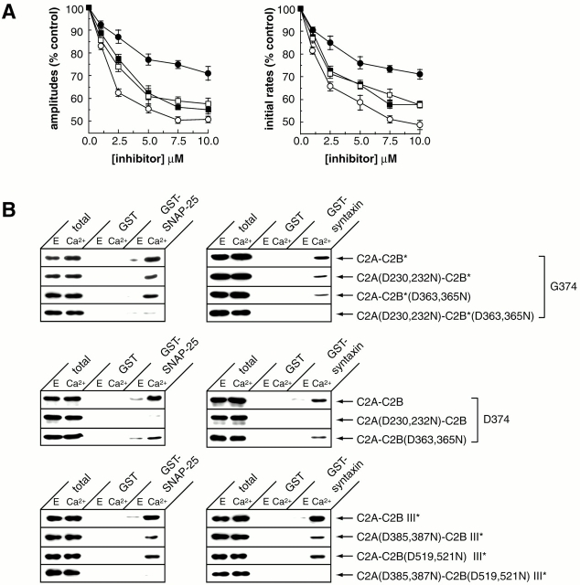
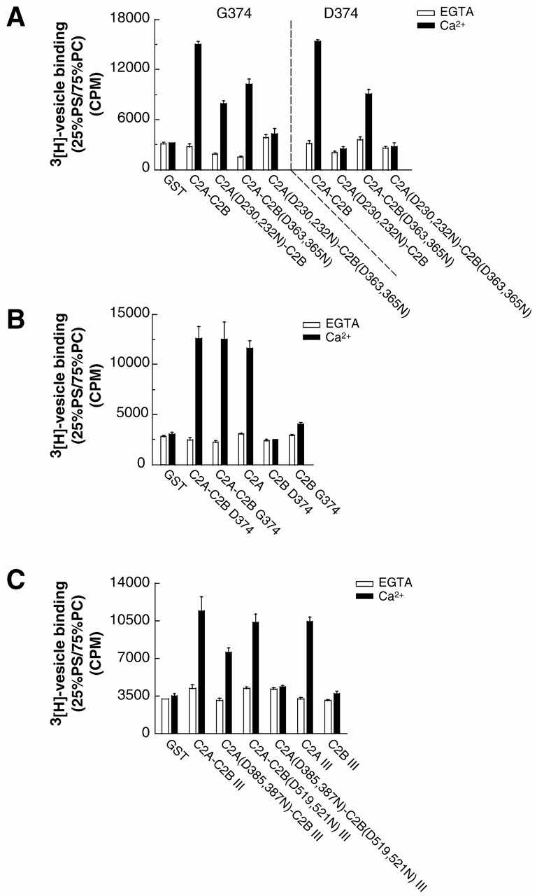
Real-time voltammetry measurements from cracked PC12 cells were used to analyze the role of synaptotagmin-SNARE interactions during Ca2+-triggered exocytosis. The isolated C2A domain of synaptotagmin I neither binds SNAREs nor inhibits norepinephrine secretion. In contrast, two C2 domains in tandem (either C2A-C2B or C2A-C2A) bind strongly to SNAREs, displace native synaptotagmin from SNARE complexes, and rapidly inhibit exocytosis. The tandem C2 domains of synaptotagmin cooperate via a novel mechanism in which the disruptive effects of Ca2+ ligand mutations in one C2 domain can be partially alleviated by the presence of an adjacent C2 domain. Complete disruption of Ca2+-triggered membrane and target membrane SNARE interactions required simultaneous neutralization of Ca2+ ligands in both C2 domains of the protein. We conclude that synaptotagmin-SNARE interactions regulate membrane fusion and that cooperation between synaptotagmin's C2 domains is crucial to its function.


Figures

Figure 1.
Figure 1.
Ca2 + and temperature dependence of NE release from cracked PC12 cells measured with a real-time assay. (A) Schematic diagram of the instrument used to measure exocytosis by RDE voltammetry (modified from Earles and Schenk, 1998). The Ag/AgCl reference (ref) and platinum auxiliary (aux) electrodes are indicated. (B) Ca2+ but not Mg2+ releases NE (top; arrow indicates the time of addition). Immediate response upon addition of NE to cell-free buffer demonstrates that Ca2+-triggered release is monitored in real time. Ca2+-dependent release profiles fit single exponential functions; 1/τ (τ = time constant) was plotted versus [Ca2+]free (bottom), the [Ca2+]1/2 was 19.7 ± 4.7 μM (n = 3–6, ± SD); τ = 45 s at saturating [Ca2+]. (C) Temperature dependence of exocytosis. NE was measured 80 s after addition of 100 μM Ca2+. (D) Arrhenius plot yields an activation energy (Ea) of 108 ± 7 kJ/mole (n = 3, ± SD).
Figure 2.
Figure 2.
Tandem C2A domains (C2A-C2A) block syt–SNARE interactions and rapidly inhibit Ca2 + -triggered exocytosis. (A) Effect of syt constructs on the rate and extent of NE release. C2A-C2B (♦; G374 version) potently inhibits secretion, presumably by blocking native syt oligomerization (Desai et al., 2000; Littleton et al., 2001). C2A-C2B* (▪), harbors a K326,327A mutation that disrupts the ability of the recombinant protein to bind native syt in the cracked cells (without causing misfolding; Desai et al., 2000). Although C2A-C2B* cannot interfere with native syt oligomerization (because it does not bind endogenous syt in the cracked cells), this construct exhibits a second mode of inhibition that occurs at higher protein concentrations. C2A, which fails to bind SNAP-25, fails to inhibit secretion (○). However, tethering two C2A domains together (C2A-C2A; □) resulted in a chimeric protein with SNAP-25 binding activity (B) and inhibitory activity that were similar to C2A-C2B*. As a control, two Ca2+ ligands in each C2A domain were neutralized D230,232N, disrupting the Ca2+ binding activity of the protein (Zhang et al., 1998). C2A(D230,232N)-C2A(D230,232N) binding to SNAREs was not detectable under our assay conditions (B), and this construct exhibited reduced inhibitory activity (•). Note the potency of inhibition of C2A-C2B (♦) and C2A-C2B* (▪) using this assay differs from a previous study using primed cells in which secretion was monitored using 3H-NE (Desai et al., 2000). The reason for this difference is not clear; however, in all experiments the rank order of inhibition by different inhibitors is the same between these two assay systems. Also note that to facilitate comparisons between the proteins used in this study, the ordinate is cut-off at 50% inhibition; at 10 μM, C2A-C2B inhibited 80–90% of release. (B) Tethering two C2A domains together results in a t-SNARE binding protein. Binding assays were carried out as described in Davis et al. (1999) except that 30 μg SNAP-25 was immobilized on beads. Eight percent of bound proteins was subjected to SDS-PAGE and visualized by immunoblotting using ECL. As a positive control, C2A-C2B* was analyzed in parallel. This mutant form was used to avoid increases in bound material that occur as a result of Ca2+-triggered syt homooligomerization (that is, the interaction of additional copies of syt with the syt that is bound to the SNAP-25 beads). As a further control, C2A(D230,232N)-C2A(D230,232N) was assayed for SNAP-25 binding activity. Standards are 10 ng C2A-C2B* and C2A-C2A, and 5 ng C2A. (C) C2A-C2A displaces native syt from SNARE complexes. Experiments were carried out by immunoprecipitation of synaptobrevin. 1-ml aliquots of rat BDE (1 mg/ml; prepared as described in Chapman et al. [1996]) were incubated in either 2 mM EGTA or 1 mM Ca2+ with the indicated concentrations of C2A-C2A for 1.5 h. SNARE complexes were immunoprecipitated with an antisynaptobrevin antibody (69.1; 10 μL) for 1.5 h followed by addition of 60 μl of protein G-Sepharose Fast Flow (Amersham Pharmacia Biotech) for 1 h. Immunoprecipitates were washed four times, and 20% of bound proteins was subjected to SDS-PAGE and visualized by ECL. Total corresponds to 1 μg BDE. Polyclonal antibodies were used to detect native syt (anti-C2B) and SNAP-25, and monoclonal antibodies were used to detect syntaxin, synaptobrevin, and C2A-C2A (anti-C2A). (D) Tethering two C2A domains together does not result in additional new protein–protein interactions. These experiments were carried out as described in Chapman et al. (1998) but expanded to include SV2. To assay for SV2, 6 μl of BDE (total) and 40% of the bound material was subjected to SDS-PAGE and immunoblotted using anti-SV2 antibodies. (E) Timing of inhibition. Addition of C2A-C2B* (final concentration 4 μM) at the times indicated (arrows) inhibits release to a similar extent at all stages in the release process and does not require preincubation; addition of Ca2+ is indicated by the vertical lines. Identical results were obtained using C2A-C2A (unpublished data).
Figure 3.
Figure 3.
Partial functional and biochemical redundancy between Ca 2+ ligands in C2A and C2B that regulate syt–t-SNARE interactions. (A) Functional redundancy of the Ca2+ ligands in the C2 domains of syt I (G374). The effects of Ca2+ ligand mutations on the ability C2A-C2B* to inhibit release was assayed as described in the legend to Fig. 2 A. Inhibition by C2A-C2B* is indicated by ○. Inhibition was only partially abrogated by muta- tions that disrupt Ca2+ binding to C2A (D230,232N; □) or C2B (D363,365N; ▪). However, inhibitory activity was largely abolished by simultaneous disruption of the Ca2+-sensing ability of both C2A and C2B (D230,232,363,365N; •). (B) Ca2+-triggered t-SNARE binding activity of syt I (G374) and III exhibits redundancy between Ca2+ ligands in C2A and C2B. Binding assays were carried out as described in the legend to Fig. 2 B except the GST-syntaxin was 15 μg. “Total” corresponds to 1% of the binding reaction. As described in the legend to Fig. 2 B, the asterisk indicates a mutation (K326,327A in syt I or R482A,K483A in syt III) that disrupts C2B-mediated syt oligomerization (Desai et al., 2000) to avoid increases in binding to t-SNARE beads that result from syt oligomerization. (Top) In syt I (G374), disruption of the Ca2+-sensing ability of either C2 domain is partially tolerated (D230,232N or D363,365N); simultaneous neutralization of Ca2+ ligands in both C2 domains (D230,232N,D363,365N) disrupts t-SNARE binding activity. (Middle) In syt I (D374), mutations that disrupt the Ca2+-sensing ability of C2A (D230,232N) abolish SNARE binding activity, whereas mutations that disrupt the Ca2+-sensing ability of C2B domain (D363,365N) have a lesser effect (Bai et al., 2000). (Bottom) Syt III exhibits Ca2+ ligand redundancy for SNARE binding activity analogous to syt I (G374). Simultaneous neutralization of Ca2+ ligands in both C2 domains (D385,387N and D519,521N) was required to disrupt Ca2+-triggered SNARE binding activity.
Figure 4.
Figure 4.
Partial redundancy between Ca 2+ ligands in C2A and C2B that regulate syt–membrane interactions. Wild-type and mutant forms of syt I and III were immobilized (0.1 nmole) on glutathione–Sepharose beads. The isolated C2 domains of these isoforms were also analyzed. 3H-labeled 25% PS/75% PC liposome binding assays were carried out as described (Bai et al., 2000). (A) In the G374 form of syt I (C2A-C2B), Ca2+-triggered liposome binding activity is abolished only when Ca2+ ligands are simultaneously neutralized in both C2A and C2B (D230,232,363,365N). In the D374 form of syt I (C2A-C2B), Ca2+-triggered liposome binding activity is abolished by mutations that neutralize Ca2+ ligands in the C2A domain (D230,232N; Bai et al., 2000). (B) C2A but not C2B derived from either G374 or D374 syt I forms an autonomous Ca2+-dependent lipid-binding module. (C) Syt III exhibits the same redundancy as G374 syt I above; complete disruption of Ca2+-triggered liposome binding activity required simultaneous neutralization of Ca2+ ligands in both C2 domains (D385,387N and D519,521N); the isolated C2B domain failed to bind membranes.

Similar articles

Cited by

References

    1. Augustine, G.J. 2001. How does calcium trigger neurotransmitter release? Curr. Opin. Neuro. 11:320–326. - PubMed
    1. Bai, J., C. Earles, J. Lewis, and E.R. Chapman. 2000. Membrane-embedded synaptotagmin penetrates cis and trans target membranes and clusters via a novel mechanism. J. Biol. Chem. 275:25427–25435. - PubMed
    1. Bark, I.C., and M.C. Wilson. 1994. Human cDNA clones encoding two different isoforms of the nerve terminal protein SNAP-25. Gene. 139:291–292. - PubMed
    1. Bennett, M.K., N. Calakos, and R.H. Scheller. 1992. Syntaxin—a synaptic protein implicated in docking of synaptic vesicles at presynaptic active zones. Science. 257:255–259. - PubMed
    1. Brose, N.A., G. Petrenko, T.C. Südhof, and R. Jahn. 1992. Synaptotagmin: a Ca2+ sensor on the synaptic vesicle surface. Science. 256:1021–1025. - PubMed

Publication types

MeSH terms