

High calcium and dobutamine positive inotropy in the perfused mouse heart: myofilament calcium responsiveness, energetic economy, and effects of protein kinase C inhibition
- PMID: 11553322
- PMCID: PMC55339
- DOI: 10.1186/1472-6793-1-12
High calcium and dobutamine positive inotropy in the perfused mouse heart: myofilament calcium responsiveness, energetic economy, and effects of protein kinase C inhibition
Abstract
Background: In perfused hearts, high calcium-induced inotropy results in less developed pressure relative to myocardial oxygen consumption compared to the beta-adrenergic agonist dobutamine. Calcium handling is an important determinant of myocardial oxygen consumption. Therefore, we hypothesized that this phenomenon was due to reduced myofilament responsiveness to calcium, related to protein kinase C activation.
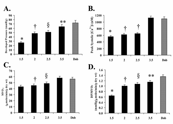
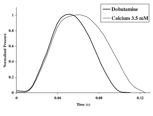
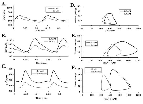
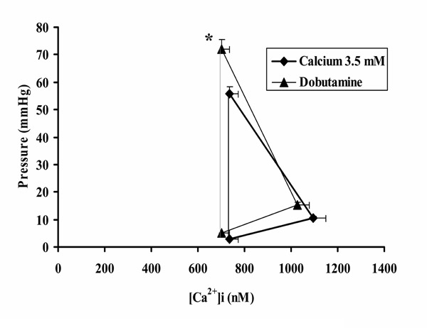
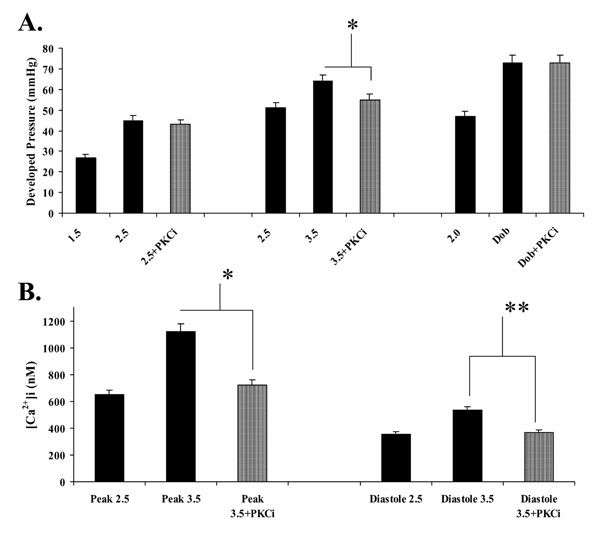
Results: Developed pressure was significantly higher with dobutamine compared to high perfusate calcium of 3.5 mM (73 +/- 10 vs 63 +/- 10 mmHg, p < 0.05), though peak systolic intracellular calcium was not significantly different, suggesting reduced myofilament responsiveness to intracellular calcium with high perfusate calcium. The ratio of developed pressure to myocardial oxygen consumption, an index of economy of contraction, was significantly increased with dobutamine compared to high perfusate calcium (1.35 +/- 0.15 vs 1.15 +/- 0.15 mmHg/micromoles/min/g dry wt, p < 0.05), suggesting energetic inefficiency with high perfusate calcium. The specific protein kinase C inhibitor, chelerythrine, significantly attenuated the expected increase in developed pressure when increasing perfusate calcium from 2.5 to 3.5 mM (3.5 mM: 64 +/- 8 vs 3.5 mM + chelerythrine: 55 +/- 5 mmHg, p < 0.05), though had no effects on dobutamine, or lower levels of perfusate calcium (1.5 to 2.5 mM).
Conclusions: By measuring intracellular calcium, developed pressures and myocardial oxygen consumption in perfused mouse hearts, these results demonstrate that high perfusate calcium positive inotropy compared to dobutamine results in reduced myofilament responsiveness to intracellular calcium, which is associated with energetic inefficiency and evidence of protein kinase C activation.
Figures
Similar articles
-
Comparison of the effects of ORG 30029, dobutamine and high perfusate calcium on function and metabolism in rat heart.J Mol Cell Cardiol. 1998 Dec;30(12):2605-12. doi: 10.1006/jmcc.1998.0817. J Mol Cell Cardiol. 1998. PMID: 9990532
-
Differential effect of troponin T mutations on the inotropic responsiveness of mouse hearts--role of myofilament Ca2+ sensitivity increase.J Physiol. 2006 Aug 15;575(Pt 1):201-13. doi: 10.1113/jphysiol.2006.107557. Epub 2006 Jun 15. J Physiol. 2006. PMID: 16777946 Free PMC article.
-
Compensatory changes in Ca(2+) and myocardial O(2) consumption in beta-tropomyosin transgenic hearts.Am J Physiol Heart Circ Physiol. 2001 Dec;281(6):H2539-48. doi: 10.1152/ajpheart.2001.281.6.H2539. Am J Physiol Heart Circ Physiol. 2001. PMID: 11709421
-
The myofilament force-calcium relationship as a target for positive inotropic therapy in congestive heart failure.Cardiovasc Drugs Ther. 2005 May;19(3):203-10. doi: 10.1007/s10557-005-2465-9. Cardiovasc Drugs Ther. 2005. PMID: 16142598 Review.
-
Inotropic and energetic effects of altering the force-calcium relationship: mechanisms, experimental results, and potential molecular targets.J Card Fail. 2000 Jun;6(2):144-56. J Card Fail. 2000. PMID: 10908089 Review.
References
-
- Ponce-Hornos JE. Energetics of Calcium Movements. Calcium and the Heart, (Ed. Langer GA), New York Raven Press. 1990. pp. 269–298.
-
- Hasenfuss G, Mulieri LA, Leavitt BJ, Allen PD, Haeberle JR, Alpert NA. Alteration of contractile function and excitation-contraction coupling in dilated cardiomyopathy. Circ Res. 1992;70:1225–1232. - PubMed
-
- Grandis DJ, DelNido PJ, Koretsky AP. Functional and energetic effects of the inotropic agents EMD-57033 and BAPTA on the isolated rat heart. Am J Physiol. 1995;269 (Cell Physiol.38):C472–C479. - PubMed
-
- Popping S, Fischer MY, Ionescu I, Kammermeier H, Rose H. Economy of contraction of cardiomyocytes as influenced by different positive inotropic interventions. Am J Physiol. 1996;271(Heart Circ. Physiol 40):H357–H364. - PubMed
Publication types
MeSH terms
Substances
Grants and funding
LinkOut - more resources
Full Text Sources
