

Silencing of oxidative stress response in Mycobacterium tuberculosis: expression patterns of ahpC in virulent and avirulent strains and effect of ahpC inactivation
- PMID: 11553532
- PMCID: PMC98723
- DOI: 10.1128/IAI.69.10.5967-5973.2001
Silencing of oxidative stress response in Mycobacterium tuberculosis: expression patterns of ahpC in virulent and avirulent strains and effect of ahpC inactivation
Abstract
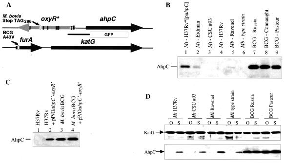
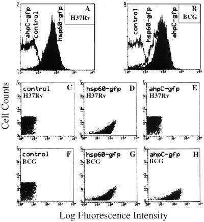
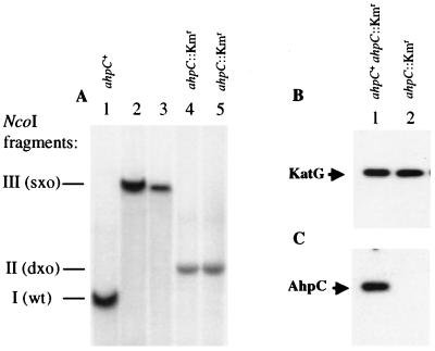
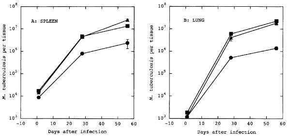
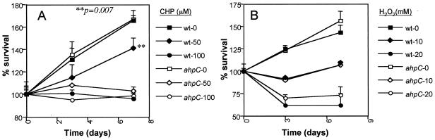
Intracellular pathogens such as Mycobacterium tuberculosis are able to survive in the face of antimicrobial products generated by the host cell in response to infection. The product of the alkyl hydroperoxide reductase gene (ahpC) of M. tuberculosis is thought to be involved in protecting the organism against both oxidative and nitrosative stress encountered within the infected macrophage. Here we report that, contrary to expectations, ahpC expression in virulent strains of M. tuberculosis and Mycobacterium bovis grown in vitro is repressed, often below the level of detection, whereas expression in the avirulent vaccine strain M. bovis BCG is constitutively high. The repression of the ahpC gene of the virulent strains is independent of the naturally occurring lesions of central regulator oxyR. Using a green fluorescence protein vector (gfp)-ahpC reporter construct we present data showing that repression of ahpC of virulent M. tuberculosis also occurred during growth inside macrophages, whereas derepression in BCG was again seen under identical conditions. Inactivation of ahpC on the chromosome of M. tuberculosis by homologous recombination had no effect on its growth during acute infection in mice and did not affect in vitro sensitivity to H2O2. However, consistent with AhpC function in detoxifying organic peroxides, sensitivity to cumene hydroperoxide exposure was increased in the ahpC::Km(r) mutant strain. The preservation of a functional ahpC gene in M. tuberculosis in spite of its repression under normal growth conditions suggests that, while AhpC does not play a significant role in establishing infection, it is likely to be important under certain, as yet undefined conditions. This is supported by the observation that repression of ahpC expression in vitro was lifted under conditions of static growth.
Figures
Similar articles
-
Oxidative stress response and its role in sensitivity to isoniazid in mycobacteria: characterization and inducibility of ahpC by peroxides in Mycobacterium smegmatis and lack of expression in M. aurum and M. tuberculosis.J Bacteriol. 1996 Jun;178(12):3641-9. doi: 10.1128/jb.178.12.3641-3649.1996. J Bacteriol. 1996. PMID: 8655566 Free PMC article.
-
Antisense RNA to ahpC, an oxidative stress defence gene involved in isoniazid resistance, indicates that AhpC of Mycobacterium bovis has virulence properties.Microbiology (Reading). 1998 Oct;144 ( Pt 10):2687-2695. doi: 10.1099/00221287-144-10-2687. Microbiology (Reading). 1998. PMID: 9802010
-
Effects of overexpression of the alkyl hydroperoxide reductase AhpC on the virulence and isoniazid resistance of Mycobacterium tuberculosis.Infect Immun. 1997 Apr;65(4):1395-401. doi: 10.1128/iai.65.4.1395-1401.1997. Infect Immun. 1997. PMID: 9119479 Free PMC article.
-
AhpC, oxidative stress and drug resistance in Mycobacterium tuberculosis.Biofactors. 1999;10(2-3):211-7. doi: 10.1002/biof.5520100219. Biofactors. 1999. PMID: 10609885 Review.
-
Peroxiredoxin systems in mycobacteria.Subcell Biochem. 2007;44:207-17. doi: 10.1007/978-1-4020-6051-9_9. Subcell Biochem. 2007. PMID: 18084895 Review.
Cited by
-
Comparative analysis of mycobacterial truncated hemoglobin promoters and the groEL2 promoter in free-living and intracellular mycobacteria.Appl Environ Microbiol. 2012 Sep;78(18):6499-506. doi: 10.1128/AEM.01984-12. Epub 2012 Jul 6. Appl Environ Microbiol. 2012. PMID: 22773641 Free PMC article.
-
The Absence of Pupylation (Prokaryotic Ubiquitin-Like Protein Modification) Affects Morphological and Physiological Differentiation in Streptomyces coelicolor.J Bacteriol. 2015 Nov;197(21):3388-99. doi: 10.1128/JB.00591-15. Epub 2015 Aug 17. J Bacteriol. 2015. PMID: 26283768 Free PMC article.
-
Synergistic Response of Rifampicin with Hydroperoxides on Mycobacterium: A Mechanistic Study.Front Microbiol. 2017 Oct 31;8:2075. doi: 10.3389/fmicb.2017.02075. eCollection 2017. Front Microbiol. 2017. PMID: 29163385 Free PMC article.
-
Host-pathogen genetic interactions underlie tuberculosis susceptibility in genetically diverse mice.Elife. 2022 Feb 3;11:e74419. doi: 10.7554/eLife.74419. Elife. 2022. PMID: 35112666 Free PMC article.
-
Methionine sulfoxide reductase A (MsrA) deficiency affects the survival of Mycobacterium smegmatis within macrophages.J Bacteriol. 2004 Jun;186(11):3590-8. doi: 10.1128/JB.186.11.3590-3598.2004. J Bacteriol. 2004. PMID: 15150247 Free PMC article.
References
-
- Behr M A, Wilson M A, Gill W P, Salamon H, Schoolnik G K, Rane S, Small P M. Comparative genomics of BCG vaccines by whole-genome DNA microarray. Science. 1999;284:1520–1523. - PubMed
-
- Bryk R, Griffin P, Nathan C. Peroxynitrite reductase activity of bacterial peroxiredoxins. Nature. 2000;407:211–215. - PubMed
Publication types
MeSH terms
Substances
Grants and funding
LinkOut - more resources
Full Text Sources
Medical
Molecular Biology Databases
