

CD85/LIR-1/ILT2 and CD152 (cytotoxic T lymphocyte antigen 4) inhibitory molecules down-regulate the cytolytic activity of human CD4+ T-cell clones specific for Mycobacterium tuberculosis
- PMID: 11553539
- PMCID: PMC98730
- DOI: 10.1128/IAI.69.10.6022-6029.2001
CD85/LIR-1/ILT2 and CD152 (cytotoxic T lymphocyte antigen 4) inhibitory molecules down-regulate the cytolytic activity of human CD4+ T-cell clones specific for Mycobacterium tuberculosis
Abstract
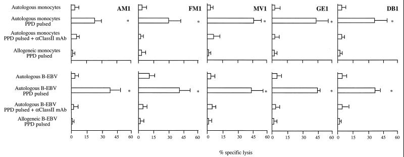
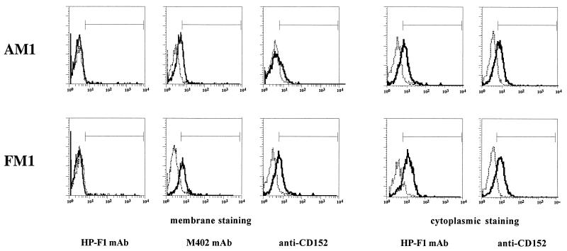
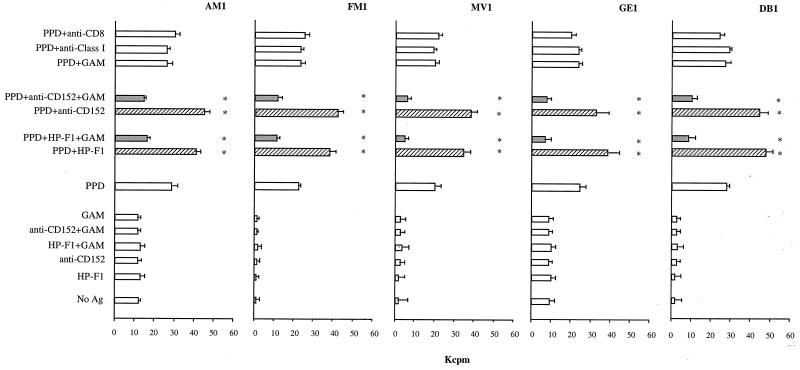
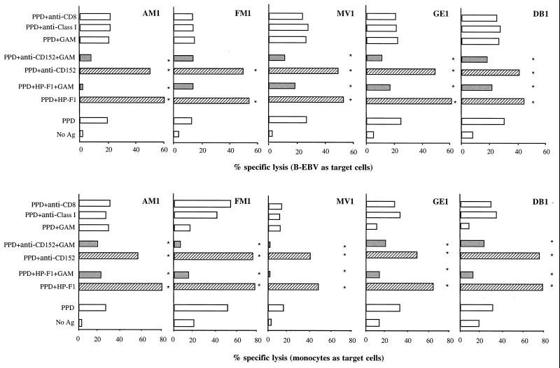
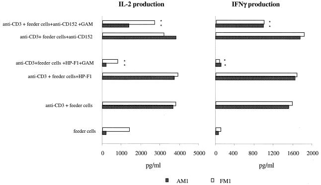
Antigen-specific cytolytic CD4+ T lymphocytes control Mycobacterium tuberculosis infection by secreting cytokines and by killing macrophages that have phagocytosed the pathogen. However, lysis of the latter cells promotes microbial dissemination, and other macrophages engulf the released bacteria. Subsequently, CD4+ T-cell-mediated killing of macrophages goes on, and this persistent process may hamper control of infection, unless regulatory mechanisms maintain a subtle balance between lysis of macrophages by cytolytic CD4+ cells and activation of cytolytic CD4+ cells by infected macrophages. We asked whether inhibitory molecules expressed by CD4+ cytolytic T lymphocytes could play a role in such a balance. To this end, human CD4+ T-cell clones specific for M. tuberculosis were produced that displayed an autologous major histocompatibility complex class II-restricted lytic ability against purified protein derivative (PPD)-pulsed antigen-presenting cells. All T-cell clones expressed CD152 (cytotoxic T-lymphocyte antigen 4 [CTLA-4]) and CD85/leukocyte immunoglobulin-like receptor 1 (LIR-1)/immunoglobulin-like transcript 2 (ILT2) inhibitory receptors, but not CD94 and the killer inhibitory receptor (or killer immunoglobulin-like receptor [KIR]) p58.2. CD3-mediated activation of the clones was inhibited in a redirected killing assay in which CD152 and CD85/LIR-1/ILT2 were cross-linked. Specific antigen-mediated proliferation of the clones was also sharply reduced when CD152 and CD85/LIR-1/ILT2 were cross-linked by specific monoclonal antibody (MAb) followed by goat anti-mouse antiserum. In contrast, blockade of the receptors by specific MAb only increased their proliferation. Production of interleukin 2 (IL-2) and gamma interferon (IFN-gamma) by the T-cell clones was also strongly reduced when CD152 and CD85/LIR-1/ILT2 were cross-linked. The lytic activity of the T-cell clones against PPD-pulsed autologous monocytes or Epstein-Barr virus-activated B cells was increased by blockade and decreased by cross-linking of the receptors. These results indicate that CD152 and CD85/LIR-1/ILT2 play a role in the regulation of the antigen-specific activity of CD4+ cytolytic T lymphocytes against PPD-presenting cells.
Figures

Similar articles
-
Dual effect of CD85/leukocyte Ig-like receptor-1/Ig-like transcript 2 and CD152 (CTLA-4) on cytokine production by antigen-stimulated human T cells.J Immunol. 2002 Jan 1;168(1):207-15. doi: 10.4049/jimmunol.168.1.207. J Immunol. 2002. PMID: 11751964
-
The CD85/LIR-1/ILT2 inhibitory receptor is expressed by all human T lymphocytes and down-regulates their functions.J Immunol. 2000 Oct 1;165(7):3742-55. doi: 10.4049/jimmunol.165.7.3742. J Immunol. 2000. PMID: 11034379
-
CTLA-4 (CD152) inhibits the specific lysis mediated by human cytolytic T lymphocytes in a clonally distributed fashion.J Immunol. 1999 Jan 15;162(2):651-8. J Immunol. 1999. PMID: 9916682
-
Anatomy of the immune system: facts and problems.Ital J Anat Embryol. 2000 Oct-Dec;105(4):97-124. Ital J Anat Embryol. 2000. PMID: 11265217 Review.
-
Molecules that inhibit T-cell functions: cytochemical localization and shuttling.Eur J Histochem. 2000;44(1):89-99. Eur J Histochem. 2000. PMID: 10868297 Review.
Cited by
-
Expression of the innate immune receptor LILRB5 on monocytes is associated with mycobacteria exposure.Sci Rep. 2016 Feb 24;6:21780. doi: 10.1038/srep21780. Sci Rep. 2016. PMID: 26908331 Free PMC article.
-
Up-regulation of circRNA-0003528 promotes mycobacterium tuberculosis associated macrophage polarization via down-regulating miR-224-5p, miR-324-5p and miR-488-5p and up-regulating CTLA4.Aging (Albany NY). 2020 Dec 9;12(24):25658-25672. doi: 10.18632/aging.104175. Epub 2020 Dec 9. Aging (Albany NY). 2020. PMID: 33318319 Free PMC article.
-
Potential of immunomodulatory agents as adjunct host-directed therapies for multidrug-resistant tuberculosis.BMC Med. 2016 Jun 15;14:89. doi: 10.1186/s12916-016-0635-1. BMC Med. 2016. PMID: 27301245 Free PMC article. Review.
-
Granulysin-expressing CD4+ T cells as candidate immune marker for tuberculosis during childhood and adolescence.PLoS One. 2011;6(12):e29367. doi: 10.1371/journal.pone.0029367. Epub 2011 Dec 27. PLoS One. 2011. PMID: 22216262 Free PMC article.
-
Downregulation of immunoglobulin-like transcript-4 (ILT4) in patients with psoriatic arthritis.PLoS One. 2014 Mar 27;9(3):e92018. doi: 10.1371/journal.pone.0092018. eCollection 2014. PLoS One. 2014. PMID: 24676037 Free PMC article.
References
-
- Alegre M L, Noel P J, Eisfelder B J, Chuang E, Clark M R, Reiner S L, Thompson C B. Regulation of surface and intracellular expression of CTLA-4 on mouse T cells. J Immunol. 1996;157:4762–4770. - PubMed
-
- Banchereau J, Steinman R M. Dendritic cells and the control of immunity. Nature. 1998;392:245–252. - PubMed
-
- Borges L, Cosman D. LIRs/ILTs/MIRs, inhibitory and stimulatory Ig-superfamily receptors expressed in myeloid and lymphoid cells. Cytokine Growth Factor Rev. 2000;11:209–217. - PubMed
-
- Brunner M C, Chambers C A, Chan F K, Hanke J, Winoto A, Allison J P. CTLA-4-mediated inhibition of early events of T cell proliferation. J Immunol. 1999;162:5813–5820. - PubMed
Publication types
MeSH terms
Substances
LinkOut - more resources
Full Text Sources
Research Materials
