. 2001 Oct;69(10):6038-43.
doi: 10.1128/IAI.69.10.6038-6043.2001.

Role of ADP-ribosyltransferase activity of pertussis toxin in toxin-adhesin redundancy with filamentous hemagglutinin during Bordetella pertussis infection

Affiliations

Role of ADP-ribosyltransferase activity of pertussis toxin in toxin-adhesin redundancy with filamentous hemagglutinin during Bordetella pertussis infection

S Alonso et al. Infect Immun. 2001 Oct.

Abstract

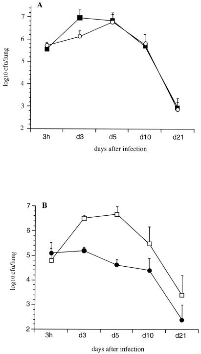
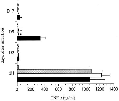
Pertussis toxin (PT) and filamentous hemagglutinin (FHA) are two major virulence factors of Bordetella pertussis. FHA is the main adhesin, whereas PT is a toxin with an A-B structure, in which the A protomer expresses ADP-ribosyltransferase activity and the B moiety is responsible for binding to the target cells. Here, we show redundancy of FHA and PT during infection. Whereas PT-deficient and FHA-deficient mutants colonized the mouse respiratory tract nearly as efficiently as did the isogenic parent strain, a mutant deficient for both factors colonized substantially less well. This was not due to redundant functions of PT and FHA as adhesins, since in vitro studies of epithelial cells and macrophages indicated that FHA, but not PT, acts as an adhesin. An FHA-deficient B. pertussis strain producing enzymatically inactive PT colonized as poorly as did the FHA-deficient, PT-deficient strain, indicating that the ADP-ribosyltransferase activity of PT is required for redundancy with FHA. Only strains producing active PT induced a local transient release of tumor necrosis factor alpha (TNF-alpha), suggesting that the pharmacological effects of PT are the basis of the redundancy with FHA, through the release of TNF-alpha. This may lead to damage of the pulmonary epithelium, allowing the bacteria to colonize even in the absence of FHA.


Figures

FIG. 1
FIG. 1
Lung colonization by B. pertussis. OF1 mice were infected i.n. with B. pertussis BPSM (black squares) or BPRA (open circles) (A) or BPGR4 (open squares) or BPDR (black circles) (B). At the indicated time points, the mice were sacrificed, and the CFU present in the lungs were counted. Each curve represents an average of independent experiments. The results are expressed as the means ± the standard errors of the means. Four mice per time point and per group were assessed for each experiment.
FIG. 2
FIG. 2
Adherence of B. pertussis to cell lines. Human pulmonary epithelial cells (A549), murine alveolar macrophages (MH-S), primary cultures of dendritic cells (dendritic), and human tracheal cells (HEp-2) were incubated with the indicated 35S-labeled B. pertussis strains for 1 h 30 min at a multiplicity of infection of 20. After washing, adherence was estimated by scintillation counting. The results are expressed as percentages of counts per minute relative to the counts per minute present in the inoculum. The data represent averages and standard deviations for quadruplicate experiments. ∗, P < 0.05.
FIG. 3
FIG. 3
Internalization of B. pertussis into MH-S cell monolayers. MH-S cell monolayers were infected with the indicated B. pertussis strains at a multiplicity of infection of 20. After 1 h of incubation, the monolayers were treated with gentamicin and lysed, and the bacteria were plated onto BG agar. The results are expressed as percentages of internalized bacteria relative to the number of bacteria present in the inoculum. The data represent averages and standard deviations for triplicate experiments. ∗, P < 0.05.
FIG. 4
FIG. 4
Lung colonization by B. pertussis mutants. OF1 mice were infected i.n. with B. pertussis BPGR4 (open squares), BPDR (black circles), or BPDR-RE (black triangles), and colonization was monitored as described for Fig. 1. The stippled line represents the limit of detection of the number of CFU present in the lungs.
FIG. 5
FIG. 5
TNF-α production in BAL fluids of B. pertussis-infected mice. OF1 mice were infected i.n. with B. pertussis BPGR4 (black bars), BPDR (white bars), or BPDR-RE (gray bars) or received PBS (stippled bars). At indicated time points, the concentration of TNF-α was measured in the BAL fluids. Five mice were analyzed per time point for each group. ∗, P < 0.05.
FIG. 6
FIG. 6
Lung colonization after coinfection with B. pertussis BPDR and B. pertussis BPDR-RE. OF1 mice were infected i.n. with a mixture of B. pertussis BPDR (black circles) and B. pertussis BPDR-RE (black triangles). The results are the means ± standard errors of the means of four mice per time point.

References

    1. Antoine R, Locht C. Molecular studies on the interaction of the S1 subunit with the B oligomer of pertussis toxin. Zentbl Bakteriol Suppl. 1992;23:292–293.
    1. Antoine R, Locht C. Roles of the disulfide bond and the carboxy-terminal region of the S1 subunit in the assembly and biosynthesis of pertussis toxin. Infect Immun. 1990;58:1518–1526. - PMC - PubMed
    1. Bassinet L, Gueirard P, Maitre B, Housset B, Gounon P, Guiso N. Role of adhesins and toxins in invasion of human tracheal epithelial cells by Bordetella pertussis. Infect Immun. 2000;68:1934–1941. - PMC - PubMed
    1. Cahill E S, O'Hagan D T, Illum L, Redhead K. Mice are protected against Bordetella pertussis infection by intra-nasal immunization with filamentous haemagglutinin. FEMS Microbiol Lett. 1993;107:211–216. - PubMed
    1. Dei-Cas E, Brun-Pascaud M, Bille-Hansen V, Allaert A, Aliouat E M. Animal models of pneumocystosis. FEMS Immunol Med Microbiol. 1998;22:163–168. - PubMed

Publication types

MeSH terms

Substances

LinkOut - more resources