. 2001 Oct;69(10):6248-55.
doi: 10.1128/IAI.69.10.6248-6255.2001.

Release of Toll-like receptor-2-activating bacterial lipoproteins in Shigella flexneri culture supernatants

Affiliations

Release of Toll-like receptor-2-activating bacterial lipoproteins in Shigella flexneri culture supernatants

A O Aliprantis et al. Infect Immun. 2001 Oct.

Abstract

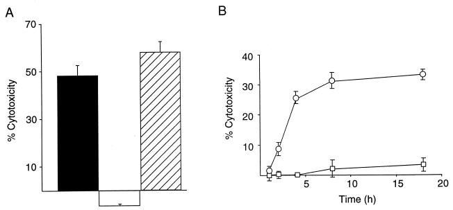
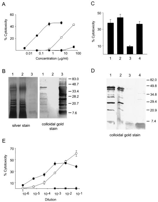
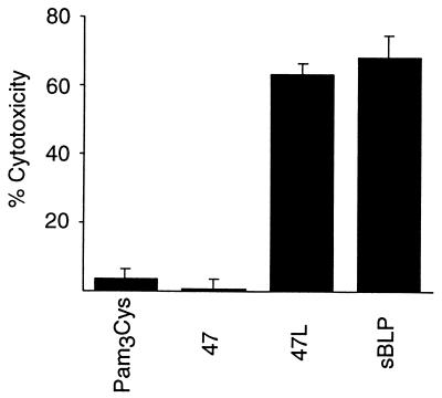
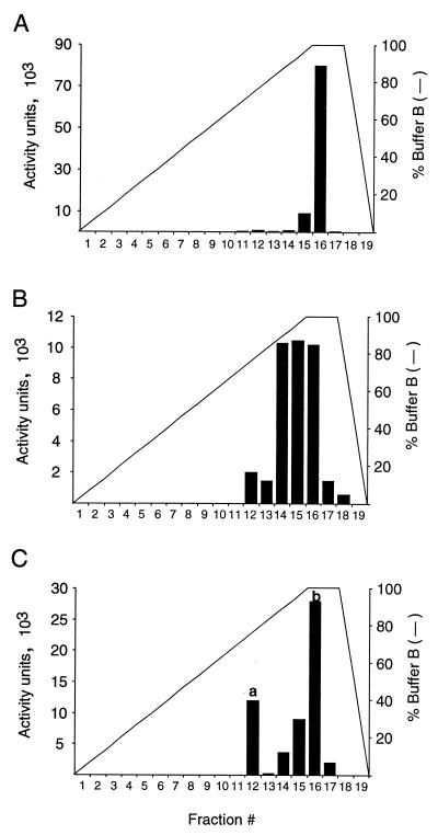
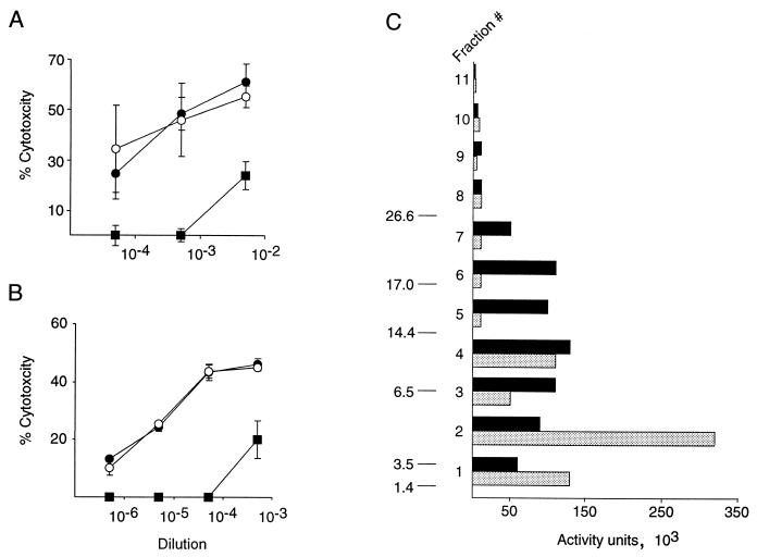
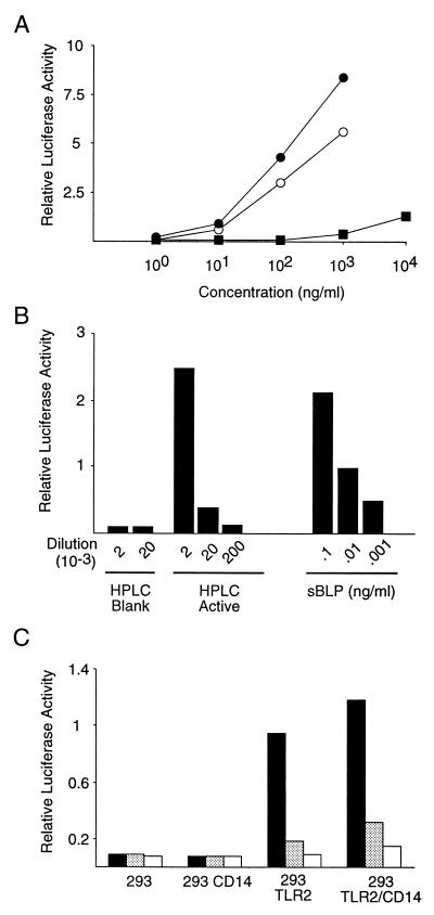
Shigella spp. cause dysentery, a severe form of bloody diarrhea. Apoptosis, or programmed cell death, is induced during Shigella infections and has been proposed to be a key event in the pathogenesis of dysentery. Here, we describe a novel cytotoxic activity in the sterile-culture supernatants of Shigella flexneri. An identical activity was identified in purified S. flexneri endotoxin, defined here as a mixture of lipopolysaccharide (LPS) and endotoxin-associated proteins (EP). Separation of endotoxin into EP and LPS revealed the activity to partition exclusively to the EP fraction. Biochemical characterization of S. flexneri EP and culture supernatants, including enzymatic deactivation, reverse-phase high-pressure liquid chromatography analysis, sodium dodecyl sulfate-polyacrylamide gel electrophoresis, and a Toll-like receptor-2 (TLR2) activation assay, indicates that the cytotoxic component is a mixture of bacterial lipoproteins (BLP). We show that biologically active BLP are liberated into culture supernatants of actively growing S. flexneri. In addition, our data indicate that BLP, and not LPS, are the component of endotoxin of gram-negative organisms responsible for activating TLR2. The activation of apoptosis by BLP shed from S. flexneri is discussed as a novel aspect of the interaction of bacteria with the host.


Figures

FIG. 1
FIG. 1
Cytotoxic activity in S. flexneri culture supernatants and infected macrophages. (A) Culture supernatants of S. flexneri type 1A-infected J774 macrophages (black bar) and S. flexneri type 1A alone (striped bar) contain similar levels of cytotoxic activity. The culture supernatants of macrophages alone (white bar) were noncytotoxic. (B) Time course of the cytotoxic activity in commercial S. flexneri type 1A LPS preparations. THP-1 cells were incubated with 10 μg of S. flexneri type 1A PW-LPS (open squares) or TCA-endotoxin (open circles) per ml and assayed for cytotoxicity at the indicated time points. Percent cytotoxicity was determined by an LDH release assay.
FIG. 2
FIG. 2
The cytotoxic activity of TCA-endotoxin partitions to the phenol phase in a phenol-water extraction. (A) PW-LPS (closed squares) and EP (closed circles) were prepared from S. flexneri type 1A TCA-endotoxin (open circles). These preparations were tested for cytotoxic activity at the indicated concentrations on THP-1 cells. Percent cytotoxicity in panels A, C, and E was determined by an LDH release assay. (B) Silver and colloidal-gold protein stain of S. flexneri type 1A TCA-endotoxin (lane 1), PW-LPS (lane 2), and EP (lane 3) resolved by SDS-PAGE (15% polyacrylamide). On the silver-stained gel, note the characteristic LPS ladders in both TCA-endotoxin and PW-LPS, indicative of repeating O-antigen units. The colloidal-gold protein stain illustrates that the proteins in TCA-endotoxin partition exclusively to the phenol phase (EP). (C) The cytotoxic activity in EP is stable when subjected to PK treatment but is depleted by PB-coated agarose beads. A 1-mg/ml solution of EP was either mock treated (1) or incubated with 50 μl of uncoupled agarose beads (2), 50 μl of PB-agarose beads (3), or 0.2 U of PK-agarose beads (4) for 4 h at 37°C. The beads were removed by centrifugation, and supernatants were tested for cytotoxic activity at a dilution of 1:500. (D) SDS–15% PAGE of samples shown in panel C. The gel was stained with the colloidal-gold protein stain. (E) The cytotoxic activity in S. flexneri type 1A culture supernatants is extracted by phenol. S. flexneri type 1A culture supernatants were prepared, concentrated 1,000-fold, and extracted with hot phenol. Ethanol precipitates of the phenol (closed circles) and aqueous phases (closed squares) were resuspended in equivalent volumes of PBS and tested for cytotoxicity on THP-1 cells at the indicated dilutions. As a reference, unextracted 1,000-fold-concentrated S. flexneri type 1A culture supernatant was also tested (open circles).
FIG. 3
FIG. 3
BLP are cytotoxic to THP-1 cells. THP-1 cells were incubated with the lipopeptides sBLP or 47L at a final concentration of 10 nM and assayed for cytotoxicity. The negative-control compounds, 47 and Pam3Cys, were tested at 1,000 nM. Percent cytotoxicity was determined by an LDH release assay.
FIG. 4
FIG. 4
The cytotoxic activities in S. flexneri culture supernatants. S. flexneri EP and sBLP coelute from an RP-HPLC column. Eight hundred microliters of a phenol extract of S. flexneri culture supernatants (A), 200 μg of EP (B), or 200 μg of sBLP (C) was loaded onto a C4 RP-HPLC column and eluted with a linear gradient of increasing ACN concentration. One-milliliter fractions were collected and assayed for cytotoxic activity by an LDH release assay on THP-1 cells by serial dilution directly into tissue culture media. Activity is defined as 1/(dilution yielding 50% maximal cytotoxic activity). HPLC conditions were as follows. Buffer A contained dH2O and 0.1% TFA, buffer B contained 95% ACN and 0.1% TFA, and the flow rate was 1 ml/min. The gradient was as follows: 0 to 15 min, 0 to 100% buffer B (linear gradient); 15 to 17 min, 100% buffer B; 17 to 19 min, 100 to 0% buffer B.
FIG. 5
FIG. 5
The cytotoxic activity in EP contains lipase and protease sensitive components. (A) A 50-μg/ml S. flexneri EP solution was incubated for 4 h at 37°C either alone (closed circles), with uncoupled agarose beads (open circles), or with 22 U of wheat germ type 1A triacylglycerol lipase-agarose beads per ml (closed squares). Beads were removed by centrifugation, and the supernatants were tested for cytotoxicity on THP-1 cells at the indicated dilutions. Percent cytotoxicity in panels A and B was determined by an LDH release assay. (B) A 200-μg/ml sBLP solution was treated and assayed as described for panel A. (C) SDS-PAGE analysis of the cytotoxic activity in S. flexneri EP and PK-treated S. flexneri EP. One hundred sixty-six micrograms of S. flexneri EP (black bars) or 166 μg of PK-treated S. flexneri EP (gray bars) was resolved by Tris-Tricine SDS–16.5% PAGE. Eleven 5-mm gel sections (fractions) were made per lane, extracted with octyl glucoside, and tested for cytotoxic activity by an LDH release assay on cycloheximide-treated THP-1 cells by serial dilution directly into tissue culture media. Activity is defined as 1/(dilution yielding 50% maximal cytotoxic activity). Peptide molecular mass markers (in kilodaltons) are shown on the left.
FIG. 6
FIG. 6
An HPLC fraction containing the cytotoxic activity in EP stimulates an NF-κB-regulated reporter gene through TLR2. (A) S. flexneri TCA-endotoxin and EP, but not PW-LPS, activate TLR2 signaling. 293 cells were transiently transfected with expression plasmids encoding TLR2 and an NF-κB-regulated luciferase reporter gene. The cells were stimulated with S. flexneri TCA-endotoxin (open circles), EP (closed circles), or PW-LPS (closed squares), and reporter gene activity was assayed. (B) The cytotoxic activity in EP was purified sequentially by preparative SDS-PAGE and RP-HPLC. RP-HPLC fractions were assayed for cytotoxic activity on THP-1 cells, and an active fraction was identified as described in Materials and Methods. 293TLR2/CD14 cells were transiently transfected with the NF-κB-regulated luciferase reporter construct as described previously (1) and stimulated with the indicated dilutions of the cytotoxic HPLC fraction (HPLC Active) or an identical HPLC fraction collected from a blank HPLC run (HPLC Blank). As a positive control for TLR2 activation, cells were stimulated with sBLP. (C) The indicated cell lines were transiently transfected with an NF-κB-regulated luciferase reporter construct and incubated with 1:2,000 (black bars), 1:20,000 (gray bars), or 1:200,000 (white bars) dilutions of the cytotoxic HPLC fraction.

References

    1. Aliprantis A O, Yang R B, Mark M R, Suggett S, Devaux B, Radolf J D, Klimpel G R, Godowski P, Zychlinsky A. Cell activation and apoptosis by bacterial lipoproteins through toll-like receptor-2. Science. 1999;285:736–739. - PubMed
    1. Aliprantis A O, Yang R B, Weiss D S, Godowski P, Zychlinsky A. The apoptotic signaling pathway activated by Toll-like receptor-2. EMBO J. 2000;19:3325–3336. - PMC - PubMed
    1. Baeuerle P, Henkel T. Function and activation of NF-κB in the immune system. Annu Rev Immunol. 1994;12:141–179. - PubMed
    1. Belisle J T, Brandt M E, Radolf J D, Norgard M V. Fatty acids of Treponema pallidum and Borrelia burgdorferi lipoproteins. J Bacteriol. 1994;176:2151–2157. - PMC - PubMed
    1. Brightbill H D, Libraty D H, Krutzik S R, Belisle J T, Maitland M, Norgard M V, Plevy S E, Smale S T, Brennan P J, Bloom B R, Godowski P J, Modlin R L. Host defense mechanisms triggered by microbial lipoproteins through toll-like receptors. Science. 1999;285:732–736. - PubMed

Publication types

MeSH terms

LinkOut - more resources