. 2001 Oct;69(10):6391-400.
doi: 10.1128/IAI.69.10.6391-6400.2001.

Macrophage nitric oxide synthase associates with cortical actin but is not recruited to phagosomes

Affiliations

Macrophage nitric oxide synthase associates with cortical actin but is not recruited to phagosomes

J L Webb et al. Infect Immun. 2001 Oct.

Abstract

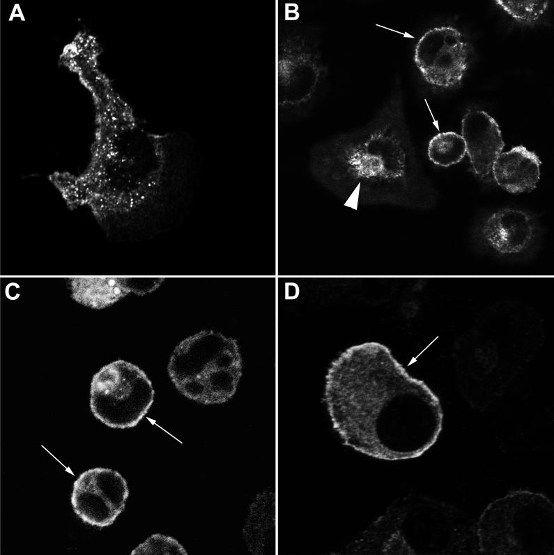
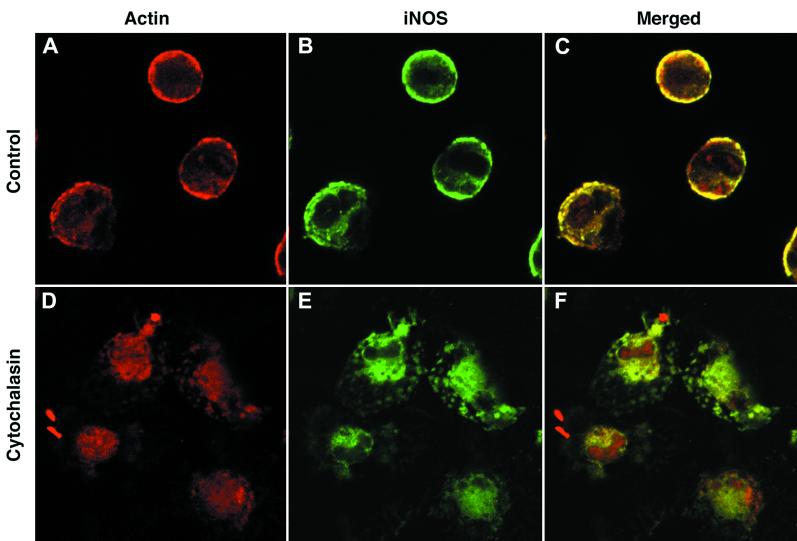
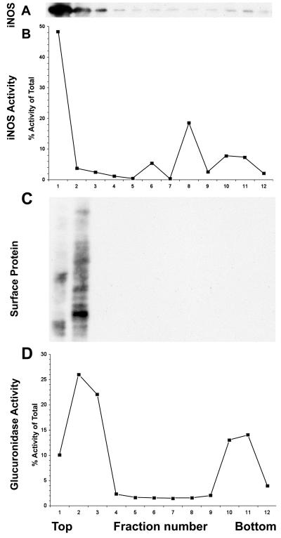
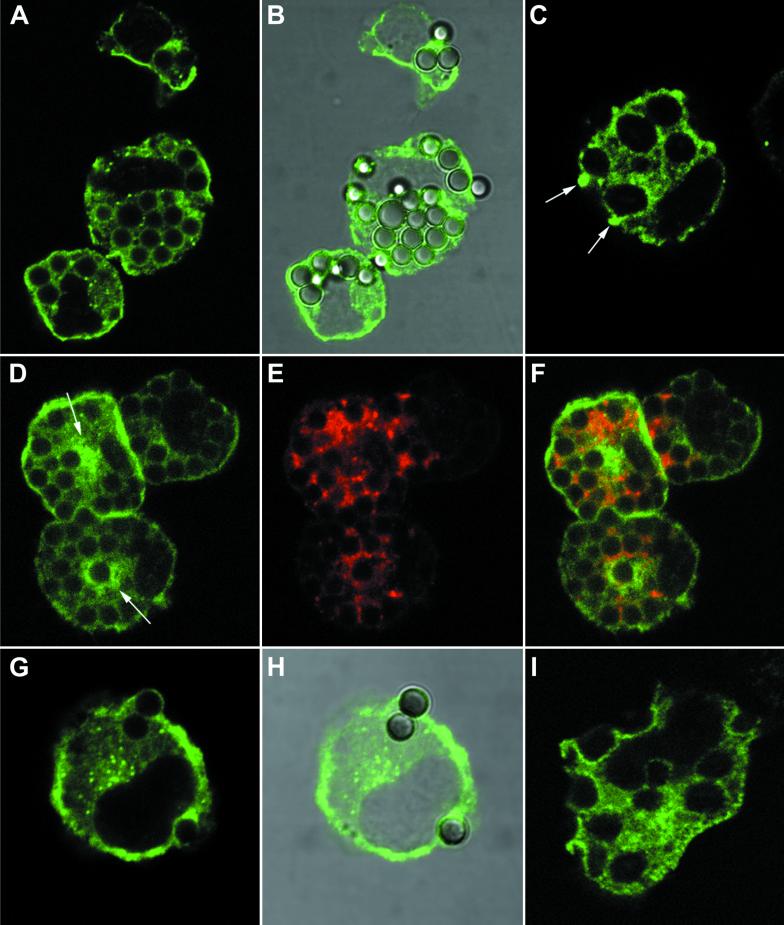
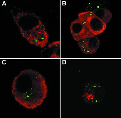
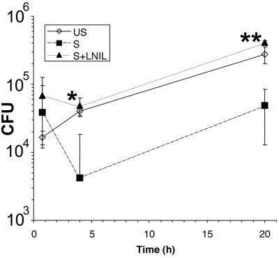
Nitric oxide (NO) produced from inducible NO synthase (iNOS) is an important component of host defense against intracellular pathogens. To understand how phagocytes deliver NO to ingested microorganisms while avoiding cytotoxicity, we set out to study the subcellular localization of iNOS within macrophages following phagocytosis. Confocal microscopy of immunostained cells showed that iNOS was located not only diffusely within cytoplasm but also in vesicles, as well as immediately adjacent to the peripheral cell membrane. This peripheral iNOS colocalized with the cortical actin cytoskeleton and was removed by the actin-depolymerizing drug cytochalasin B. Biochemical fractionation of RAW 264 macrophages showed that 32.75% (+/-5.11%; n = 3) of iNOS was present in a particulate fraction, which cosedimented with low-density cellular vesicles. Following phagocytosis of latex beads, zymosan, immunoglobulin G-coated beads, or complement-coated zymosan, submembranous cortical iNOS was not recruited to phagosomes, nor was there any relocalization of intracellular iNOS. Similarly, following phagocytosis of Salmonella enterica serovar Typhimurium there was no recruitment of iNOS to the Salmonella vacuole at any stage after internalization. NO mediated significant killing of intracellular S. enterica serovar Typhimurium in RAW macrophages treated with lipopolysaccharide and gamma interferon; this was evident 4 h after infection. Although not recruited to phagosomes, iNOS association with the submembranous cortical actin cytoskeleton is ideally suited to deliver NO to microbes in contact with the cell surface and may contribute to early killing of ingested Salmonella.


Figures

FIG. 1
FIG. 1
Localization of iNOS within macrophages. Primary mouse peritoneal macrophages (A to C) or RAW 264 cells (D) were stimulated with IFN-γ alone (A) or with IFN-γ and LPS (B to D) and immunostained for iNOS, using the mouse monoclonal antibody. All images were obtained using laser confocal microscopy. Magnification, ×2,000 (A), ×600 (B to C), or ×6,000 (D). Arrows indicate peripheral plasma membrane staining of iNOS. The solid arrowhead in panel B indicates iNOS staining in the perinuclear area.
FIG. 2
FIG. 2
iNOS can be found in the Golgi apparatus. RAW 264 cells were stimulated with LPS and IFN-γ and dually stained for iNOS using a rabbit polyclonal antibody, with red staining (A), and the intrinsic Golgi protein GM130, with green staining (B). (C) Merged images of panels A and B, with areas of colocalization in yellow. Magnification, ×1,000 for all panels.
FIG. 3
FIG. 3
iNOS associates with the cortical actin cytoskeleton. Primary peritoneal macrophages were stimulated with LPS and IFN-γ and immunostained for iNOS with the mouse monoclonal antibody in green (B and E) or polymerized actin in red (A and D). Cells were either untreated before staining (A to C) or treated with cytochalasin B (D to F). Panels C and F show the merged actin and iNOS images from panels A and B and panels D and E, respectively; areas of colocalization are shown in yellow. Magnification, ×600 for all panels.
FIG. 4
FIG. 4
Density gradient centrifugation analysis of the particulate fraction of IFN-γ- and LPS-treated RAW 264 cells. Each panel shows an assay performed on each of 12 fractions from the density gradient, with the lighter fractions to the left (top) and the denser ones to the right (bottom). The experiment was repeated with similar results. (A) iNOS protein, detected by Western blotting. The band shown is the major band detected, at 135 kDa. So that the bands in lanes 6 to 11 could be seen, the blot was overexposed for lanes 1 to 3. (B) iNOS activity, as assayed by arginine-to-citrulline conversion. (C) Surface proteins of the cell, labeled by surface biotinylation, identified by probing the Western blot with horseradish peroxidase-streptavidin. The gel shows proteins ranging in size from 200 kDa at the top of the panel to 29 kDa at the bottom. (D) β-glucuronidase activity.
FIG. 5
FIG. 5
iNOS distribution following phagocytosis in mouse primary peritoneal macrophages. Cells treated with IFN-γ and LPS were used in all panels. (A and B) Uptake of latex beads at 30 min after addition. iNOS staining (mouse monoclonal) in green is shown alone (A) and also merged with the differential interference contrast (DIC) image of the same cell (B). (C) A cell, 2 min after addition of zymosan particles, stained for iNOS (mouse monoclonal) in green. Accumulation of iNOS at the tips of pseudopodia closing around zymosan just entering the cell is shown by arrows. (D to F) Cells, 2 h after being fed latex beads for 10 min, which were then removed by washing. The panels show the pattern of iNOS staining (mouse monoclonal) in green (D), LAMP-1 staining in red (E), and the merged images of panels D and E (F), where areas of colocalization are shown in yellow. Two areas showing apparent accumulation of iNOS around the outside of phagocytosed beads are indicated by arrows in panel D. (G and H) Uptake of IgG-coated magnetic beads at 5 min after addition. iNOS staining (rabbit polyclonal antibody) in green is shown alone (G) and merged with the DIC image of the same cell (H). (I) A cell, 10 min after the addition of complement-coated zymosan particles stained for iNOS (mouse monoclonal) in green. Magnification, ×2,000 for all panels.
FIG. 6
FIG. 6
iNOS and nitrotyrosine in macrophages following phagocytosis of serovar Typhimurium. In all cases, the bacteria are labeled green. (A) iNOS (red; mouse monoclonal) in a primary macrophage 2 h after phagocytosis of wild-type stationary phase serovar Typhimurium. (B) iNOS (red; rabbit polyclonal) in a RAW 264 cell 90 min after phagocytosis of wild-type stationary phase serovar Typhimurium. (C) iNOS (red; rabbit polyclonal) in a RAW 264 cell 4 h after phagocytosis of a SPI-2 mutant stationary-phase serovar Typhimurium. (D) Nitrotyrosine staining (red) in a RAW 264 cell 4 h after phagocytosis of wild-type stationary-phase serovar Typhimurium. Magnification, ×1,000 (B and D) or ×2,000 (A and C).
FIG. 7
FIG. 7
Survival of serovar Typhimurium within RAW macrophages. Cells were infected at a multiplicity of infection of 10 with 107 bacteria at time zero. The graph shows the mean intracellular bacterial numbers found in RAW 264 cells either unstimulated (US), treated with LPS and IFN-γ (S), or treated with LPS and IFN-γ plus L-NIL (S+LNIL). Error bars, ± 1 standard deviation. A significant difference was found in intracellular Salmonella survival between cells treated with LPS and IFN-γ and cells treated with LPS, IFN-γ, and L-NIL at 4 and 20 h after infection. ∗, P < 0.05; ∗∗ P < 0.01 (two-tailed t test).

Similar articles

Cited by

References

    1. Allen L A, Aderem A. Molecular definition of distinct cytoskeletal structures involved in complement- and Fc receptor-mediated phagocytosis in macrophages. J Exp Med. 1996;184:627–637. - PMC - PubMed
    1. Beckman J S, Siedow J N. Bactericidal agents generated by the peroxidase-catalyzed oxidation of para-hydroquinones. J Biol Chem. 1985;260:14604–14609. - PubMed
    1. Beuzon C R, Meresse S, Unsworth K E, Ruiz-Albert J, Garvis S, Waterman S R, Ryder T A, Boucrot E, Holden D W. Salmonella maintains the integrity of its intracellular vacuole through the action of SifA. EMBO J. 2000;19:3235–3249. - PMC - PubMed
    1. Borregaard N, Heiple J M, Simons E R, Clark R A. Subcellular localization of the b-cytochrome component of the human neutrophil microbicidal oxidase: translocation during activation. J Cell Biol. 1983;97:52–61. - PMC - PubMed
    1. Brune B, von Knethen A, Sandau K B. Nitric oxide and its role in apoptosis. Eur J Pharmacol. 1998;351:261–272. - PubMed

Publication types

LinkOut - more resources