. 2001 Oct;69(10):6427-33.
doi: 10.1128/IAI.69.10.6427-6433.2001.

Bartonella henselae-specific cell-mediated immune responses display a predominantly Th1 phenotype in experimentally infected C57BL/6 mice

Affiliations

Bartonella henselae-specific cell-mediated immune responses display a predominantly Th1 phenotype in experimentally infected C57BL/6 mice

M Arvand et al. Infect Immun. 2001 Oct.

Abstract

Immune responses of the immunocompetent host to Bartonella henselae infection were investigated in the murine infection model using C57BL/6 mice. Following intraperitoneal infection with human-derived B. henselae strain Berlin-1, viable bacteria could be recovered from livers and spleens during the first week postinfection, while Bartonella DNA remained detectable by PCR in the liver for up to 12 weeks after infection. Granulomatous lesions developed in livers of infected mice, reached maximal density at 12 weeks after infection, and persisted for up to 20 weeks, indicating that B. henselae induced a chronic granulomatous hepatitis in the immunocompetent murine host. T-cell-mediated immune responses were analyzed in vitro by means of spleen cell proliferation and cytokine release assays as well as analysis of immunoglobulin G (IgG) isotypes. Spleen cells from infected mice proliferated specifically upon stimulation with heat-killed Bartonella antigen. Proliferative responses were mainly mediated by CD4+ T cells, increased during the course of infection, peaked at 8 weeks postinfection, and decreased thereafter. Gamma interferon, but not interleukin-4, was produced in vitro by spleen cells from infected animals upon stimulation with Bartonella antigens. Bartonella-specific IgG was detectable in serum of infected mice by 2 weeks, and the antibody concentration peaked at 12 weeks postinfection. IgG2b was the prominent isotype among the Bartonella-specific serum IgG antibodies. These data indicate that B. henselae induces cell-mediated immune responses with a Th1 phenotype in immunocompetent C57BL/6 mice.


Figures

FIG. 1
FIG. 1
Granulomatous lesion in the liver tissue of B. henselae-infected mice. C57BL/6 mice were infected i.p. with 2 × 10⁸ CFU of B. henselae for 2 (A) or 12 weeks (B). Sections were stained with hematoxylin and eosin. Original magnification, ×400.
FIG. 2
FIG. 2
Kinetics of cellular inflammatory reactions in the livers of B. henselae-infected mice. C57BL/6 mice were injected i.p. with 2 × 10⁸ CFU of B. henselae or PBS. At indicated time points, five animals per group were euthanized, liver tissue was stained for histology with hematoxylin and eosin, and lesions were counted. Results are presented as means ± standard errors.
FIG. 3
FIG. 3
Proliferative responses of spleen cells from B. henselae-infected mice. C57BL/6 mice were injected i.p. with 2 × 10⁸ CFU of B. henselae or PBS. At 8 weeks p.i., four animals per group were euthanized and spleen cells were isolated. Splenocytes were stimulated in vitro with 104 to 107 CFU of HKBH/ml or medium for 3 days, and proliferation was determined by [3H]TdR uptake. Results are means ± standard errors. Single asterisk, P < 0.05; double asterisks, P < 0.001.
FIG. 4
FIG. 4
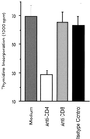
Proliferative responses of splenocytes from infected mice in the presence of anti-CD4 or anti-CD8 antibodies. C57BL/6 mice were infected i.p. with 2 × 10⁸ CFU of B. henselae for 8 weeks. Spleen cells were stimulated with 106 CFU of HKBH/ml for 3 days in the presence of 20 μg of MAbs against CD4, CD8, or an isotype-matched control/ml or in the absence of antibodies. Results represent mean counts per minute ± standard errors (three animals). The mean spontaneous proliferation rate without stimulation was 11,170 ± 2,708 cpm.
FIG. 5
FIG. 5
Kinetics of Bartonella-specific proliferation of spleen cells from infected mice. Spleen cell proliferation assays were performed as described for Fig. 3. SIs were calculated by dividing the mean counts per minute of wells (triplicates) containing antigen (HKBH at the indicated concentrations) by the mean counts per minute for wells without antigen. SRs were calculated by dividing the SI of each infected mouse by the mean SI of three uninfected control mice tested simultaneously. Results represent mean counts per minute ± standard errors.
FIG. 6
FIG. 6
IFN-γ secretion by spleen cells from B. henselae-infected mice. C57BL/6 mice were injected i.p. with 2 × 10⁸ CFU of B. henselae or PBS. At 12 weeks p.i., five animals per group were euthanized and spleens were collected. Spleen cells were stimulated in vitro for 48 h with 104 to 107 CFU of HKBH/ml or medium only, and IFN-γ release was determined by ELISA. Results are means ± standard errors. Asterisk, P < 0.05.
FIG. 7
FIG. 7
Production of B. henselae-specific IgG antibodies in infected mice. C57BL/6 mice were injected i.p. with 2 × 10⁸ CFU of B. henselae or PBS. At indicated time points, five animals per group were euthanized and serum samples were subjected to OMP ELISA. Results are means ± standard errors. OD 450, optical density at 450 nm.
FIG. 8
FIG. 8
Analysis of IgG isotypes among Bartonella-specific serum antibodies in infected mice. C57BL/6 mice were infected i.p. with 2 × 10⁸ CFU of B. henselae for 4 weeks. Serum samples were serially diluted and analyzed by OMP ELISA. Results are means ± standard errors (five animals per group). OD 450, optical density at 450 nm.

Similar articles

Cited by

References

    1. Anderson B, Sims K, Regnery R, Robinson L, Schmidt M J, Goral S, Hager C, Edwards K. Detection of Rochalimaea henselae DNA in specimens from cat scratch disease patients by PCR. J Clin Microbiol. 1994;32:942–948. - PMC - PubMed
    1. Anderson B E, Neuman M A. Bartonella spp. as emerging human pathogens. Clin Microbiol Rev. 1997;10:203–219. - PMC - PubMed
    1. Arvand M, Klose A J, Schwartz-Porsche D, Hahn H, Wendt C. Genetic variability and prevalence of Bartonella henselae in cats in Berlin, Germany, and analysis of its genetic relatedness to a strain from Berlin that is pathogenic for humans. J Clin Microbiol. 2001;39:743–746. - PMC - PubMed
    1. Arvand M, Wendt C, Regnath T, Ullrich R, Hahn H. Characterization of Bartonella henselae isolated from bacillary angiomatosis lesions in a human immunodeficiency virus-infected patient in Germany. Clin Infect Dis. 1998;26:1296–1299. - PubMed
    1. Batterman H J, Peek J A, Loutit J S, Falkow S, Tompkins L S. Bartonella henselae and Bartonella quintana adherence to and entry into cultured human epithelial cells. Infect Immun. 1995;63:4553–4556. - PMC - PubMed

MeSH terms