. 2001 Sep;12(9):2567-77.
doi: 10.1091/mbc.12.9.2567.

ASH1 mRNA localization in three acts

Affiliations
Free PMC article

ASH1 mRNA localization in three acts

D L Beach et al. Mol Biol Cell. 2001 Sep.
Free PMC article

Abstract

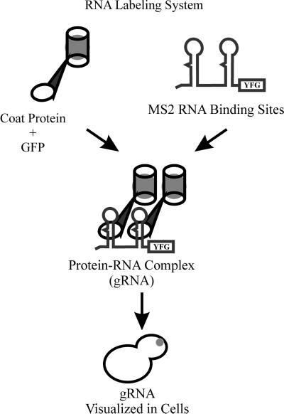
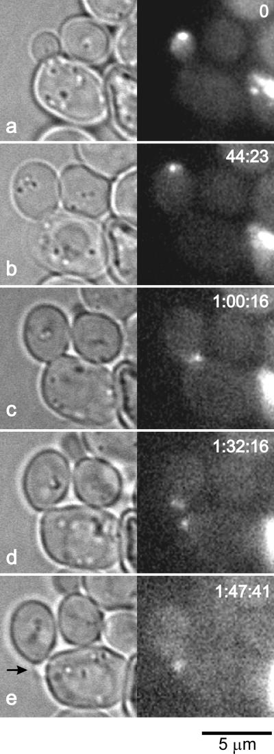
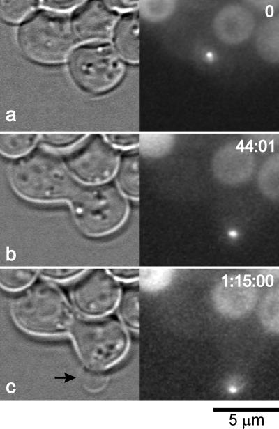
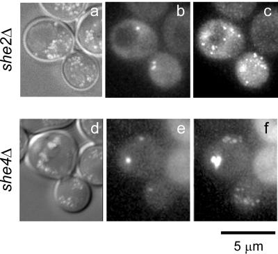
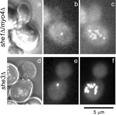
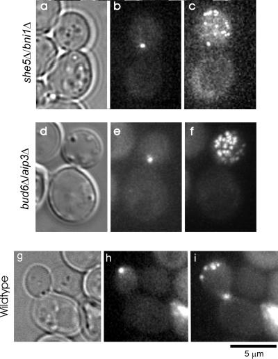
Novel green fluorescent protein (GFP) labeling techniques targeting specific mRNA transcripts reveal discrete phases of mRNA localization in yeast: packaging, transport, and docking. In budding yeast, ASH1 mRNA is translocated via actin and myosin to the tip of growing cells. A GFP-decorated reporter transcript containing the ASH1 3' untranslated region gRNA(ASH1) forms spots of fluorescence localized to a cortical domain at the bud tip, relocates to the mother-bud neck before cell separation, and finally migrates to the incipient bud site before the next budding cycle. The correct positioning of the mRNA requires at least six proteins: She1p-5p and Bud6p/Aip3p. gRNA(ASH1) localization in mutant strains identified three functional categories for the She proteins: mRNA particle formation (She2p and She4p), mRNA transport into the bud (She1p/Myo4p and She3p), and mRNA tethering at the bud tip (She5p/Bni1p and Bud6p/Aip3p). Because localization of the mRNA within the bud does not a priori restrict the translated protein, we examine the distribution of a mother-specific protein (Yta6p) translated from a mRNA directed into the bud. Yta6p remains associated with the mother cortex despite localization of the mRNA to the bud. This video essay traces the life history of a localized mRNA transcript, describes the roles of proteins required to polarize and anchor the mRNA, and demonstrates at least one instance where mRNA localization does not effect protein localization.


Figures

Figure 1
Figure 1
RNA labeling system. The RNA labeling system is comprised of two components: a site-specific RNA binding protein fused to GFP and an mRNA transcript containing the target sequence for the RNA binding protein. This system uses the MS2 coat protein and the associated RNA binding site. A mRNA transcript including “Your Favorite Gene” (YFG) and the MS2 coat protein binding site is fluorescently labeled when coexpressed with the MS2 coat protein fused to GFP. Localized transcripts can be visualized by standard fluorescence microscopy.
Figure 2
Figure 2
gRNAASH1 localization in vegetative, haploid cells. gRNAASH1 localization is dynamic, changing as the cell proceeds through the cell cycle. In small- (a) to large (b)-sized buds the gRNAASH1 is localized to the bud tip, moving on the cortex within a region at the bud tip termed the cortical bud cap. (c) gRNAASH1 migrates to the neck of large budded cells 25 min before cell separation. (d) After cell separation, the gRNAASH1 remains at the old bud site in both the mother and the bud. (e) Approximately 10 min before bud emergence (arrow) the gRNAASH1 moves to the incipient bud site then remains at the bud tip during early growth. Numbers in the upper right corner in each frame represent an elapsed time corresponding with video sequence 1. Bar, 5 μm.
Figure 3
Figure 3
gRNAASH1 localization in mating cells. gRNAASH1 localizes to regions of polarized growth throughout the mating cycle. The two cells centered in the frame (MATa with gRNAASH1 is left, top and MATα is right, bottom) proceed through mating and generate a diploid bud. (a) Before cell fusion, the gRNAASH1 is at the mating projection tip nearest the cell of opposite mating type. The GFP signal in the left, top (MATa) cell is elevated compared with levels in the bottom, right cell (MATα). (b) After cell fusion and before bud emergence, the gRNAASH1 spot is localized at the incipient bud site (compare b and c). (c) gRNAASH1 is at the tip of the first diploid bud. Numbers in the upper right corner of each frame represent an elapsed time corresponding with video sequence 3. Bar, 5 μm.
Figure 4
Figure 4
She2p and She4p are required for gRNAASH1 spot assembly. gRNAASH1 localization in she2Δ (a–c, g, and h), and she4Δ (d-f, i, and j) cells. (a–c) gRNAASH1 aggregates into spots in both the mother and the bud in she2Δ cells. A single she2Δ cell is shown imaged via DIC (a) and GFP fluorescence (b). The composite image (c) represents 20 consecutive time points at a 1-min interval to illustrate dynamic spot movements not apparent within single images (video sequence 3). (d–f) Several spots distributed between mother and but were also observed in she4Δ cells. A single she4Δ cell is shown imaged via Nomarski (a) and GFP fluorescence (b). The composite image (c) represents 20 consecutive time points at a 1-min interval to illustrate dynamic spot movements not apparent within single images (video sequence 4). Bar, 5 μm.
Figure 5
Figure 5
She1p/Myo4p and She3p facilitate gRNAASH1 transport. In the absence of either She1p/Myo4p or She3p, gRNAASH1 remains in the mother cell. (a–c) Nomarski (a), single time point (b), and composite image (c) of a she1Δ/myo4Δ cell are shown (video sequence 5). The single gRNAASH1 spot is centrally located within the mother. The composite image (c) demonstrates the spot motility in the absence of the myosin motor. Twenty sequential images >20 min are presented in the single composite image (see text). (d–f) Nomarski (d), single time point (e), and composite image (f) of a she3Δ cell are shown (video sequence 6). As seen for she1 cells, the single gRNAASH1 spot is localized in the mother domain (below the bud). The composite image (f) represents 20 sequential images and the movement of a single spot >20 min. Bar, 5 μm.
Figure 6
Figure 6
She5p/Bni1p and Bud6p/Aip3p anchor the gRNAASH1 at the bud tip. The retention of the gRNAASH1 at the bud tip is lost in she5Δ/bni1Δ and bud6Δ/aip3Δ cells. (a–c) In she5Δ/bni1Δ cells, the gRNAASH1 is transported into the bud and moves freely throughout the bud. (a) Brightfield image of the cell shown in b and c. (b) A single frame from the time lapse (video sequence 7) is shown, demonstrating a position of the gRNAASH1 proximal to the neck. (c) Composite time-lapse image is shown representing 20 sequential images, showing the distribution of the gRNAASH1 spot >20 min. (d–f) As observed for she5Δ/bni1Δ cells, gRNAASH1 is transported into the bud in bud6Δ/aip3Δ cells, and moves freely throughout the bud. (d) Brightfield image of the cell shown in e and f. (e) Single frame from the time lapse (video sequence 8) demonstrating a position of the gRNAASH1 proximal to the neck. (f) Composite time-lapse image is shown representing 20 sequential images showing the distribution of the gRNAASH1 spot >20 min. (g–i) Wild-type (YEF473a) cells are shown to compare gRNAASH1 localization distal from the neck (h) and as a composite image (i) representing 20 sequential images >20 min. (g) Brightfield image of the wild-type cell shown in h and i. Bar, 5 μm.
Figure 7
Figure 7
GFP-Yta6 localizes to the maternal cortex. (a–e) Several cells are shown with GFP-Yta6 spots on the mother cell cortex. Medium budded cells (a–d) contain GFP-Yta6 only in the mother cell, while the large budded cell (e) contains GFP-Yta6 in both mother and bud domains. (a′–e′) The same cells 150 min later. Cells b–e have completed cell separation with Yta6p present in the daughter cells. Cell a′ has not completed cell separation, yet Yta6p is localized in the bud. White lines denote the cell outline as determined from the brightfield image. Video sequence 9 demonstrates the accumulation of YTA6p in the bud late in the cell cycle and includes panels showing the nuclear cycle (Histone H4-CFP). (f and g) The localization of Yta6-ASH1 fusion protein and mRNA is shown in a cell expressing GFP-Yta6-ASH1 3′ UTR fusion and CP-GFP (video sequence 10). The mRNA is localized to the bud tip throughout the time lapse (f and g), whereas the Yta6-Ash1 fusion protein is only in the mother in small budded cells (f) and in both mother and bud in large budded cells (g). Numbers in the lower right corner (f and g) represent an elapsed time (hh:mm) corresponding with video sequence 10. Bars (top and bottom), 5 μm.

Similar articles

Cited by

References

    1. Adams AEM, Pringle JR. Relationship of actin and tubulin distribution to bud growth in wild-type and morphogenetic-mutant Saccharomyces cerevisiae. J Cell Biol. 1984;98:934–945. - PMC - PubMed
    1. Ainger K, Avossa D, Morgan F, Hill SJ, Barry C, Barbarese E, Carson JH. Transport and localization of exogenous myelin basic protein mRNA microinjected into oligodendrocytes. J Cell Biol. 1993;123:431–441. - PMC - PubMed
    1. Amberg DC, Zahner JE, Mulholland JW, Pringle JR, Botstein D. Aip3p/Bud6p, a yeast actin-interacting protein that is involved in morphogenesis and the selection of bipolar budding sites. Mol Biol Cell. 1997;8:729–753. - PMC - PubMed
    1. Barral Y, Mermall V, Mooseker M, Snyder M. Compartmentalization of the cell cortex by septins is required for maintenance of cell polarity in yeast. Mol Cell. 2000;5:841–851. - PubMed
    1. Beach DL, Salmon ED, Bloom K. Localization and anchoring of mRNA in budding yeast. Curr Biol. 1999;9:569–578. - PubMed

Publication types

MeSH terms