. 2001 Sep;12(9):2578-89.
doi: 10.1091/mbc.12.9.2578.

Dynamin GTPase domain mutants block endocytic vesicle formation at morphologically distinct stages

Affiliations
Free PMC article

Dynamin GTPase domain mutants block endocytic vesicle formation at morphologically distinct stages

H Damke et al. Mol Biol Cell. 2001 Sep.
Free PMC article

Abstract

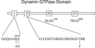
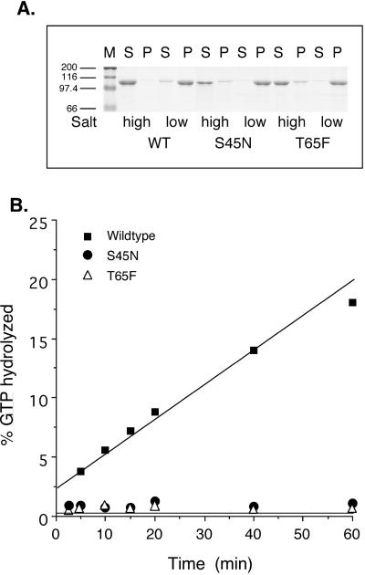
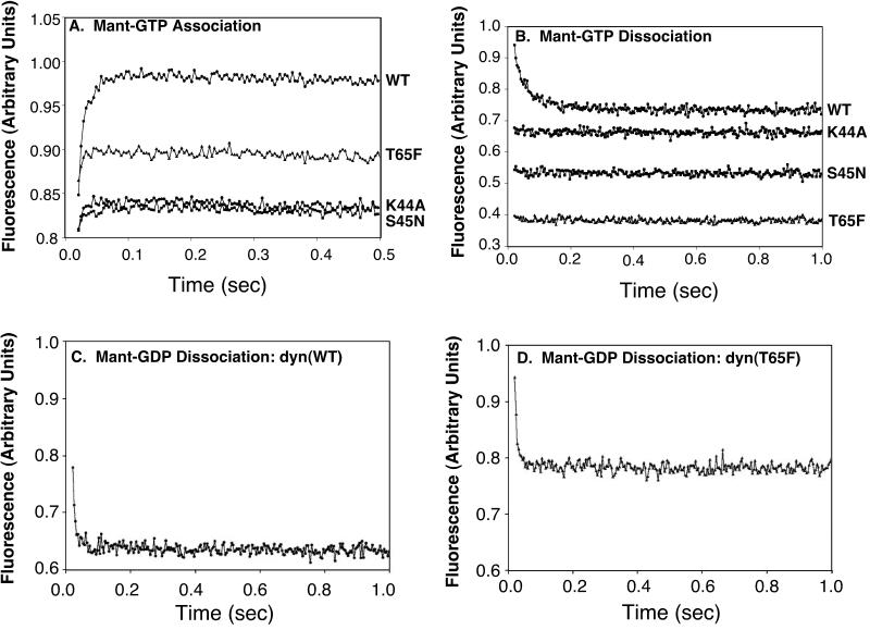
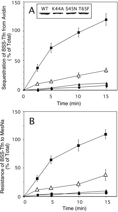
Abundant evidence has shown that the GTPase dynamin is required for receptor-mediated endocytosis, but its exact role in endocytic clathrin-coated vesicle formation remains to be established. Whereas dynamin GTPase domain mutants that are defective in GTP binding and hydrolysis are potent dominant-negative inhibitors of receptor-mediated endocytosis, overexpression of dynamin GTPase effector domain (GED) mutants that are selectively defective in assembly-stimulated GTPase-activating protein activity can stimulate the formation of constricted coated pits and receptor-mediated endocytosis. These apparently conflicting results suggest that a complex relationship exists between dynamin's GTPase cycle of binding and hydrolysis and its role in endocytic coated vesicle formation. We sought to explore this complex relationship by generating dynamin GTPase mutants predicted to be defective at distinct stages of its GTPase cycle and examining the structural intermediates that accumulate in cells overexpressing these mutants. We report that the effects of nucleotide-binding domain mutants on dynamin's GTPase cycle in vitro are not as predicted by comparison to other GTPase superfamily members. Specifically, GTP and GDP association was destabilized for each of the GTPase domain mutants we analyzed. Nonetheless, we find that overexpression of dynamin mutants with subtle differences in their GTPase properties can lead to the accumulation of distinct intermediates in endocytic coated vesicle formation.


Figures

Figure 1
Figure 1
Mutations within dyn GTPase domain. Point mutations were introduced within the nucleotide-binding region to interfere with dyn's nucleotide-binding properties and biological function. Shown are the positions of the mutations within the first of three GTP-binding elements conserved among GTPase family members.
Figure 2
Figure 2
Biochemical characteristics of dyn GTPase domain mutants. (A) A sedimentation assay, described in MATERIALS AND METHODS, was used to analyze the ability of S45N and T65F mutant dyn compared with WT dyn to self-assemble into structures that remain soluble (S) or pelletable (P) after dilution into low salt conditions. The self-assembly activity of dyn(S45N) and dyn(T65F) was indistinguishable from dyn(WT). M=marker. (B) The assembly-stimulated GTPase activity of WT and mutant dyns was measured, as described in MATERIALS AND METHODS, with the use of taxol-stabilized microtubules (2 μg) as a template for assembly. GTPase activity of dyn(K44A), dyn(S45N), and dyn(T65F) was severely impaired relative to dyn(WT).
Figure 3
Figure 3
Guanine nucleotide association and dissociation from WT and mutant dyns. Mant-GTP association (A) and dissociation (B) are plotted in arbitrary units for fluorescence over time. In A, WT and mutant dyn-1 (1 μM final concentration) were mixed with Mant-GTP (60 μM final concentration). In B, 1 μM dyn-1 and 4.0 μM Mant-GTP were mixed with 100 μM unlabeled GTP (final concentrations). Mant-GDP dissociation rates were measured by mixing 1 μM dyn(WT) (C) and dyn(T65F) (D) loaded with 4 μM Mant-GDP with 100 μM GTP (final concentrations). See MATERIALS AND METHODS for experimental details.
Figure 4
Figure 4
Tfn internalization is inhibited in cells stably expressing dyn(S45N) and dyn(T65F). Stable tTA-HeLa cells expressing WT dyn (▪), dyn(K44A) (▴), dyn(S45N) (●), or dyn(T65F) (▵) were induced for 48 h by removal of tet. The inset in A indicates the expression levels for exogenous dyn from a single representative assay as analyzed by immunoblotting. Internalization of Tfn was measured by incubating cells with 8 μg/ml BSS-Tfn for the indicated times at 37°C before measuring intracellular Tfn based on its inaccessibility to avidin (A) or MesNa (B) (see MATERIALS AND METHODS). Results are the averages ± SD of five independent experiments with comparable expression levels for dyn-1.
Figure 5
Figure 5
At equal expression levels dyn(T65F) is a weaker inhibitory mutant than dyn(S45N). tTAHela cells were infected with Ad (10 MOI/cell) for WT dyn, dyn(S45N), or dyn(T65F) in the absence or presence of low amounts (2–10 ng/ml) of tet. Cells were processed for internalization of BSS-Tfn and avidin accessibility 16 h after infection as described previously. The expression levels for exogenous dyn were controlled by immunoblot analysis of equal number of cells and quantified by densitometric analysis. Shown are data from four independent experiments. Matching expression levels for HA-dynS45N and T65F of these experiments were plotted versus the percentage of endocytosis. Endocytosis in dyn WT cells at equal expression level is 100%.
Figure 6
Figure 6
EM analysis of coated profiles in cells expressing WT and mutant dyns. Shown is a gallery of representative deep coated pits from tTA-HeLa cells infected with Ad encoding for dyn(WT), dyn(K44A), dyn(S45N), and dyn(T65F). tTA HeLa cells were infected with 10 MOI of virus in the absence of tet resulting in ≥ 95% of cells expressing dyn. Cells were incubated 16 h after infection with gold-conjugated monoclonal anti-human TfnR antibody (D65-gold, 10 μg/ml) for 2 min at 37°C before fixation, staining, and embedding in Epon for thin-section analysis as described in MATERIALS AND METHODS. Scale bar, 100 nm. The images for WT represent deep pits (a-d). For K44A, the first three images (a–c) are scored as deep pits, whereas the rightmost (D) was scored as elongated, i.e., the neck is longer than the diameter of the pit. For S45N, a and b are deep and c and d are elongated pits. For T65F a is a deep pit, and b–d are elongated pits.
Figure 7
Figure 7
Differential accumulation of endocytic intermediates in cells expressing dyn(K44A), dyn(S45N), or dyn(T65F). Data for various structures derived from the quantitation shown in Table 1 are expressed as percentages of total coated profiles for each condition.
Figure 8
Figure 8
(A) Coated pits with elongated necks accumulate in cells expressing GTPase domain mutant dyns. Ruthenium red staining shows elongated necks that accumulate in mutant cells. For labeling of invaginated membranes with Ruthenium red, tTA-HeLa cells were infected with Ad for WT or mutant dyn-1 and incubated in the absence of tet for 16 h. The cells were fixed in the presence of 0.5 mg/ml Ruthenium red and processed as described in MATERIALS AND METHODS. Scale bar, 500 nm. (B) Shown are some rarely observed examples of necks of elongated coated pits encircled by an electron-dense collar-like material. Whereas such structures were never detected in WT cells, they were observed in cells expressing each of the dyn GTPase domain mutants. These images are taken from stably transformed tTA-HeLa cells induced to express the indicated mutant dyns by removal of tet. Scale bar, 100 nm.

References

    1. Altschuler Y, Barbas S, Terlecky L, Mostov K, Schmid SL. Common and distinct functions for dynamin-1 and dynamin-2 isoforms. J Cell Biol. 1998;143:1871–1881. - PMC - PubMed
    1. Barylko B, Binns D, Lin KM, Atkinson MA, Jameson DM, Yin HL, Albanesi JP. Synergistic activation of dynamin GTPase by Grb2 and phosphoinositides. J Biol Chem. 1998;273:3791–3797. - PubMed
    1. Binns DD, Barylko B, Grichine N, Adkinson AL, Helms MK, Jameson DM, Eccleston JF, Albanesi JP. Correlation between self-association modes and GTPase activation of dynamin. J Protein Chem. 1999;18:277–290. - PubMed
    1. Binns DD, Helms MK, Barylko B, Davis CT, Jameson DM, Albanesi JP, Eccleston JF. The mechanism of GTP hydrolysis by dynamin II: a transient kinetic study. Biochemistry. 2000;39:7188–7196. - PubMed
    1. Bourne HR, Sanders DA, McCormick F. The GTPase superfamily: a conserved switch for diverse cell functions. Nature. 1990;348:125–132. - PubMed

Publication types

MeSH terms

LinkOut - more resources